
南方医科大学学报 ›› 2026, Vol. 46 ›› Issue (9): 2023-2034.doi: 10.12122/j.issn.1673-4254.2026.09.03
• • 上一篇
叶丽旋1( ), 廖雨欣2, 刘琼2, 张佳2, 高磊2, 董钊扬1()
收稿日期:2026-01-20
出版日期:2026-09-20
发布日期:2026-09-30
通讯作者:
董钊扬
E-mail:2669556676@qq.com;021092@ gzucm.edu.cn
作者简介:叶丽旋,在读硕士研究生,E-mail: 2669556676@qq.com
基金资助:
Lixuan YE1(), Yuxin LIAO2, Qiong LIU2, Jia ZHANG2, Lei GAO2, Zhaoyang DONG1()
Received:2026-01-20
Online:2026-09-20
Published:2026-09-30
Contact:
Zhaoyang DONG
E-mail:2669556676@qq.com;021092@ gzucm.edu.cn
Supported by:摘要:
目的 探讨逍遥散(XYS)在秀丽隐杆线虫(C. elegans)帕金森病(PD)模型中的神经保护作用及其分子机制。 方法 采用6-羟基多巴胺(6-OHDA)诱导建立PD线虫模型,并结合α-突触核蛋白(α-syn)过表达转基因虫株(NL5901)进行验证。将线虫分为对照组、模型组及XYS低(25 μg/mL)、中(50 μg/mL)、高(100 μg/mL)剂量组。通过生存曲线分析评估寿命;采用20 s内身体弯曲次数及基础减慢反应分别评价线虫的运动功能和食物相关行为;利用GFP标记的转基因虫株观察多巴胺能神经元损伤及α-syn聚集水平;采用二氢乙啶荧光探针(DHE)检测体内活性氧(ROS)水平,并结合RT-qPCR分析dat-1、cat-2、ced-9、gst-4及sod-3等相关基因表达;通过RNA干扰(RNAi)沉默skn-1表达,以验证XYS作用是否依赖于SKN-1/Nrf2信号通路。 结果 XYS可显著延长PD模型线虫寿命(P<0.001),增加第5天和第10天的20 s内身体弯曲次数(P<0.05),并改善基础减慢反应(P<0.05);XYS可减轻6-OHDA诱导的多巴胺能神经元损伤,提高dat-1、cat-2及ced-9表达水平(P<0.05),并降低α-syn聚集水平(P<0.001)。此外,XYS处理后降低ROS水平(P<0.05),sod-3及gst-4等抗氧化相关基因表达上调(P<0.05)。机制研究显示,XYS可增强skn-1::GFP及sod-3::GFP荧光信号(P<0.05)。在skn-1 RNAi条件下,脂质含量下降(P<0.05),且XYS对上述改变无明显改善作用(P>0.05);同时,XYS对sod-3::GFP荧光降低的恢复作用消失(P>0.05)。 结论 XYS可减轻氧化应激并抑制α-syn异常聚集,从而改善秀丽隐杆线虫PD模型神经功能,其作用可能与SKN-1/Nrf2相关抗氧化通路有关。
叶丽旋, 廖雨欣, 刘琼, 张佳, 高磊, 董钊扬. 逍遥散通过SKN-1抗氧化通路改善秀丽隐杆线虫帕金森病模型α-突触核蛋白聚集和神经元损伤[J]. 南方医科大学学报, 2026, 46(9): 2023-2034.
Lixuan YE, Yuxin LIAO, Qiong LIU, Jia ZHANG, Lei GAO, Zhaoyang DONG. Xiaoyao San alleviates abnormal α‑syn aggregation and neuronal injury in a Caenorhabditis elegans model of Parkinson disease via the SKN-1 antioxidative pathway[J]. Journal of Southern Medical University, 2026, 46(9): 2023-2034.
| Time (min) | Flow (mL/min) | % B |
|---|---|---|
| 0.00 | 0.30 | 5.0 |
| 2.00 | 0.30 | 5.0 |
| 18.00 | 0.30 | 95.0 |
| 20.00 | 0.30 | 95.0 |
表1 色谱梯度程序
Tab.1 Chromatography gradient program
| Time (min) | Flow (mL/min) | % B |
|---|---|---|
| 0.00 | 0.30 | 5.0 |
| 2.00 | 0.30 | 5.0 |
| 18.00 | 0.30 | 95.0 |
| 20.00 | 0.30 | 95.0 |
| Gene | Forward primer (5'-3') | Reverse primer (5'-3') |
|---|---|---|
| Dat-1 | TTGGTGCCTACAGACGATCC | GCTGTTCCGTCTTCTGACCA |
| Cat-2 | ACTGAAGAACCTGCGGTACG | CCGATGGTGATTGCTCCTGC |
| Gst-4 | GATGCTCGTGCTCTTGCTGA | GGAGTCGTTGGCTTCAGCTT |
| Ced-9 | TGCTCAGGACTTGCCATCAC | GCGTGAAATAGTCGACCACA |
| Sod-3 | ATCTACTGCTCGCACTGCTT | ACGTAGGTGGCATGATGCTT |
| β-actin | ACGACGAGTCCGGCCCATCC | GAAAGCTGGTGGTGACGATGGTT |
表2 目的基因引物序列
Tab.2 Primer sequences of target genes
| Gene | Forward primer (5'-3') | Reverse primer (5'-3') |
|---|---|---|
| Dat-1 | TTGGTGCCTACAGACGATCC | GCTGTTCCGTCTTCTGACCA |
| Cat-2 | ACTGAAGAACCTGCGGTACG | CCGATGGTGATTGCTCCTGC |
| Gst-4 | GATGCTCGTGCTCTTGCTGA | GGAGTCGTTGGCTTCAGCTT |
| Ced-9 | TGCTCAGGACTTGCCATCAC | GCGTGAAATAGTCGACCACA |
| Sod-3 | ATCTACTGCTCGCACTGCTT | ACGTAGGTGGCATGATGCTT |
| β-actin | ACGACGAGTCCGGCCCATCC | GAAAGCTGGTGGTGACGATGGTT |
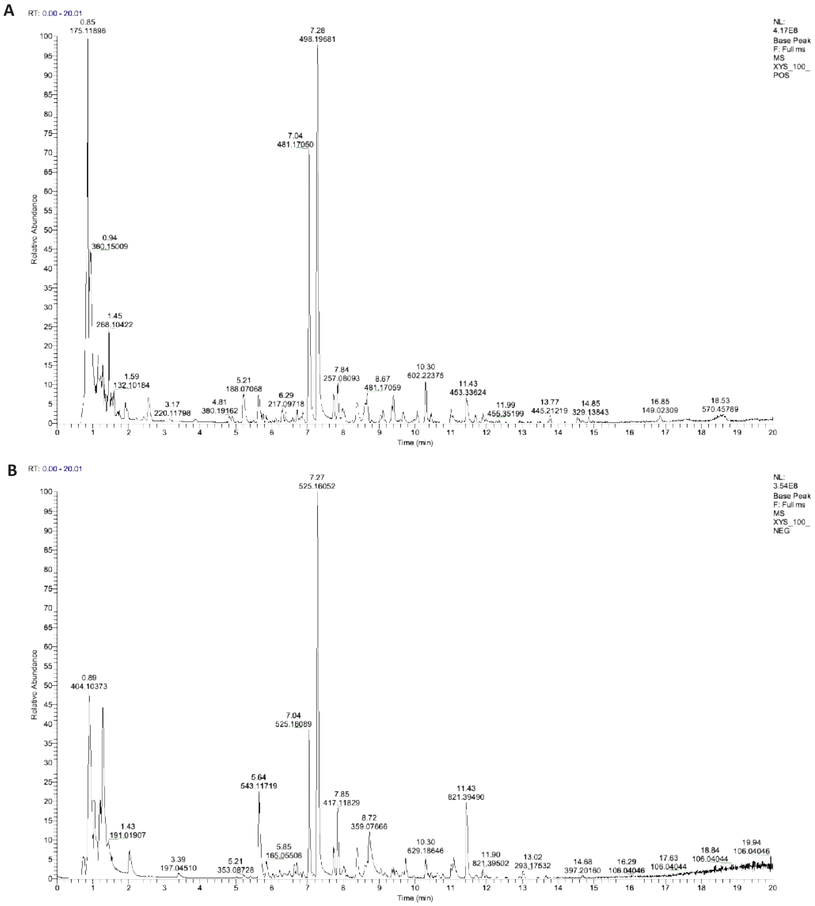
图1 UHPLC-HRMS总离子流图
Fig.1 UHPLC-HRMS total ion chromatogram (TIC) of Xiaoyao San (XYS). A: Positive ion mode (POS). B: Negative ion mode (NEG).
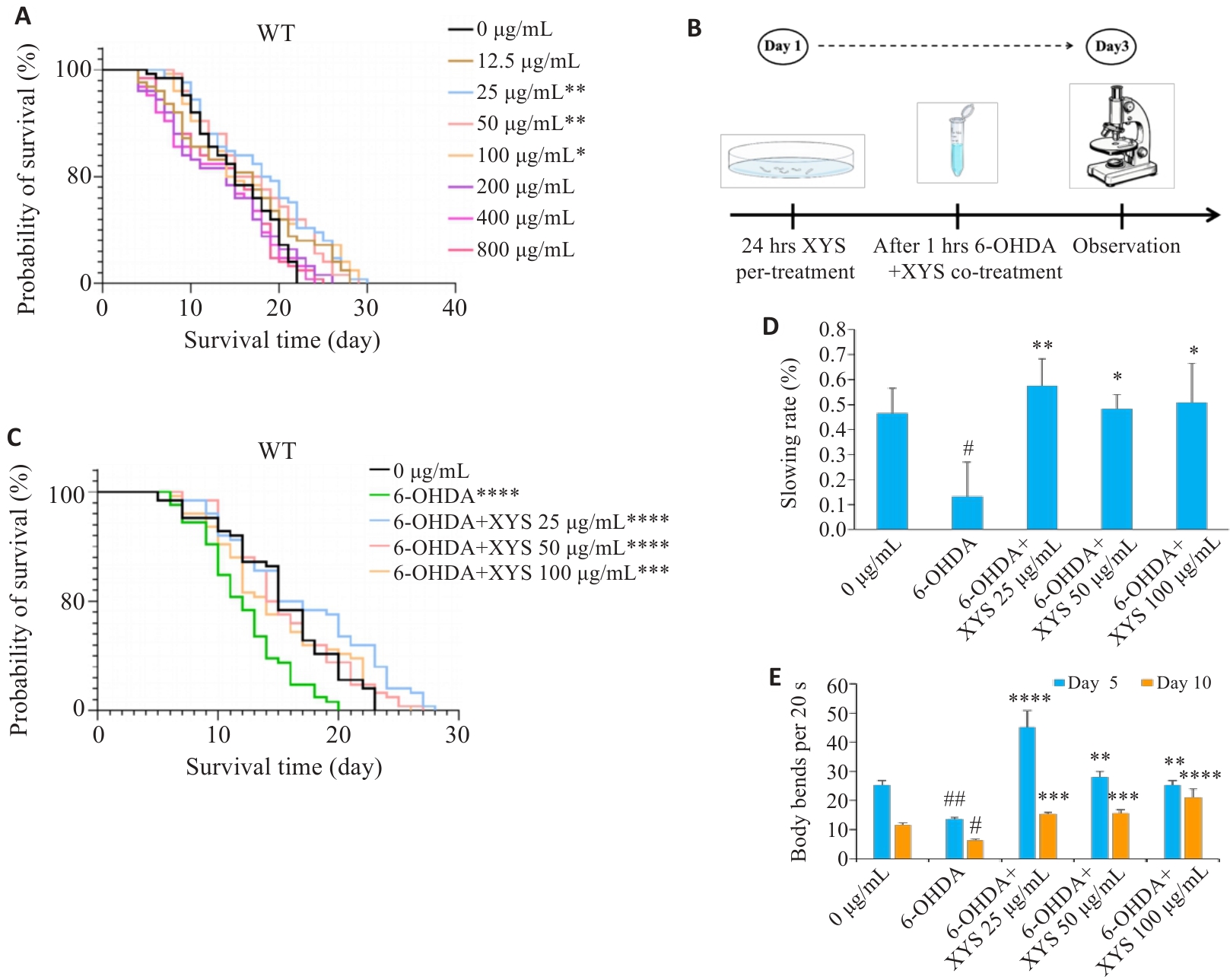
图2 XYS干预能够延长PD线虫寿命并改善其活动能力
Fig.2 XYS extends lifespan and improves locomotor activity of C. elegans PD models. A: Survival curves of C. elegans exposed to different concentrations of XYS. B: Schematic diagram of the experimental design for combined 6-OHDA and XYS treatment. C: Survival curves of 6-OHDA-induced PD model worms with or without XYS treatment. D: Quantification of the basal slowing response. E: Quantification of body bends within 20 s on days 5 and 10 post-treatment. Data represent Mean±SD from at least 3 independent biological replicates (n=50 for lifespan assays and n=30 for other assays). #P<0.05, ##P<0.01 vs control group; *P<0.05, **P<0.01, ***P<0.001, ****P<0.0001 vs model group.
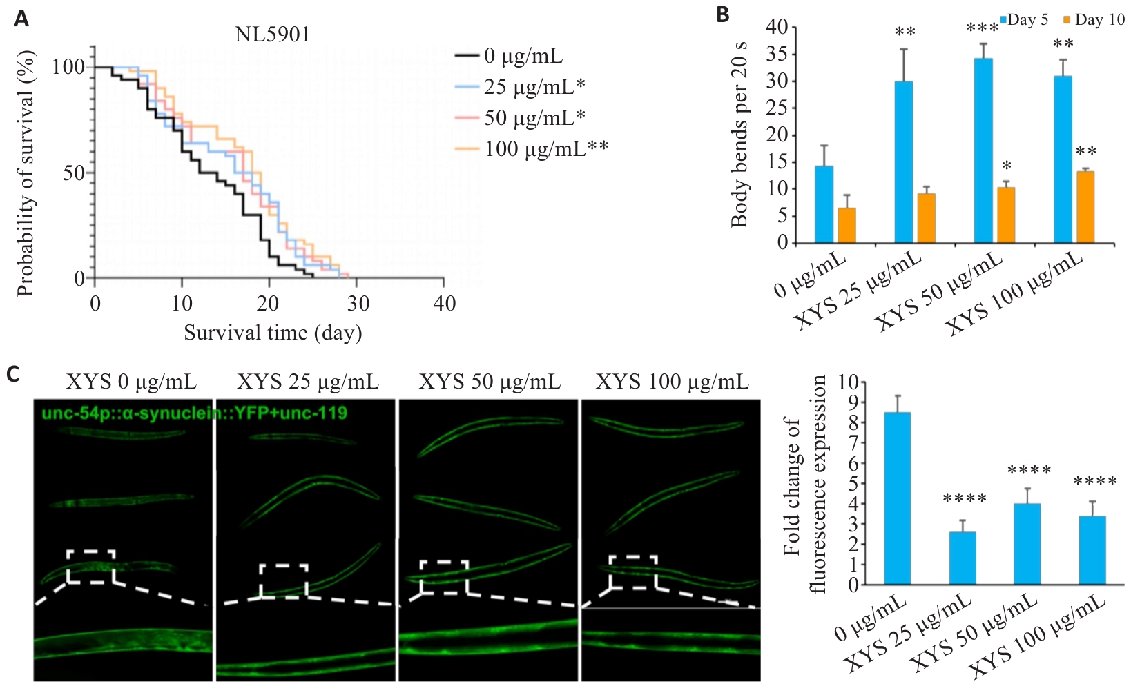
图3 XYS干预能够延长PD转基因模型NL5901线虫寿命并改善其体内的α-syn聚集
Fig.3 XYS extends lifespan and reduces α-syn aggregation in NL5901 worms. A: Survival curves of NL5901 worms with or without XYS treatment. B: Quantification of body bends within 20 s on days 5 and 10 post-treatment. C: Representative images of α‑syn::YFP fluorescence in body wall muscle cells and quantification of fluorescence intensity (Scale bar=100 μm). Data represent Mean±SD from at least 3 independent biological replicates (n=50 for lifespan assays and n=30 for other assays). *P<0.05, **P<0.01, ***P<0.001, ****P<0.0001 vs control group.
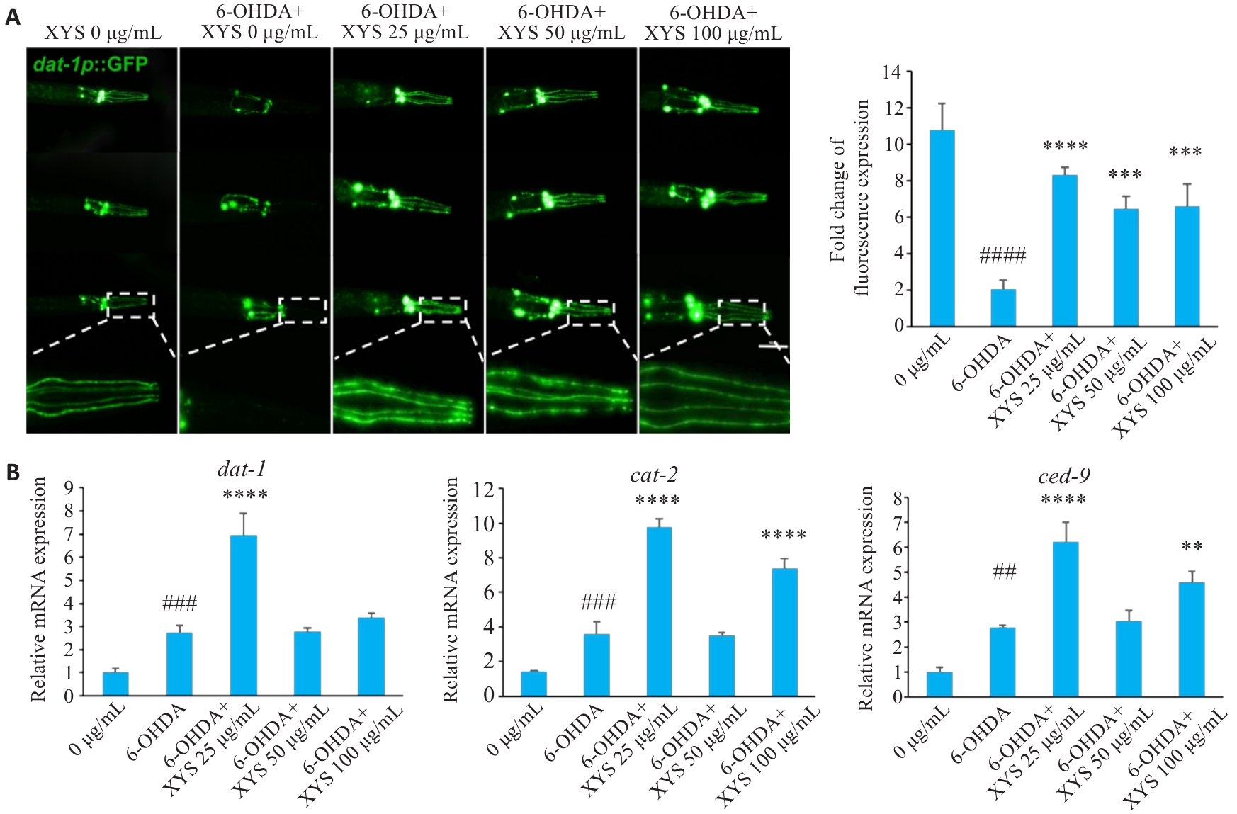
图4 XYS干预显著改善6-OHDA诱导的PD线虫模型多巴胺能神经元变性
Fig.4 XYS protects against 6-OHDA-induced dopaminergic neurodegeneration in C. elegans. A: Representative GFP images of dopaminergic neurons with or without XYS treatment and quantification of fluorescence intensity (Scale bar=100 μm). B: Relative mRNA expression levels of dat-1, cat-2, and ced-9. Data represent Mean±SD from at least three independent biological replicates (n=30 for fluorescence analysis and n=3 for qPCR analysis). ##P<0.01, ###P<0.001, ####P<0.0001 vs control group; **P<0.01, ***P<0.001, ****P<0.0001 vs model group.
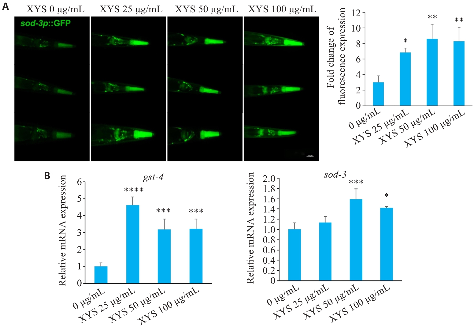
图5 XYS干预能够显著改善线虫体内抗氧化效应
Fig.5 XYS significantly enhances antioxidant capacity in C. elegans. A: Representative fluorescence images of sod-3::GFP worms and quantification of sod-3::GFP fluorescence intensity (Scale bar=100 μm). B: Relative mRNA expression levels of sod-3 and gst-4. Data represent Mean±SD from at least 3 independent biological replicates (n=30 for fluorescence analysis and n=3 for qPCR analysis). *P<0.05, **P<0.01, ***P<0.001, ****P<0.0001 vs control group.
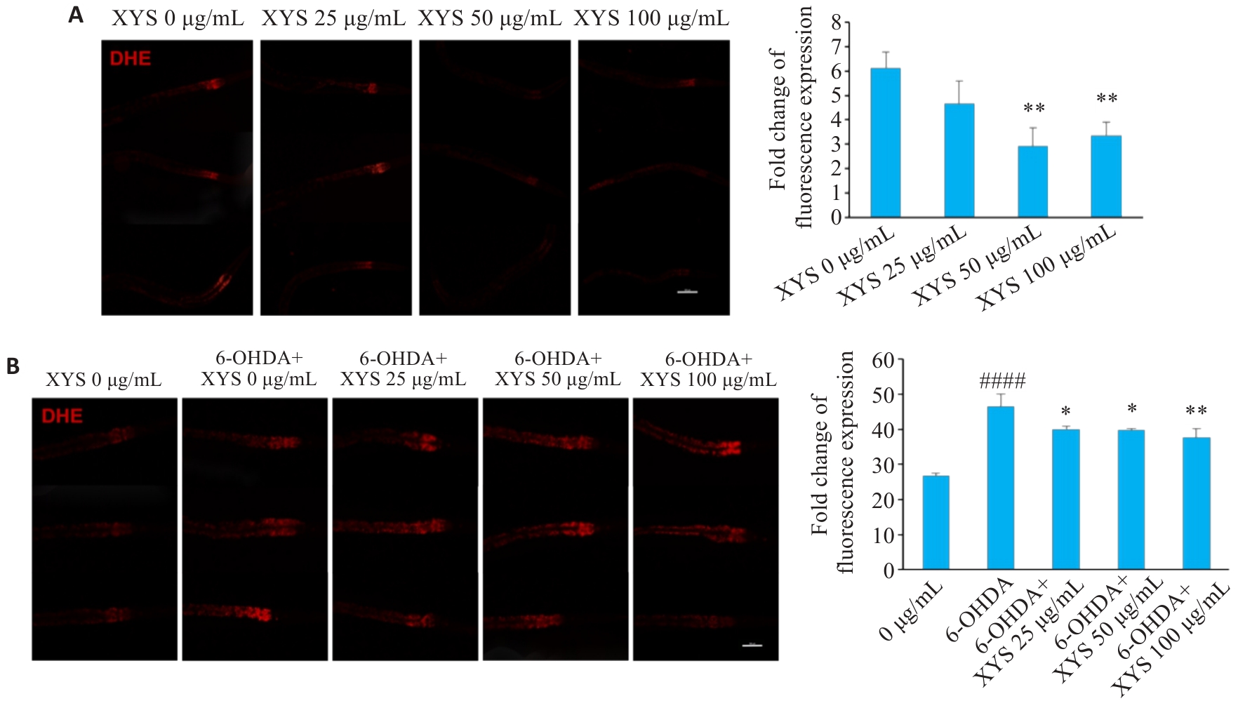
图6 XYS干预能够显著降低不同PD线虫模型体内氧化应激水平
Fig.6 XYS significantly reduces oxidative stress levels in different C. elegans models of PD. A: Representative DHE staining images of NL5901 worms with or without XYS treatment and quantification of DHE fluorescence intensity. B: Representative DHE staining images of 6-OHDA-treated N2 worms with or without XYS treatment and quantification of DHE fluorescence intensity. Scale bar=100 μm. Data represent Mean±SD from at least 3 independent biological replicates (n=30). ####P<0.0001 vs control group; *P<0.05, **P<0.01 vs model group.
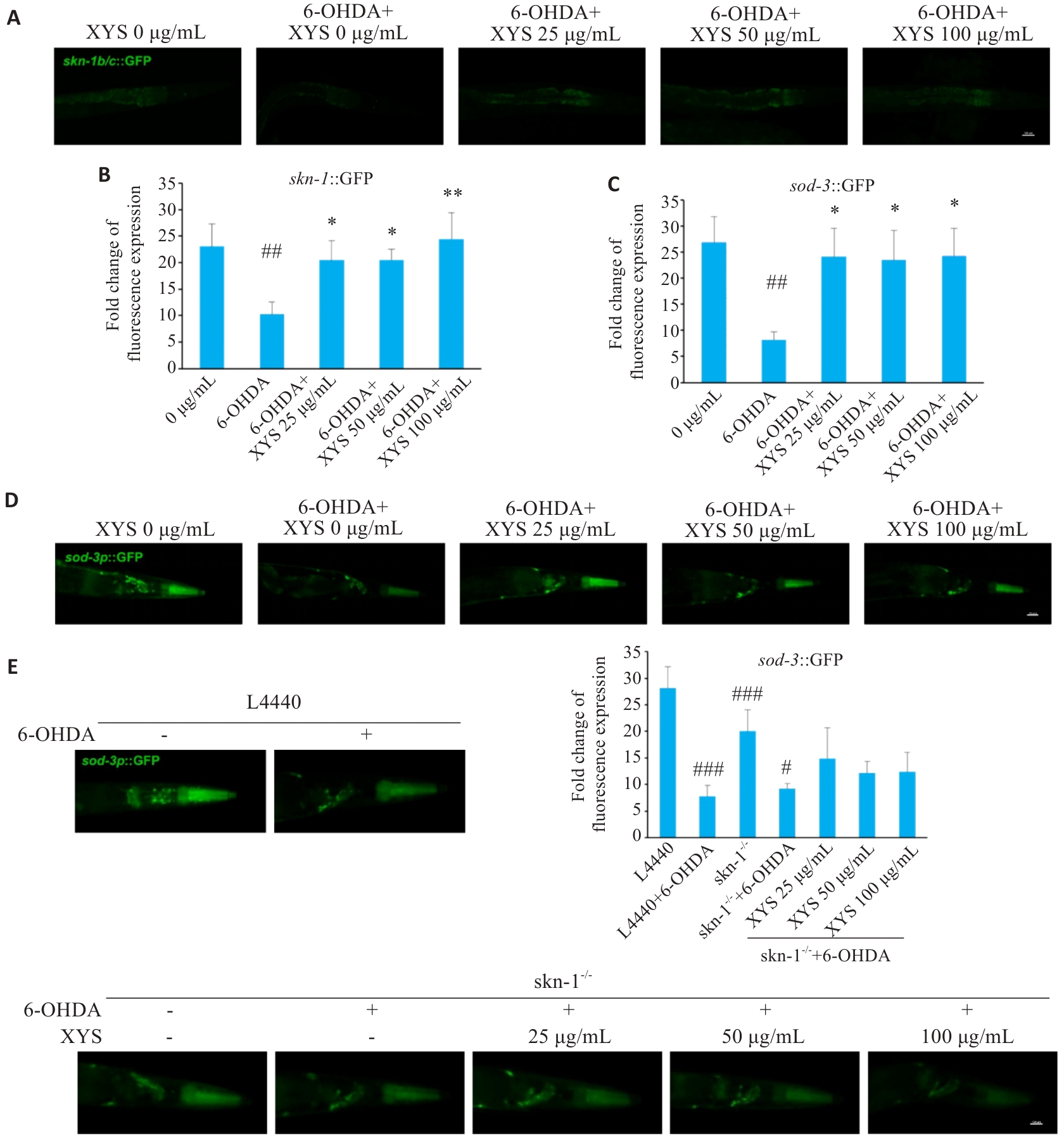
图7 XYS通过上调SKN-1改善PD线虫模型抗氧化效应
Fig.7 XYS improves antioxidant responses in PD C. elegans models through upregulation of SKN-1. A: Representative fluorescence images of LD1 worms. B: Quantification of fluorescence intensity in LD1 worms. C: Quantification of fluorescence intensity in CF1553 worms. D: Representative fluorescence images of CF1553 worms. E: Representative fluorescence images of skn-1 RNAi-treated worms and quantification of fluorescence intensity. Scale bar=100 μm. Data represent Mean±SD from at least 3 independent biological replicates (n=30). ##P<0.01, ###P<0.001 vs control group; #P<0.05 vs skn-1 RNAi group; *P<0.05, **P<0.01 vs model group.
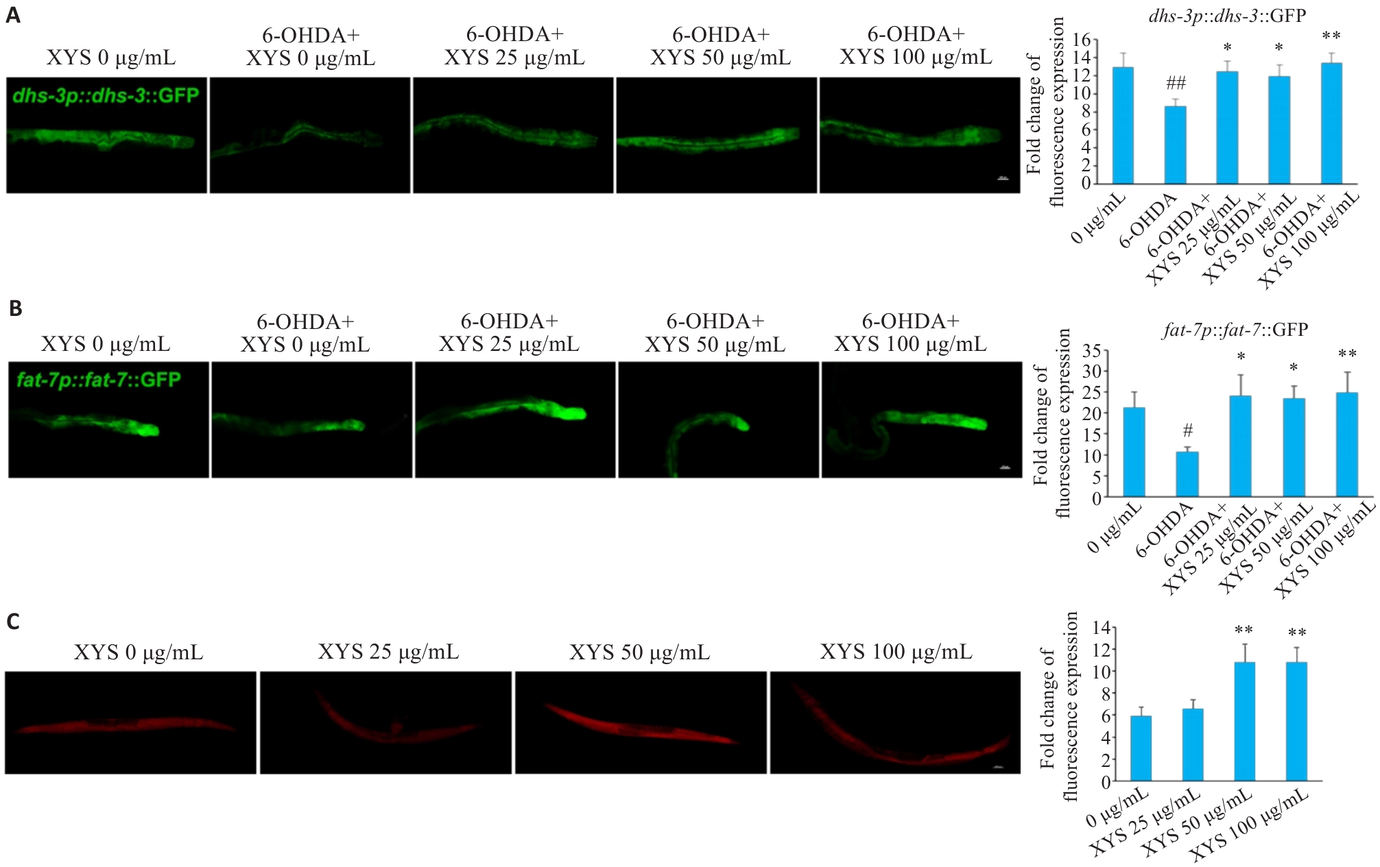
图8 XYS干预能够改善PD线虫模型脂质代谢异常
Fig.8 XYS ameliorates lipid metabolic dysfunction in PD C. elegans models. A: Representative fluorescence images of LIU104 worms and quantification of fluorescence intensity. B: Representative fluorescence images of DMS303 worms and quantification of fluorescence intensity. C: Representative Nile Red staining images of NL5901 worms and quantification of fluorescence intensity. Scale bar=100 μm. Data represent Mean±SD from at least 3 independent biological replicates (n=30). #P<0.05, ##P<0.01 vs control group; *P<0.05, **P<0.01 vs model group.
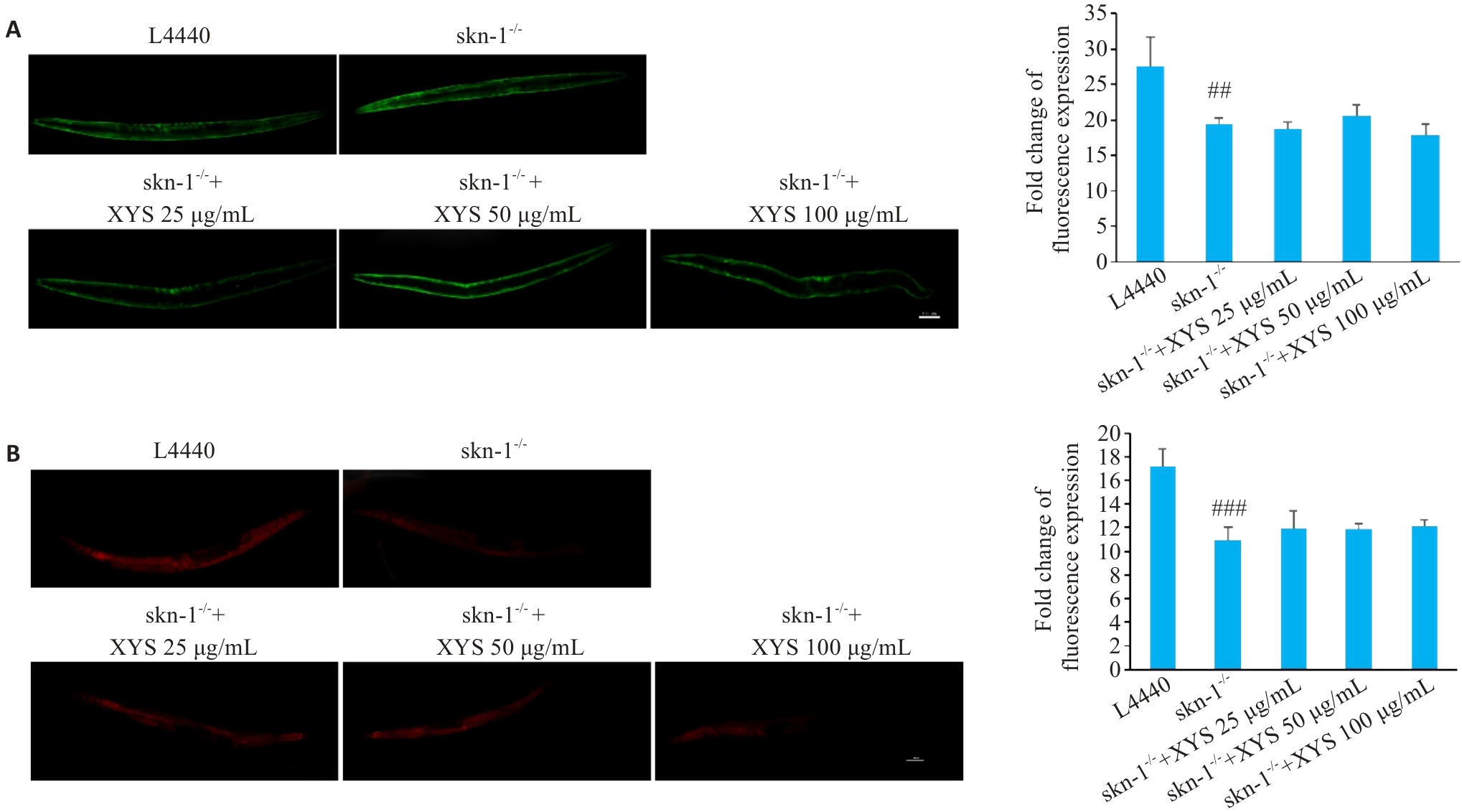
图9 XYS通过skn-1调节α-syn蓄积及脂质积累
Fig.9 XYS regulates α-synuclein accumulation and lipid deposition via skn-1. A: Representative fluorescence images of skn-1 RNAi worms and quantification of fluorescence intensity. B: Representative Nile Red staining images of skn-1 RNAi worms and quantification of lipid accumulation based on Nile Red staining. Scale bar=100 μm. Data represent Mean±SD from at least 3 independent biological replicates (n=30). ##P<0.01, ###P<0.001 vs control group.
| [1] | Tanner CM, Ostrem JL. Parkinson's disease[J]. N Engl J Med, 2024, 391(5): 442-52. doi:10.1056/nejmra2401857 |
| [2] | Chen Y, Luo XX, Yin YK, et al. The interplay of iron, oxidative stress, and α-synuclein in Parkinson's disease progression[J]. Mol Med, 2025, 31(1): 154. doi:10.1186/s10020-025-01208-3 |
| [3] | Blackwell TK, Steinbaugh MJ, Hourihan JM, et al. SKN-1/Nrf, stress responses, and aging in Caenorhabditis elegans [J]. Free Radic Biol Med, 2015, 88: 290-301. doi:10.1016/j.freeradbiomed.2015.06.008 |
| [4] | Chen JB, Lei CF, Li XJ, et al. Research progress on classical traditional Chinese medicine formula Xiaoyaosan in the treatment of depression[J]. Front Pharmacol, 2022, 13: 925514. doi:10.3389/fphar.2022.925514 |
| [5] | Liu XY, Yan YZ, Jiang AJ, et al. Xiaoyao San against depression via Glia-mediated neuroinflammation using network pharmacology and experimental validation[J]. Sci Rep, 2025, 15: 39708. doi:10.1038/s41598-025-23296-1 |
| [6] | Wei YY, Lin WF, Zhang TH, et al. Effectiveness of traditional Chinese medicineas as an adjunct therapy for refractory schizophrenia: a systematic review and meta analysis[J]. Sci Rep, 2018, 8: 6230. doi:10.1038/s41598-018-24547-0 |
| [7] | Chen JX, Xu JK, Huang P, et al. The potential applications of traditional Chinese medicine in Parkinson's disease: a new opportunity[J]. Biomed Pharmacother, 2022, 149: 112866. doi:10.1016/j.biopha.2022.112866 |
| [8] | Zhang Y, Xu XM. Chinese herbal medicine in the treatment of depression in Parkinson's disease: from molecules to systems[J]. Front Pharmacol, 2022, 13: 879459. doi:10.3389/fphar.2022.879459 |
| [9] | Xu Y, Chen WY, Chen ZR, et al. Mechanism of action of Xiaoyao San in treatment of ischemic stroke is related to anti-apoptosis and activation of PI3K/Akt pathway[J]. Drug Des Devel Ther, 2021, 15: 753-67. doi:10.2147/dddt.s280217 |
| [10] | Magalingam KB, Radhakrishnan A, Haleagrahara N. Protective effects of quercetin glycosides, rutin, and isoquercetrin against 6-hydroxydopamine (6-OHDA)‑induced neurotoxicity in rat pheo-chromocytoma (PC-12) cells[J]. Int J Immunopathol Pharmacol, 2016, 29(1): 30-9. doi:10.1177/0394632015613039 |
| [11] | Singh S, Kumar P. Piperine in combination with quercetin halt 6-OHDA induced neurodegeneration in experimental rats: Biochemical and neurochemical evidences[J]. Neurosci Res, 2018, 133: 38-47. doi:10.1016/j.neures.2017.10.006 |
| [12] | Hu K, Zhu SS, Wu FY, et al. Aureusidin ameliorates 6-OHDA-induced neurotoxicity via activating Nrf2/HO-1 signaling pathway and preventing mitochondria-dependent apoptosis pathway in SH-SY5Y cells and Caenorhabditis elegans [J]. Chem Biol Interact, 2024, 387: 110824. doi:10.1016/j.cbi.2023.110824 |
| [13] | Tamuli R, Mellick GD, Schirra HJ, et al. Mode of action of toxin 6-hydroxydopamine in SH-SY5Y using NMR metabolomics[J]. Molecules, 2025, 30(16): 3352. doi:10.3390/molecules30163352 |
| [14] | Kimble J, Nüsslein-Volhard C. The great small organisms of developmental genetics: Caenorhabditis elegans and Drosophila melanogaster [J]. Dev Biol, 2022, 485: 93-122. doi:10.1016/j.ydbio.2022.02.013 |
| [15] | Ma JY, Li KL, Shi SL, et al. The application of UHPLC-HRMS for quality control of traditional Chinese medicine[J]. Front Pharmacol, 2022, 13: 922488. doi:10.3389/fphar.2022.922488 |
| [16] | 陈周雯, 任雨敏, 唐 鲜, 等. 西南手参醇提物通过抗氧化途径延缓秀丽隐杆线虫衰老的机制[J]. 食品科学, 2026, 47(6): 214-25. doi:10.7506/spkx1002-6630-20250910-079 |
| [17] | Offenburger SL, Ho XY, Tachie-Menson T, et al. 6-OHDA-induced dopaminergic neurodegeneration in Caenorhabditis elegans is promoted by the engulfment pathway and inhibited by the transthyretin-related protein TTR-33[J]. PLoS Genet, 2018, 14(1): e1007125. doi:10.1371/journal.pgen.1007125 |
| [18] | Tsai RT, Tsai CW, Liu SP, et al. Maackiain ameliorates 6-hydroxydopamine and SNCA pathologies by modulating the PINK1/parkin pathway in models of Parkinson's disease in Caenorhabditis elegans and the SH-SY5Y cell line[J]. Int J Mol Sci, 2020, 21(12): 4455. doi:10.3390/ijms21124455 |
| [19] | Palikaras K, SenGupta T, Nilsen H, et al. Assessment of dopaminergic neuron degeneration in a C. elegans model of Parkinson's disease[J]. STAR Protoc, 2022, 3(2): 101264. doi:10.1016/j.xpro.2022.101264 |
| [20] | Balkrishna A, Pathak N, Singh R, et al. Neurogrit gold attenuates 6-OHDA-induced dopaminergic neurodegeneration in Parkinson's model of Caenorhabditis elegans by reducing α‑synuclein accumulation and pink/pdr-1 driven mitochondrial dysfunction[J]. CNS Neurosci Ther, 2025, 31(5): e70401. doi:10.1111/cns.70401 |
| [21] | Sawin ER, Ranganathan R, Horvitz HR. C. elegans locomotory rate is modulated by the environment through a dopaminergic pathway and by experience through a serotonergic pathway[J]. Neuron, 2000, 26(3): 619-31. doi:10.1016/s0896-6273(00)81199-x |
| [22] | Picca A, Calvani R, Coelho-Junior HJ, et al. Mitochondrial dysfunction, oxidative stress, and neuroinflammation: intertwined roads to neurodegeneration[J]. Antioxidants, 2020, 9(8): 647. doi:10.3390/antiox9080647 |
| [23] | Fu RH, Tsai CW, Liu SP, et al. Neuroprotective capability of narcissoside in 6-OHDA-exposed Parkinson's disease models through enhancing the MiR200a/nrf-2/GSH axis and mediating MAPK/Akt associated signaling pathway[J]. Antioxidants, 2022, 11(11): 2089. doi:10.3390/antiox11112089 |
| [24] | Kwon TG, Kim YJ, Hong JY, et al. A review of antidepressant and anxiolytic effects of Soyo-San (Xiaoyao-San) and modified Soyo-San in animal models[J]. Phytomedicine, 2024, 135: 155387. doi:10.1016/j.phymed.2024.155387 |
| [25] | Bastien BL, Haury WR, Smisko WR, et al. Nlr-1/CNTNAP regulates dopamine circuit structure and foraging behaviors in C. elegans[J]. Commun Biol, 2024, 7: 1248. doi:10.1038/s42003-024-06936-6 |
| [26] | Lee I, Knickerbocker AC, Depew CR, et al. Effect of altered production and storage of dopamine on development and behavior in C. elegans[J]. Front Toxicol, 2024, 6: 1374866. doi:10.3389/ftox.2024.1374866 |
| [27] | Jeong H, Park JY, Lee JH, et al. Deficiency in RCAT-1 function causes dopamine metabolism related behavioral disorders in Caenorhabditis elegans [J]. Int J Mol Sci, 2022, 23(4): 2393. doi:10.3390/ijms23042393 |
| [28] | Maulik M, Mitra S, Bult-Ito A, et al. Behavioral phenotyping and pathological indicators of Parkinson's disease in C. elegans models[J]. Front Genet, 2017, 8: 77. doi:10.3389/fgene.2017.00077 |
| [29] | Abidar S, Nhiri M, Hritcu L. Phytochemicals against the 6-hydroxydopamine-induced Parkinson's disease in rodents: a systematic review[J]. Next Res, 2025, 2(3): 100473. doi:10.1016/j.nexres.2025.100473 |
| [30] | Kim KH, Lee SY, Shin J, et al. Dose-dependent neuroprotective effect of standardized bee venom phospholipase a(2) against MPTP-induced Parkinson's disease in mice[J]. Front Aging Neurosci, 2019, 11: 80. doi:10.3389/fnagi.2019.00080 |
| [31] | Liu XJ, Zheng ZJ, Xue C, et al. LRRK2 mediates α‑synuclein-induced neuroinflammation and ferroptosis through the p62-Keap1-Nrf2 pathway in Parkinson's disease[J]. Inflammation, 2025, 48(5): 3666-91. doi:10.1007/s10753-025-02291-8 |
| [32] | Chakraborty S, Bornhorst J, Nguyen TT, et al. Oxidative stress mechanisms underlying Parkinson's disease-associated neuro-degeneration in C. elegans[J]. Int J Mol Sci, 2013, 14(11): 23103-28. doi:10.3390/ijms141123103 |
| [33] | Phan HD, Nguyen TTM, Lee S, et al. The metabolic contribution of SKN-1/Nrf2 to the lifespan of Caenorhabditis elegans [J]. Metabolomics, 2023, 19(6): 58. doi:10.1007/s11306-023-02022-w |
| [34] | Pang SS, Lynn DA, Lo JY, et al. SKN-1 and Nrf2 couples proline catabolism with lipid metabolism during nutrient deprivation[J]. Nat Commun, 2014, 5: 5048. doi:10.1038/ncomms6048 |
| [35] | Zhao CY, Tu J, Wang CC, et al. Lysophosphatidylcholine binds α‑synuclein and prevents its pathological aggregation[J]. Natl Sci Rev, 2024, 11(6): nwae182. doi:10.1093/nsr/nwae182 |
| [36] | Lastres-Becker I, Ulusoy A, Innamorato NG, et al. α‑Synuclein expression and Nrf2 deficiency cooperate to aggravate protein aggregation, neuronal death and inflammation in early-stage Parkinson's disease[J]. Hum Mol Genet, 2012, 21(14): 3173-92. doi:10.1093/hmg/dds143 |
| [37] | Idi W, Sheta R, Oueslati A. Interplay between lipid droplets and alpha-synuclein: implication in Parkinson's disease pathogenesis[J]. Front Mol Neurosci, 2025, 18: 1681039. doi:10.3389/fnmol.2025.1681039 |
| [38] | Coles NP, Elsheikh S, Quesnel A, et al. Alpha-synuclein aggregation induces prominent cellular lipid changes as revealed by Raman spectroscopy and machine learning analysis[J]. Brain Commun, 2025, 7(2): fcaf133. doi:10.1093/braincomms/fcaf133 |
| [1] | 石科林, 齐霄川, 徐世祥, 高缘圆, 房明哲, 胡俊诚, 王春景, 刘长青, 郭俣. CHIR99021通过抑制GSK-3β/NF-κB信号通路减轻帕金森病小鼠的神经炎症[J]. 南方医科大学学报, 2026, 46(7): 1593-1600. |
| [2] | 侯玲, 唐莉莉, 陈清, 孙径汇, 陈小娟, 李炜, 赵杨. 温肾养肝方通过调节肠道生物活性物质改善帕金森病模型小鼠的肠道功能[J]. 南方医科大学学报, 2026, 46(5): 1131-1138. |
| [3] | 陈丹丹, 任乾千, 吕梦林, 张宝文, 刘醒然, 张蒙, 王阳, 寇现娟. 天麻钩藤饮通过抑制坏死性凋亡通路改善帕金森病小鼠的运动功能障碍[J]. 南方医科大学学报, 2025, 45(8): 1571-1580. |
| [4] | 王飞霞, 张政, 孙艳, 杨柳菁, 郭桐彤, 潘夜厅, 丁嵩涛, 蒋林, 刘含登. Bmal1可能介导了丁酸钠对帕金森病小鼠的神经保护作用[J]. 南方医科大学学报, 2024, 44(5): 876-884. |
| [5] | 桂建军, 孙晓东, 温 舒, 刘 欣, 覃冰清, 桑 明. 白藜芦醇对帕金森病模型小鼠多巴胺能神经元的保护作用:基于抑制TLR4/MyD88/NF-κB通路[J]. 南方医科大学学报, 2024, 44(2): 270-279. |
| [6] | 姜诚诚, 李洋洋, 段可欣, 詹婷婷, 陈子龙, 王永雪, 赵蕊, 马彩云, 郭俣, 刘长青. Parkin通过介导PINK 1/Parkin线粒体自噬信号通路加速小鼠帕金森病发展及加剧神经炎症发生[J]. 南方医科大学学报, 2024, 44(12): 2359-2366. |
| [7] | 任自敬, 周佩洋, 田静. 基于ceRNA芯片研究帕金森病患者血浆长链非编码RNA表达谱并验证lnc-CTSD-5∶1在PD细胞模型中的作用[J]. 南方医科大学学报, 2024, 44(11): 2146-2155. |
| [8] | 徐佳佳, 李洋洋, 仲光尚, 方祝玲, 刘淳博, 马彩云, 王春景, 郭 俣, 刘长青. 人诱导多能干细胞体外定向分化为多巴胺能神经元祖细胞[J]. 南方医科大学学报, 2023, 43(2): 175-182. |
| [9] | 石 皓, 张 超, 赵嘉敏, 李逸文, 李允家, 李俊杰, 曾志云, 高 磊. 寒冷胁迫可降低秀丽隐杆线虫的寿命和活动能力:通过介导脂代谢紊乱和应激反应异常[J]. 南方医科大学学报, 2022, 42(8): 1159-1165. |
| [10] | 黄河灵, 高玉元, 聂 坤, 王丽娟. 巨噬细胞移动抑制因子介导MPP+/MPTP诱导的小胶质细胞NLRP3炎症小体的激活[J]. 南方医科大学学报, 2021, 41(7): 972-979. |
| [11] | 牟斐斐, 陈 曦, 杜希恂, 焦 倩, 毕明霞, 姜 宏. 帕金森病模型中α-突触核蛋白对干扰素调节因子1的调控及其机制[J]. 南方医科大学学报, 2021, 41(11): 1641-1648. |
| [12] | 郑小斌,余良宏,万欣龙,王惠清,俞 挺,何 秋,林章雅,康德智. 颅内电极重建在帕金森病患者脑深部刺激治疗中的应用价值[J]. 南方医科大学学报, 2019, 39(12): 1461-1468. |
| [13] | 赵茸,王天仲,狄政莉,杨军乐,徐敏,刘志勤,朱旭蓉,邬小平,高晓宇. 基于体素分析的动脉自旋标记技术在帕金森病脑血流量中的应用[J]. 南方医科大学学报, 2018, 38(01): 117-. |
| [14] | 周溦,林敬明,王素丽,林海桢. 半枝莲总黄酮对延缓秀丽隐杆线虫和人脐静脉内皮细胞衰老的影响[J]. 南方医科大学学报, 2017, 37(06): 821-. |
| [15] | 范玲玲,邓博,闫君宝,胡志红,任爱红,胡咏梅,杨东伟. 5-羟色胺-7受体激动剂对帕金森病模型大鼠内侧前额叶皮层锥体神经元兴奋性的影响[J]. 南方医科大学学报, 2016, 36(06): 756-. |
| 阅读次数 | ||||||
|
全文 |
|
|||||
|
摘要 |
|
|||||
