南方医科大学学报 ›› 2026, Vol. 46 ›› Issue (4): 880-889.doi: 10.12122/j.issn.1673-4254.2026.04.16
• • 上一篇
徐陈陈( ), 韩永升, 程楠, 董健健()
收稿日期:2025-10-24
出版日期:2026-04-20
发布日期:2026-04-24
通讯作者:
董健健
E-mail:senorita@ ahtcm.edu.cn;jjdong@ahtcm.edu.cn
作者简介:徐陈陈,主治医师,硕士,E-mail: senorita@ ahtcm.edu.cn
基金资助:
Chenchen XU(), Yongsheng HAN, Nan CHENG, Jianjian DONG()
Received:2025-10-24
Online:2026-04-20
Published:2026-04-24
Contact:
Jianjian DONG
E-mail:senorita@ ahtcm.edu.cn;jjdong@ahtcm.edu.cn
Supported by:摘要:
目的 通过建立PKR沉默Wilson病模型毒牛奶(TX)小鼠,探讨通腑养髓治法(MGDD)通过PKR/eIF2α通路介导的突触损伤对TX小鼠认知功能的改善机制。 方法 将36只TX小鼠随机分为TX组、TX+MGDD组、C16组(PKR抑制剂干预组)及C16+MGDD组,9只/组。C16组与C16+MGDD组先腹腔注射PKR抑制剂C16(300 μg/kg,持续30 d),之后分别灌胃生理盐水或MGDD;TX组与TX+MGDD组则分别灌胃等体积生理盐水或MGDD,持续4周。行为学测试(巴恩斯迷宫与旷场实验)后取脑组织,进行免疫荧光、TUNEL染色、透射电镜(TEM)、RT-qPCR及Western blotting分析。 结果 行为学方面,与C16组相比,C16+MGDD组在边上路程减少(P<0.05),但在中央时间、中央路程、边上时间及目标洞口潜伏期等方面差异无统计学意义。免疫荧光显示,C16+MGDD组阳性细胞数较C16组减少,与TX+MGDD组相近;同时,MGDD治疗的TX小鼠在C16干预前后8-OHdG阳性细胞数差异无统计学意义。TEM观察显示,MGDD或C16干预均能增加突触与囊泡数量,改善突触膜结构清晰度,但MGDD联合C16未产生叠加效应。Western blotting结果显示,C16+MGDD组突触相关蛋白PSD93、PSD95、Synapsin1和Synaptophysin表达较C16组上调(P<0.05);与TX+MGDD组相比,除PSD93外,其余蛋白表达有差异(P<0.05)。在PKR/eIF2α通路方面,RT-qPCR显示C16+MGDD组Pkr、eIF2alpha等mRNA表达与C16组差异无统计学意义;Western blotting提示C16+MGDD组P-eIF2α与CHOP蛋白水平下降,而P-CREB上升,P-PKR与ATF4变化差异无统计学意义。 结论 通腑养髓治法可能通过抑制PKR/eIF2α通路活性,促进突触相关蛋白表达,改善突触结构与功能,从而缓解Wilson病TX小鼠的认知功能障碍。
徐陈陈, 韩永升, 程楠, 董健健. 通腑养髓治法通过抑制PKR介导的突触损伤改善Wilson病TX小鼠认知功能[J]. 南方医科大学学报, 2026, 46(4): 880-889.
Chenchen XU, Yongsheng HAN, Nan CHENG, Jianjian DONG. Modified Gandou Decoction improves cognitive function of TX mice with Wilson's disease by inhibiting the PKR/eIF2α pathway[J]. Journal of Southern Medical University, 2026, 46(4): 880-889.
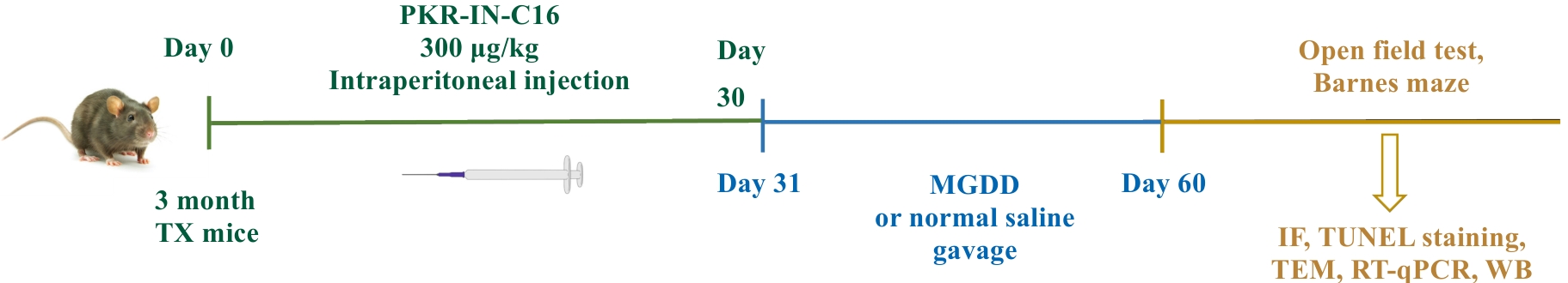
图1 PKR沉默Wilson病模型构建及实验流程图
Fig.1 Construction of the PKR-silenced Wilson's disease model and the experimental flow chart.
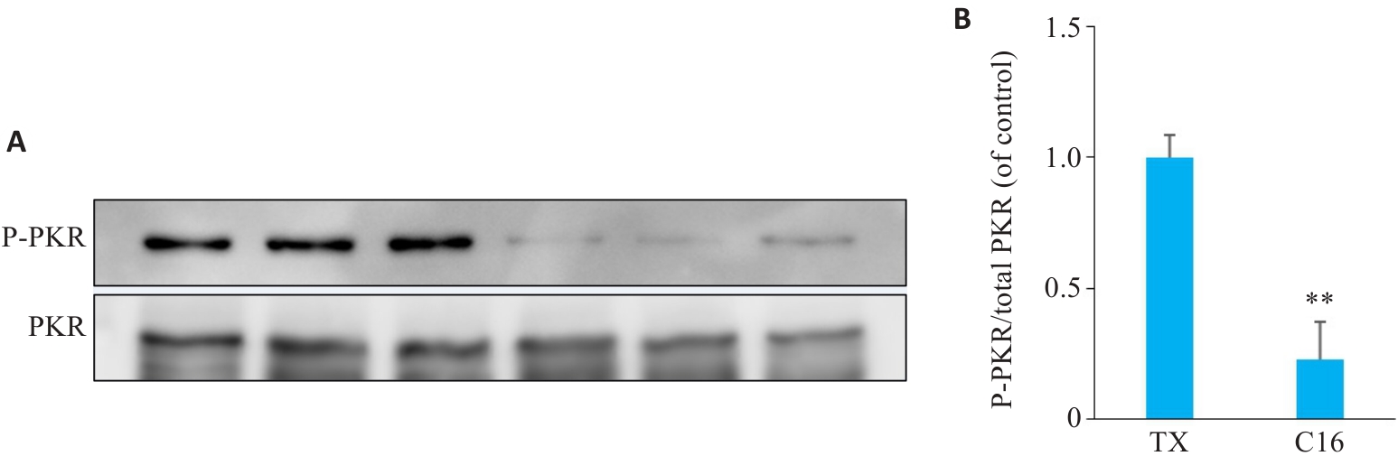
图2 PKR抑制剂C16对TX小鼠海马PKR磷酸化的抑制作用
Fig.2 Inhibitory effect of C16, a PKR inhibitor, on phosphorylation of PKR in the hippocampus of TX mice. A: Gray-scale band images of phosphorylated PKR and total PKR in the hippocampus of mice. B: Expression levels of phosphorylated PKR and total PKR in the different groups quantified using Western blotting (Mean±SD, n=3). **P<0.01 vs TX group.
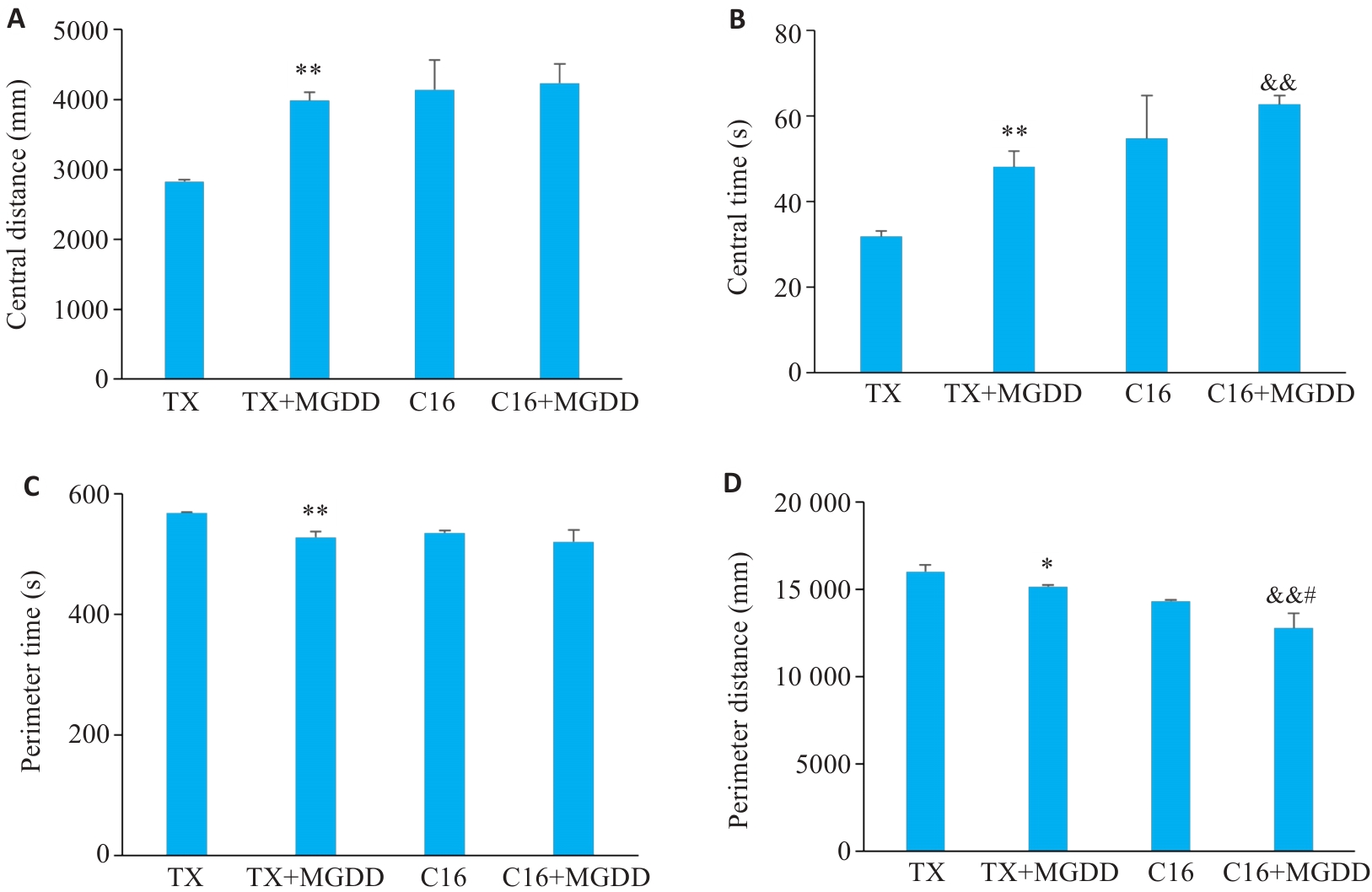
图3 PKR抑制背景下"通腑养髓"法对TX小鼠旷场实验中焦虑样行为的影响
Fig.3 Effect of the Modified Gandou Decoction (MGDD) on anxiety-like behaviors of TX mice in open field test. A: Distance traveled in the center zone. B: Time spent in the center zone. C: Distance traveled in the peripheral zone. D: Time spent in the peripheral zone (Mean±SD, n=3). *P<0.05, **P<0.01 vs TX group; #P<0.05 vs C16 group; &&P<0.05 vs TX+MGDD group.
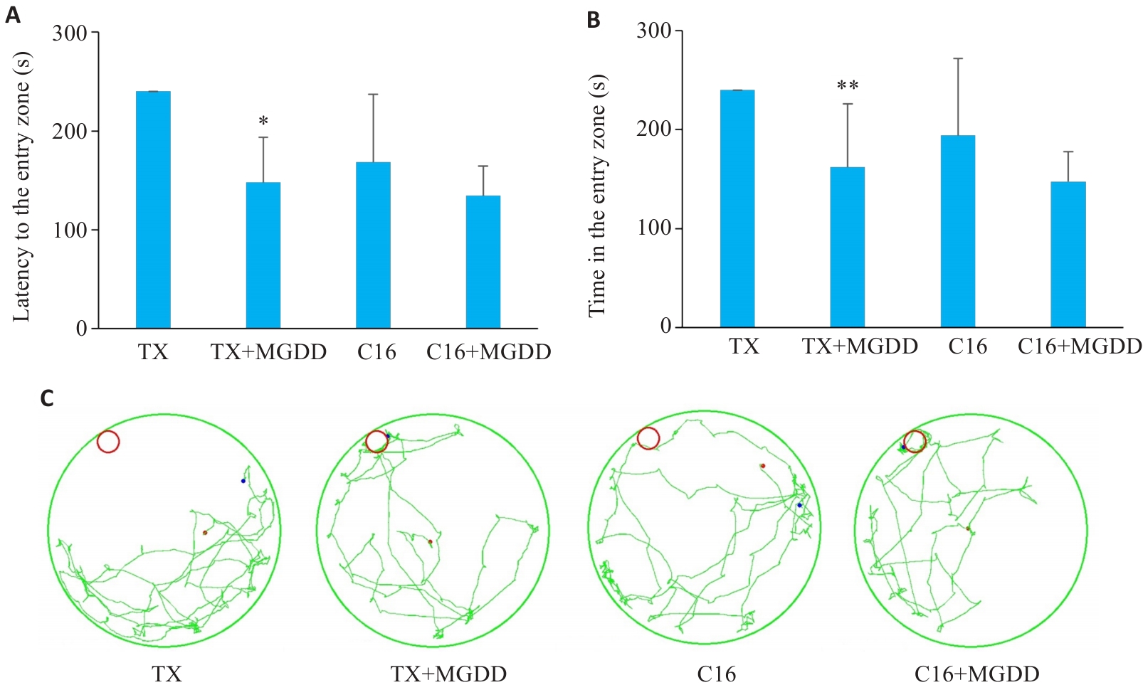
图4 PKR抑制背景下"通腑养髓"法对TX小鼠巴恩斯迷宫空间学习记忆的影响
Fig.4 Effect of MGDD on spatial learning and memory of TX mice. A: Barnes maze test results showing latency to the entry zone of mice in each group. B: Time in the entry zone in each group. C: Representative trajectory of the mice in each group in the test. Data are presented as Mean±SD (n=3-6). *P<0.05, **P<0.01 vs TX group.
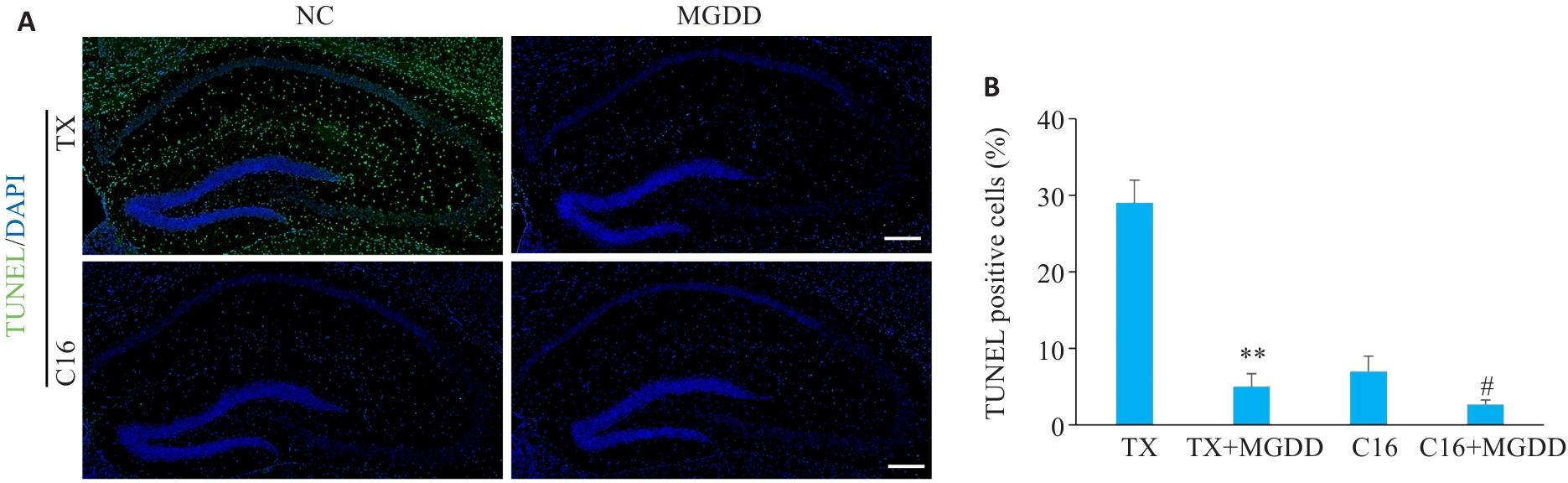
图5 PKR抑制背景下"通腑养髓"法对TX小鼠海马神经元凋亡的影响
Fig.5 Effect of the MGDD on the apoptosis of the hippocampus neurons in TX mice. A: Representative immunofluorescence staining images for TUNEL in the hippocampus of mice in each group. Green: TUNEL. Blue: DAPI. Scale bar=200 µm. B: The TUNEL-positive cells of hippocampal neurons in each group of mice (Mean±SD, n=3). **P<0.01 vs TX group; #P<0.05 vs C16 group.
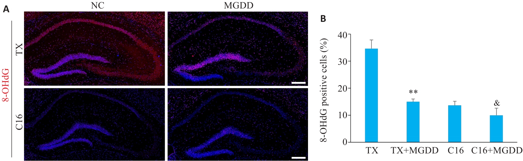
图6 PKR抑制背景下"通腑养髓"法对TX小鼠海马氧化应激损伤的影响
Fig.6 Effect of MGDD on oxidative stress injury in the hippocampus of TX mice. A: Representative immunofluo-rescence staining images for 8-OHdG in the CA1 region of the hippocampus of mice in each group. Red: 8-OHdG. Blue: DAPI. Scale bar=200 µm. B: OHdG-positive cells in the CA1 region of the hippocampus of mice in each group (Mean±SD, n=3). **P<0.01 vs TX group; &P<0.05 vs TX+MGDD group.
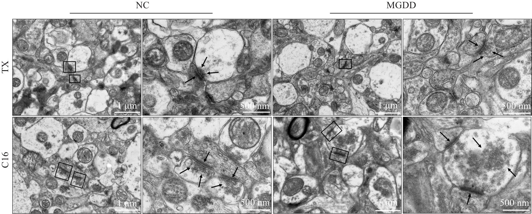
图7 PKR抑制背景下"通腑养髓"法对TX小鼠海马突触超微结构的影响
Fig.7 Representative TEM images of synapses in the CA1 region of the hippocampus in each group of mice. The right-side images are enlarged versions of the black boxes on the left images, and the arrows indicate to synaptic vesicles and the pre- and post-synaptic membranes. Scale bars on the left represent 1 µm, and those on the right represent 500 nm.
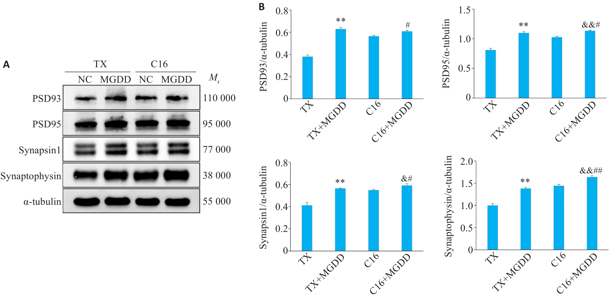
图8 PKR抑制背景下"通腑养髓"法对TX小鼠海马突触相关蛋白表达的影响
Fig.8 Effect of MGDD on synapse-related protein in the hippocampus of TX mice. A: Gray-scale band images of PSD95, PSD93, Synapsin1, Synaptophysin and α-tubulin in the hippocampus of mice. B: Expression level of synapse-related protein quantified using Western blotting in the different groups (Mean±SD, n=3). **P<0.01 vs TX group; #P<0.05, ##P<0.05 vs C16 group; &P<0.05, &&P<0.05 vs TX+MGDD group.
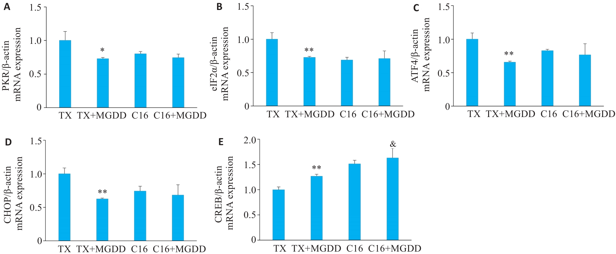
图9 PKR抑制背景下"通腑养髓"法对TX小鼠海马PKR/eIF2α通路相关基因mRNA表达的影响
Fig.9 Effect of MGDD on PKR/eIF2α‑related mRNA in the hippocampus of TX mice. A-E: Relative expression levels of Pkr, eIF2a, Atf4, Chop and Creb mRNAs (Mean±SD, n=3). *P<0.05, **P<0.01 vs TX group; &P<0.05 vs TX+MGDD group.
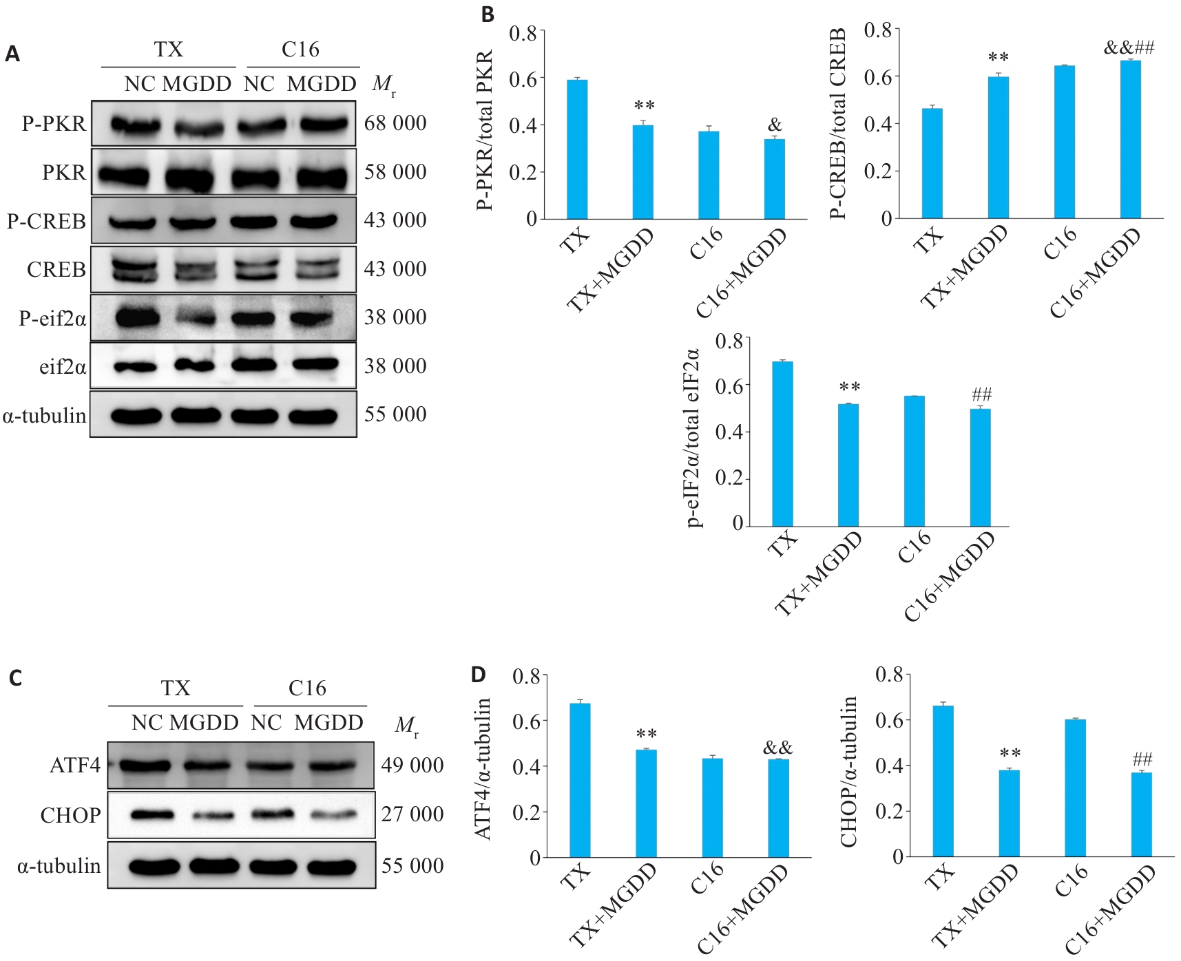
图10 PKR抑制背景下"通腑养髓"法对TX小鼠海马PKR/eIF2α/CREB信号通路的关键蛋白表达及磷酸化水平的影响
Fig.10 Effect of MGDD on expression levels of key proteins and phosphorylation in the PKR/eIF2α/CREB signaling pathway in the hippocampus of TX mice. A: Gray-scale band images of PKR, P-PKR, eIF2α, P-eIF2α, CREB, P-CREB and α‑tubulin in the hippocampus of mice. B: Expression levels of phosphorylated protein or total protein. C: Gray-scale band images of ATF4, CHOP and α‑tubulin in the hippocampus of the mice. D: Expression levels of ATF4 and CHOP. Data are presented as Mean±SD (n=3). **P<0.01 vs TX group; ##P<0.05 vs C16 group; &P<0.05, &&P<0.05 vs TX+MGDD group.
| [1] | Rodriguez-Castro KI, Hevia-Urrutia FJ, Sturniolo GC. Wilson's disease: a review of what we have learned[J]. World J Hepatol, 2015, 7(29): 2859-70. doi:10.4254/wjh.v7.i29.2859 |
| [2] | Petruzzelli R, Catalano F, Crispino R, et al. Prion protein promotes copper toxicity in Wilson disease[J]. Nat Commun, 2025, 16(1): 1468. doi:10.1038/s41467-025-56740-x |
| [3] | Członkowska A, Litwin T, Dusek P, et al. Wilson disease[J]. Nat Rev Dis Primers, 2018, 4: 21. doi:10.1038/s41572-018-0018-3 |
| [4] | Wang LY, Wu LM, Wang TT, et al. Gandou Bushen decoction ameliorates cognitive impairment in Wilson disease model TX mice by regulating melatonin synthesis via the SIRT3/FOXO3α pathway[J]. J Sichuan Univ Med Sci Ed, 2025, 56(1): 102-11. |
| [5] | Wang X, Chen H, Zhang XY, et al. Therapeutic targets and natural product screening for cognitive impairments associated with ferroptosis in Wilson's disease[J]. Am J Chin Med, 2024, 52(8): 2423-52. doi:10.1142/s0192415x24500927 |
| [6] | 中华医学会神经病学分会神经遗传学组, 吴志英, 李洵桦, 等. 中国肝豆状核变性诊治指南2021[J]. 中华神经科杂志, 2021, 54(4): 310-9. doi:10.3760/cma.j.cn113694-20200826-00661 |
| [7] | Schilsky ML, Roberts EA, Bronstein JM, et al. A multidisciplinary approach to the diagnosis and management of Wilson disease: Executive summary of the 2022 Practice Guidance on Wilson disease from the American Association for the Study of Liver Diseases[J]. Hepatology, 2023, 77(4): 1428-55. doi:10.1002/hep.32805 |
| [8] | 中华医学会肝病学分会遗传代谢性肝病协作组, 段钟平, 郑素军, 等. 肝豆状核变性诊疗指南(2022年版)[J]. 中华肝脏病杂志, 2022(1): 9-20. doi:10.3760/cma.j.cn501113-20211217-00603 |
| [9] | Hu YY, Wei WB, Liu SY, et al. Changes in neurotransmitter metabolic profiles in copper-loaded rats as assessed by mass spectrometry imaging and the effect of Gandouling intervention[J]. Exp Neurol, 2025, 395: 115460. doi:10.1016/j.expneurol.2025.115460 |
| [10] | Kirk FT, Munk DE, Laursen TL, et al. Cognitive impairment in stable Wilson disease across phenotype[J]. Metab Brain Dis, 2021, 36(7): 2173-7. doi:10.1007/s11011-021-00804-6 |
| [11] | Ma Q, Ying M, Sui XJ, et al. Chronic copper exposure causes spatial memory impairment, selective loss of hippocampal synaptic proteins, and activation of PKR/eIF2α pathway in mice[J]. J Alzheimers Dis, 2015, 43(4): 1413-27. doi:10.3233/jad-140216 |
| [12] | Hugon J, Paquet C. The PKR/P38/RIPK1 signaling pathway as a therapeutic target in Alzheimer's disease[J]. Int J Mol Sci, 2021, 22(6): 3136. doi:10.3390/ijms22063136 |
| [13] | Xu CC, Liu SY, Cheng N, et al. PKR downregulation prevents copper-induced synaptic dysfunction and cognitive impairment in a murine model of Wilson's disease[J]. Front Neurosci, 2024, 18: 1447304. doi:10.3389/fnins.2024.1447304 |
| [14] | 刘松杨, 程 楠, 徐陈陈, 等. 肝豆汤改良方调控PKR/eIF2α通路改善Wilson病模型TX小鼠突触功能障碍的机制研究[J]. 安徽中医药大学学报, 2021, 40(6): 75-81. doi:10.3969/j.issn.2095-7246.2021.06.017 |
| [15] | 饶志红, 杨文明, 杨玉龙, 等. 肝豆状核变性“肝-肾-脑” 轴病机阐释及中医辨治策略[J]. 北京中医药大学学报, 2025, 48(9): 1270-7. |
| [16] | 钱南南, 杨文明, 魏涛华, 等. 肝豆状核变性伏毒阻络病因病机探要[J]. 中国实验方剂学杂志, 2022, 28(12): 133-40. |
| [17] | 杨任民, 韩咏竹, 任明山, 等. 中药治疗肝豆状核变性107例疗效观察[J]. 中医杂志, 1993, 34(11): 676-7. |
| [18] | 徐陈陈, 董健健, 程 楠, 等. 肝豆汤改良方对Wilson’s病模型TX乳鼠神经元内Cyt C/Caspase信号通路的分子调控机制[J]. 中国实验方剂学杂志, 2017, 23(6): 143-8. doi:10.13422/j.cnki.syfjx.2017060143 |
| [19] | 许子夜, 汪 瀚. 基于伏毒理论论治肝豆状核变性的治疗[J]. 临床医学进展, 2025, 15(4), 2002-8. doi:10.12677/acm.2025.1541147 |
| [20] | 徐陈陈. Wilson病炎症因子表达谱及肝豆汤改良方对TX小鼠神经元Cer信号通路调控机制的研究[D]. 合肥: 安徽中医药大学, 2017. |
| [21] | 杨任民. 肝豆状核变性[M]. 北京: 人民卫生出版社, 2015. |
| [22] | Shanaki Bavarsad M, Spina S, Oehler A, et al. Comprehensive mapping of synaptic vesicle protein 2A (SV2A) in health and neurodegenerative diseases: a comparative analysis with synaptophysin and ground truth for PET-imaging interpretation[J]. Acta Neuropathol, 2024, 148(1): 58. doi:10.1007/s00401-024-02816-9 |
| [23] | Yan PP, Liu HC, Zhou T, et al. Crosstalk of Synapsin1 palmitoylation and phosphorylation controls the dynamicity of synaptic vesicles in neurons[J]. Cell Death Dis, 2022, 13(9): 786. doi:10.1038/s41419-022-05235-4 |
| [24] | De Los Reyes DA, Karkoutly MY, Zhang YH. Synapse-associated protein 102-a highly mobile MAGUK predominate in early synaptogenesis[J]. Front Mol Neurosci, 2023, 16: 1286134. doi:10.3389/fnmol.2023.1286134 |
| [25] | Fan X, Wang H, Ping JC, et al. Synaptic scaffold protein PSD-95: a therapeutic target for Alzheimer's disease[J]. Biochem Pharmacol, 2025, 242(Pt 3): 117401. doi:10.1016/j.bcp.2025.117401 |
| [26] | Chung WS, Welsh CA, Barres BA, et al. Do Glia drive synaptic and cognitive impairment in disease[J]? Nat Neurosci, 2015, 18(11): 1539-45. doi:10.1038/nn.4142 |
| [27] | Hu J, Hua Y, Li CQ, et al. cTBS enhanced synaptic plasticity in the affected and unaffected motor cortex after cerebral ischemia via astrocyte-mediated TSP1 pathway[J]. Exp Neurol, 2025, 393: 115409. doi:10.1016/j.expneurol.2025.115409 |
| [28] | Kleidonas D, Kirsch M, Andrieux G, et al. Microglia modulate TNFα-mediated synaptic plasticity[J]. Glia, 2023, 71(9): 2117-36. doi:10.1002/glia.24383 |
| [29] | Greenough MA, Camakaris J, Bush AI. Metal dyshomeostasis and oxidative stress in Alzheimer's disease[J]. Neurochem Int, 2013, 62(5): 540-55. doi:10.1016/j.neuint.2012.08.014 |
| [30] | Mouton-Liger F, Paquet C, Dumurgier J, et al. Oxidative stress increases BACE1 protein levels through activation of the PKR-eIF2α pathway[J]. Biochim Biophys Acta BBA Mol Basis Dis, 2012, 1822(6): 885-96. doi:10.1016/j.bbadis.2012.01.009 |
| [31] | Wang Q, Wang ZW, Li YT, et al. Baicalein improves motor dysfunction and cognitive impairment while promoting remyelination in an animal model of multiple sclerosis through the antioxidant mechanism[J]. Front Pharmacol, 2025, 16: 1659631. doi:10.3389/fphar.2025.1659631 |
| [32] | Liu XQ, Deng YX, Dai Z, et al. Sodium tanshinone IIA sulfonate protects against Aβ1-42-induced cellular toxicity by modulating Aβ-degrading enzymes in HT22 cells[J]. Int J Biol Macromol, 2020, 151: 47-55. doi:10.1016/j.ijbiomac.2020.02.040 |
| [33] | Chen CW, Papadopoli D, Szkop KJ, et al. Plasticity of the mammalian integrated stress response[J]. Nature, 2025, 641(8065): 1319-28. doi:10.1038/s41586-025-08794-6 |
| [34] | Costa-Mattioli M, Walter P. The integrated stress response: From mechanism to disease[J]. Science, 2020, 368(6489): eaat5314. doi:10.1126/science.aat5314 |
| [35] | Sharma V, Sood R, Khlaifia A, et al. eIF2α controls memory consolidation via excitatory and somatostatin neurons[J]. Nature, 2020, 586(7829): 412-6. doi:10.1038/s41586-020-2805-8 |
| [36] | Wek RC, Jiang HY, Anthony TG. Coping with stress: eIF2 kinases and translational control[J]. Biochem Soc Trans, 2006, 34(Pt 1): 7-11. doi:10.1042/bst0340007 |
| [37] | Kolac UK, Goker Bagca B, Donmez Yalcin G, et al. Thymoquinone attenuates poly(I: C)-induced cellular stress via PKR/ATF4/CHOP signaling and autophagy modulation in human alveolar epithelial cells[J]. Toxicol In Vitro, 2025, 111: 106165. doi:10.1016/j.tiv.2025.106165 |
| [38] | Martínez NW, Gómez F, Tapia-Godoy A, et al. PKR-driven ISR signaling controls synaptic translation and structural plasticity in an age-dependent manner[J]. Neurobiol Dis, 2025, 216: 107113. doi:10.1016/j.nbd.2025.107113 |
| [39] | Feng WJ, Lv CH, Cheng L, et al. Targeting ERS-mitophagy in hippocampal neurons to explore the improvement of memory by tea polyphenols in aged type 2 diabetic rats[J]. Free Radic Biol Med, 2024, 213: 293-308. doi:10.1016/j.freeradbiomed.2024.01.044 |
| [40] | You CC, Zhang ZL, Ying HY, et al. Blockage of calcium-sensing receptor improves chronic intermittent hypoxia-induced cognitive impairment by PERK-ATF4-CHOP pathway[J]. Exp Neurol, 2023, 368: 114500. doi:10.1016/j.expneurol.2023.114500 |
| [41] | Wang YH, Wu D, Li DN, et al. The role of PERK-eIF2α‑ATF4-CHOP pathway in sevoflurane induced neuroapoptosis and cognitive dysfunction in aged mice[J]. Cell Signal, 2023, 110: 110841. doi:10.1016/j.cellsig.2023.110841 |
| [42] | Jiang ZH, Belforte JE, Lu Y, et al. eIF2alpha Phosphorylation-dependent translation in CA1 pyramidal cells impairs hippocampal memory consolidation without affecting general translation[J]. J Neurosci, 2010, 30(7): 2582-94. doi:10.1523/jneurosci.3971-09.2010 |
| [43] | Li YF, Cheng YF, Huang Y, et al. Phosphodiesterase-4D knock-out and RNA interference-mediated knock-down enhance memory and increase hippocampal neurogenesis via increased cAMP signaling[J]. J Neurosci, 2011, 31(1): 172-83. doi:10.1523/jneurosci.5236-10.2011 |
| [44] | Smith SG, Haynes KA, Hegde AN. Degradation of transcriptional repressor ATF4 during long-term synaptic plasticity[J]. Int J Mol Sci, 2020, 21(22): 8543. doi:10.3390/ijms21228543 |
| [1] | 王庆阁, 赵晓慧, 何宇轩, 刘飞祥, 张运克. 芪芎左归颗粒通过上调BDNF/TrkB通路提高衰老大鼠突触可塑性[J]. 南方医科大学学报, 2025, 45(8): 1589-1598. |
| [2] | 张梦影, 赵晨玲, 田丽伟, 余郭芳, 杨文明, 董婷. 肝豆扶木汤通过GPX4/ACSL4/ALOX15通路抑制铁死亡改善Wilson病小鼠的肝脏脂肪变性[J]. 南方医科大学学报, 2025, 45(7): 1471-1478. |
| [3] | 刘露玉, 公茂伟, 廖国松, 赵维星, 傅强. 高血压通过UCP2下调介导的线粒体功能障碍加重大鼠术后学习记忆损伤[J]. 南方医科大学学报, 2025, 45(4): 725-735. |
| [4] | 李明明, 何梁超, 李天雨, 鲍岩, 徐祥, 陈光. 小鼠顶叶皮层反复轻度创伤性脑损伤抑制延髓NLG-1和PSD-95的表达[J]. 南方医科大学学报, 2024, 44(5): 960-966. |
| [5] | 张笑颜, 王 谢, 王 杰, 邵 楠, 蔡 标, 谢道俊. 黄蒲通窍胶囊改善Wilson病铜负荷大鼠的认知损害:基于抑制内质网应激介导的凋亡途径[J]. 南方医科大学学报, 2024, 44(3): 447-454. |
| [6] | 林如辉, 夏金言, 马小涵, 李钻芳. 电针通过促进突触再生改善脑缺血再灌注损伤大鼠的学习记忆功能[J]. 南方医科大学学报, 2024, 44(12): 2317-2326. |
| [7] | 银苗朱, 陈奎玉, 吴丽敏, 江鹏宇, 籍志慧, 张念, 周欢, 韩辉. 肝豆补肾汤通过激活ERK信号通路减少Wilson病TX小鼠异常精子生成并促进生精细胞增殖[J]. 南方医科大学学报, 2024, 44(11): 2063-2073. |
| [8] | 贺舒凝, 张佳豪, 杨若男, 袁 萍. 我国45岁及以上人群认知功能障碍的空间分布及其影响因素[J]. 南方医科大学学报, 2023, 43(4): 611-619. |
| [9] | 赵晨玲, 董 婷, 孙伦燕, 胡慧冰, 王 琼, 田丽伟, 江张胜. Wilson病脂代谢异常患者发生肝纤维化的列线图预测模型的建立与验证[J]. 南方医科大学学报, 2022, 42(11): 1720-1725. |
| [10] | 张雪卫, 傅 强. 脑脊液中β淀粉样蛋白42和神经丝轻链蛋白水平与术后神经认知功能障碍的相关性:基于90例66~78岁患者[J]. 南方医科大学学报, 2021, 41(4): 574-578. |
| [11] | 李 智, 李 虎, 姚尚龙, 程明华, 陈建颜. 右美托咪定剂量对老年脊柱手术患者术后认知功能障碍发生率及Aβ和细胞因子水平的影响:120例随机对照试验[J]. 南方医科大学学报, 2021, 41(4): 600-606. |
| [12] | 葛鑫宇, 郭 芳, 范 俊, 陈宝田, 于 林, 任 静, 李纪强, 卢成林, 莫嘉文, 李树基, 袁乐欣, 胡号应, 刘 赟, 周 晓, 崔 娟, 朱志敏, 曹 雄. 柴胡桂枝汤通过sirt1-p53信号通路产生抗抑郁作用[J]. 南方医科大学学报, 2021, 41(3): 399-405. |
| [13] | 魏 巍, 郑 曦, 谷 宇, 唐春林, 尧永华. 术后镇痛策略对老年单肺通气患者术后神经认知功能及早期康复的影响:90例随机对照临床试验[J]. 南方医科大学学报, 2020, 40(12): 1821-1825. |
| [14] | 陈小慧,任晓强,马亚兵,葛莉,胡钟元,阎文军. 术后疼痛引起老年患者术后认知功能障碍的相关机制研究进展[J]. 南方医科大学学报, 2019, 39(09): 1122-. |
| [15] | 张津玮,张小宝,钱海涛,崔吉正,顾小萍. 右美托咪定可减轻大鼠胫骨骨折手术所致的术后认知功能障碍[J]. 南方医科大学学报, 2019, 39(03): 292-. |
| 阅读次数 | ||||||
|
全文 |
|
|||||
|
摘要 |
|
|||||