
南方医科大学学报 ›› 2026, Vol. 46 ›› Issue (8): 1870-1877.doi: 10.12122/j.issn.1673-4254.2026.08.14
• • 上一篇
吴浩松1( ), 蒋易1, 张晓林2, 汪盛1()
收稿日期:2025-10-21
出版日期:2026-08-20
发布日期:2026-08-01
通讯作者:
汪盛
E-mail:whs06052024@163.com;bydoctorw@163.com
作者简介:吴浩松,硕士,E-mail: whs06052024@163.com
基金资助:
Haosong WU1(), Yi JIANG1, Xiaolin ZHANG2, Sheng WANG1()
Received:2025-10-21
Online:2026-08-20
Published:2026-08-01
Contact:
Sheng WANG
E-mail:whs06052024@163.com;bydoctorw@163.com
摘要:
目的 探讨依维莫司通过调控p70S6K/PI3K/MAPK信号通路对膀胱癌耐药细胞RT-112-RS生物学行为的影响。 方法 采用梯度法筛选厄达替尼浓度,选取5 μmol/L作为终浓度并持续诱导16周以建立厄达替尼耐药的RT-112-RS细胞模型。设对照组(RT-112-RS常规培养)、PDGF组(+PDGF-BB)、PDGF+0.01 μmol/L依维莫司组、PDGF+0.1 μmol/L依维莫司组及PDGF+1 μmol/L依维莫司组。CCK-8法测定细胞毒性并计算IC50;划痕和Transwell实验评估迁移与侵袭能力;RT-qPCR检测0.001~1 μmol/L依维莫司处理下相关基因表达;流式细胞术分析凋亡率;Western blotting检测mTORC1/p70S6K、PI3K/Akt及Raf1/MEK/ERK通路关键蛋白磷酸化水平。 结果 依维莫司在0.001~0.1 μmol/L范围内呈剂量依赖性抑制RT-112-RS增殖(P<0.05)。相比RT-112细胞,RT-112-RS迁移与侵袭能力增强(819.83±71.39 vs 1579±136.30,P<0.05)。1 μmol/L依维莫司显著抑制p70S6K磷酸化(P<0.05),但未进一步增强抗增殖效应,并特异性激活Raf1/MEK/ERK通路,对PI3K/Akt(Thr308)或mTORC2/Akt(Ser473)无明显影响。MEK抑制剂U-0126可逆转依维莫司诱导的ERK1/2活化(5.52±0.40 vs 0.74±0.07,P<0.05),PI3K抑制剂LY294002则减弱ERK1/2激活(7.58±1.54 vs 11.2±0.24,P<0.05)。 结论 依维莫司通过抑制p70S6K活性抑制RT-112-RS增殖。而高浓度时可能因减弱负反馈抑制而激活PI3K-Raf1/MEK/ERK通路,从而部分抵消其抗增殖作用。
吴浩松, 蒋易, 张晓林, 汪盛. 依维莫司通过p70S6K/PI3K/MAPK信号通路抑制厄达替尼耐药的膀胱癌细胞生物学行为[J]. 南方医科大学学报, 2026, 46(8): 1870-1877.
Haosong WU, Yi JIANG, Xiaolin ZHANG, Sheng WANG. Everolimus inhibits malignant phenotype of erdafitinib-resistant bladder cancer cells through the p70S6K/PI3K/MAPK signaling pathway[J]. Journal of Southern Medical University, 2026, 46(8): 1870-1877.
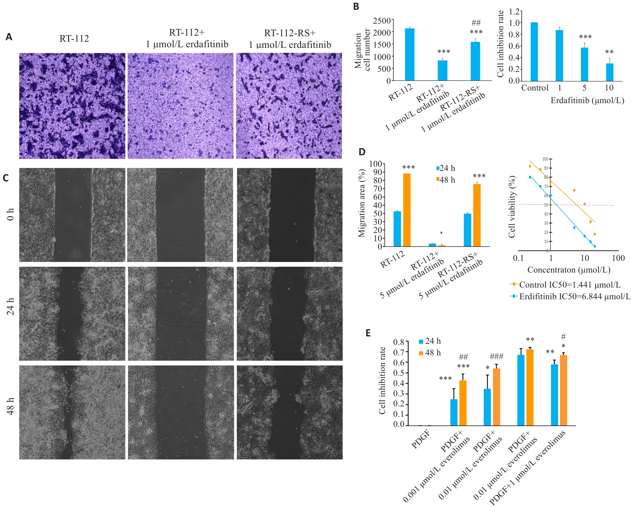
图1 RT-112和RT-112-RS在含有厄达替尼培养基中的迁移和侵袭能力
Fig.1 Migration and invasion abilities of RT-112 and erdafitinib-resistant RT-112-RS cells in the medium containing erdafitinib. A: Migration ability of RT-112 and RT-112-RS cells determined by Transwell assay. B: Migration ability of RT-112 cells treated with erdafitinib. C: Migration ability of RT-112 and RT-112-RS cells determined by scratch assay. D: IC50 values of erdafitinib in RT-112 and RT-112-RS cells determined by CCK-8 assay. E: CCK-8 assay for assessing cell proliferation (Original magnification: ×40). *P<0.05, **P<0.01, ***P<0.001 vs RT-112; #P<0.05, ##P<0.01, ###P<0.001 vs RT-112+1 μmol/L erdafitinib (Mean±SD, n=6).
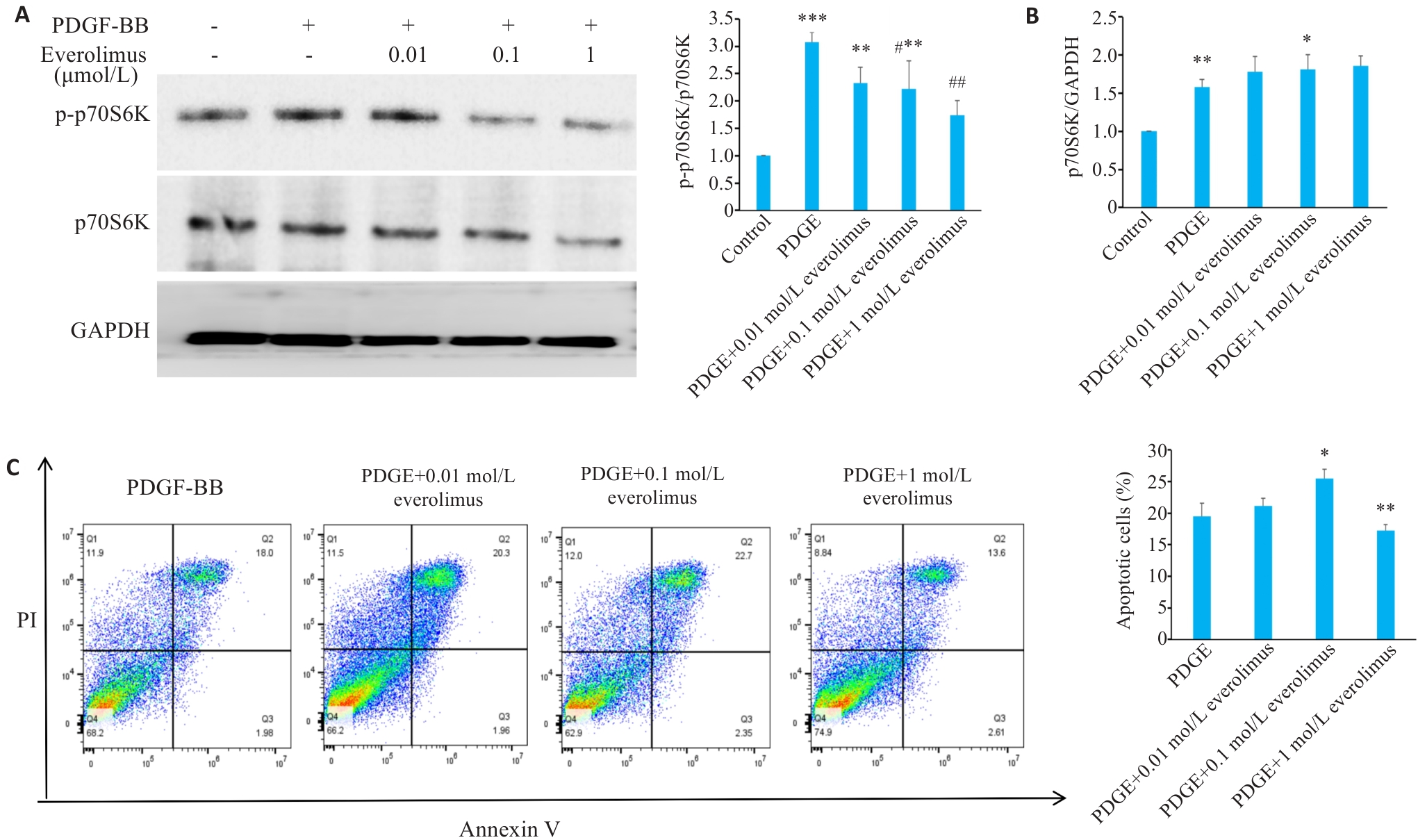
图2 依维莫司对 p-p70S6K和细胞凋亡的影响
Fig.2 Effect of PDGF-BB alone or in combination with everolimus on p-p70S6K expression and apoptosis in RT-112-RS cells. A: Everolimus significantly inhibits phosphorylation of p70S6K detected by Western blotting. B: Detection of p70S6k mRNA. C: Analysis of apoptosis of RT-112-RS cells following treatment. *P<0.05, **P<0.01, ***P<0.001 vs control; #P<0.05, ##P<0.01 vs PDGF-BB (Mean±SD, n=3).
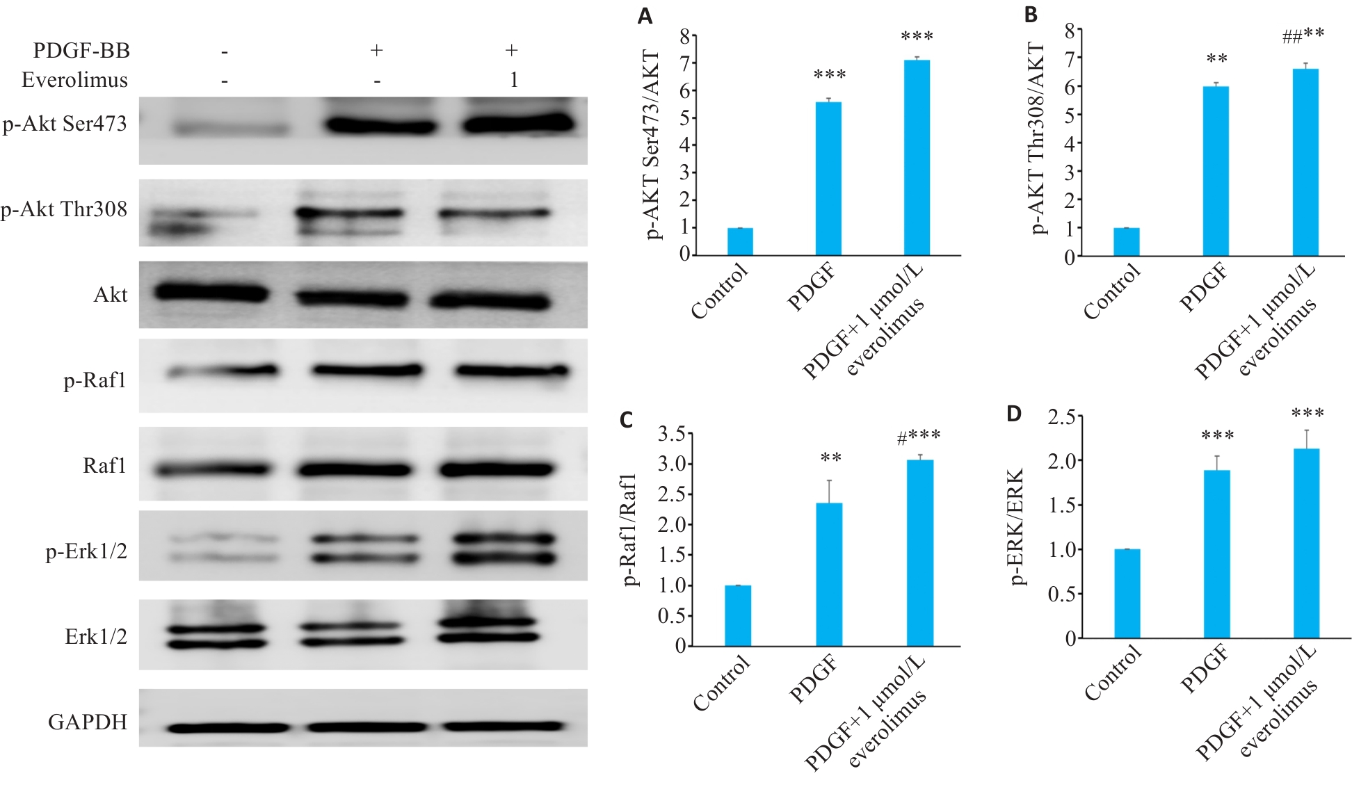
图3 Raf1-MEK-ERK通路蛋白表达分析
Fig.3 Analysis of expressions of Raf1-MEK-ERK pathway proteins using Western blotting in RT-112-RS cells following treatment with PDGF-BB alone or in combination with everolimus. A: p-Akt (Ser473)/Akt expressions. B: p-Akt (Thr308)/Akt expressions. C: p-Raf1/Raf1 expressions. D: p-ERK1/2/ERK1/2 expressions. **P<0.01, ***P<0.001 vs control; #P<0.05, ##P<0.01 vs PDGF-BB (Mean±SD, n=3).
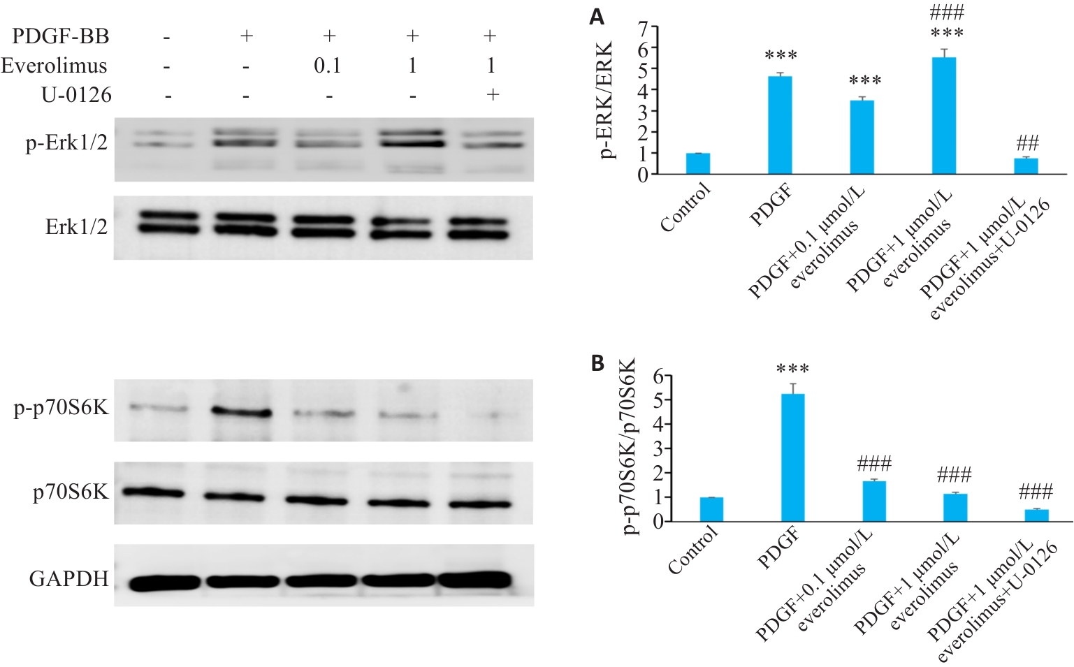
图4 依维莫司和U-0126对 RT-112- RS中ERK和p70S6K磷酸化的影响
Fig.4 Effects of PDGF-BB, everolimus and U-0126 on phosphorylation of ERK and p70S6K in RT-112-RS cells. A: p-ERK1/2/ERK1/2. B: p-p70S6K/p70S6K. ***P<0.001 vs control; ##P<0.01, ###P<0.001 vs PDGF-BB (Mean±SD, n=3).
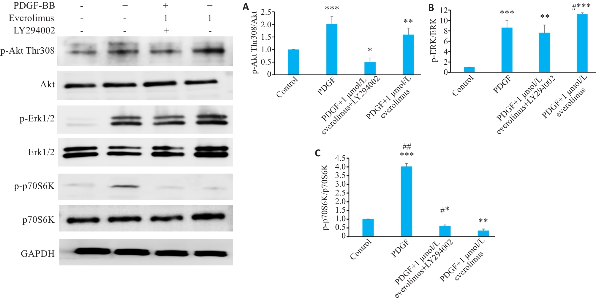
图5 依维莫司和LY294002对RT-112- RS中p70S6K/PI3K/ERK磷酸化的影响
Fig.5 Effects of everolimus and LY294002 on phosphorylation of p70S6K/PI3K/ERK in RT-112-RS cells. A: p-AKT (Thr308)/AKT expressions. B: p-ERK1/2/ERK1/2 expressions. C: p-p70S6K/p70S6K expressions. *P<0.05, **P<0.01, ***P<0.001 vs control; #P<0.05, ##P<0.01 vs PDGF-BB (Mean±SD, n=3).
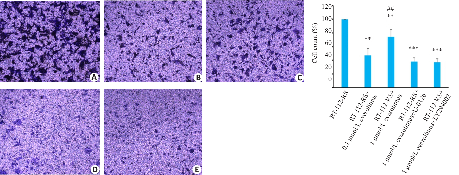
图6 U-0126和LY294002对依维莫司诱导的RT-112- RS迁移与侵袭能力的影响
Fig.6 Effects of U-0126 and LY294002 on migration and invasion capabilities of RT-112-RS cells treated with everolimus ( ×40). A: RT-112-RS group. B: RT-112-RS+0. 1 μmol/L everolimus group. C: RT-112-RS+1 μmol/L everolimus group. D: RT-112-RS+0.1 μmol/L everolimus+U-0126 group. E: RT-112-RS+0.1 μmol/L everolimus+LY294002 group. **P<0.01, ***P<0.001 vs RT-112-RS; ##P<0.01 vs RT-112-RS+PDGF (Mean±SD, n=3).
| [1] | Sung H, Ferlay J, Siegel RL, et al. Global cancer statistics 2020: GLOBOCAN estimates of incidence and mortality worldwide for 36 cancers in 185 countries[J]. CA Cancer J Clin, 2021, 71(3): 209-49. doi:10.3322/caac.21660 |
| [2] | Paner GP, Kamat A, Netto GJ, et al. International society of urological pathology (ISUP) consensus conference on current issues in bladder cancer. working group 2: grading of mixed grade, invasive urothelial carcinoma including histologic subtypes and divergent differentiations, and non-urothelial carcinomas[J]. Am J Surg Pathol, 2024, 48(1): e11-23. doi:10.1097/pas.0000000000002077 |
| [3] | Cathomas R, Lorch A, Bruins HM, et al. The 2021 updated European association of urology guidelines on metastatic urothelial carcinoma[J]. Eur Urol, 2022, 81(1): 95-103. doi:10.1016/j.eururo.2021.09.026 |
| [4] | Pan ZR, Zhang HX, Dokudovskaya S. The role of mTORC1 pathway and autophagy in resistance to platinum-based chemotherapeutics[J]. Int J Mol Sci, 2023, 24(13): 10651. doi:10.3390/ijms241310651 |
| [5] | Singh A, Osbourne AS, Koshkin VS. Perioperative immunotherapy in muscle-invasive bladder cancer[J]. Curr Treat Options Oncol, 2023, 24(9): 1213-30. doi:10.1007/s11864-023-01113-z |
| [6] | Wu QS, Wong JPC, Kwok HF. Putting the brakes on tumorigenesis with natural products of plant origin: insights into the molecular mechanisms of actions and immune targets for bladder cancer treatment[J]. Cells, 2020, 9(5): 1213. doi:10.3390/cells9051213 |
| [7] | Hao XP, Fan HX, Yang J, et al. Network pharmacology research and dual-omic analyses reveal the molecular mechanism of natural product nodosin inhibiting muscle-invasive bladder cancer in vitro and in vivo [J]. J Nat Prod, 2022, 85(8): 2006-17. doi:10.1021/acs.jnatprod.2c00400 |
| [8] | Su QL, Tao T, Tang L, et al. Down-regulation of PKM2 enhances anticancer efficiency of THP on bladder cancer[J]. J Cell Mol Med, 2018, 22(5): 2774-90. doi:10.1111/jcmm.13571 |
| [9] | Tate T, Xiang TN, Wobker SE, et al. Pparg signaling controls bladder cancer subtype and immune exclusion[J]. Nat Commun, 2021, 12: 6160. doi:10.1038/s41467-021-26421-6 |
| [10] | Guo CC, Lee S, Lee JG, et al. Molecular profile of bladder cancer progression to clinically aggressive subtypes[J]. Nat Rev Urol, 2024, 21(7): 391-405. doi:10.1038/s41585-023-00847-7 |
| [11] | Eisner JR, de Jong FC, Shibata Y, et al. Characterization of FGFR alterations and activation in patients with high-risk non–muscle-invasive bladder cancer[J]. Clin Cancer Res, 2024, 30(23): 5374-84. doi:10.1158/1078-0432.ccr-24-2015 |
| [12] | Komori T. Regulation of proliferation, differentiation and functions of osteoblasts by Runx2[J]. Int J Mol Sci, 2019, 20(7): 1694. doi:10.3390/ijms20071694 |
| [13] | Zhang Y, Ling L, Ajay D/O Ajayakumar A, et al. FGFR2 accommodates osteogenic cell fate determination in human mesenchymal stem cells[J]. Gene, 2022, 818: 146199. doi:10.1016/j.gene.2022.146199 |
| [14] | Robertson AG, Kim J, Al-Ahmadie H, et al. Comprehensive molecular characterization of muscle-invasive bladder cancer[J]. Cell, 2018, 174(4): 1033. |
| [15] | Hou CY, Lei YJ, Li N, et al. Collagen from Iris squid grafted with polyethylene glycol and collagen peptides promote the proliferation of fibroblast through PI3K/AKT and Ras/RAF/MAPK signaling pathways[J]. Int J Biol Macromol, 2023, 247: 125772. doi:10.1016/j.ijbiomac.2023.125772 |
| [16] | Casadei C, Dizman N, Schepisi G, et al. Targeted therapies for advanced bladder cancer: new strategies with FGFR inhibitors[J]. Ther Adv Med Oncol, 2019, 11: 1758835919890285. doi:10.1177/1758835919890285 |
| [17] | Peng Y, Wang YY, Zhou C, et al. PI3K/Akt/mTOR pathway and its role in cancer therapeutics: are we making headway [J]. Front Oncol, 2022, 12: 819128. doi:10.3389/fonc.2022.819128 |
| [18] | Loriot Y, Matsubara N, Park SH, et al. Erdafitinib or chemotherapy in advanced or metastatic urothelial carcinoma[J]. N Engl J Med, 2023, 389(21): 1961-71. doi:10.1056/nejmoa2308849 |
| [19] | Franza A, Pirovano M, Giannatempo P, et al. Erdafitinib in locally advanced/metastatic urothelial carcinoma with certain FGFR genetic alterations[J]. Future Oncol, 2022, 18(19): 2455-64. doi:10.2217/fon-2021-1151 |
| [20] | Nishina T, Takahashi S, Iwasawa R, et al. Safety, pharmacokinetic, and pharmacodynamics of erdafitinib, a pan-fibroblast growth factor receptor (FGFR) tyrosine kinase inhibitor, in patients with advanced or refractory solid tumors[J]. Investig New Drugs, 2018, 36(3): 424-34. doi:10.1007/s10637-017-0514-4 |
| [21] | Szwed A, Kim E, Jacinto E. Regulation and metabolic functions of mTORC1 and mTORC2[J]. Physiol Rev, 2021, 101(3): 1371-426. doi:10.1152/physrev.00026.2020 |
| [22] | Yang K, Tang xiang-jun, Xu feng-fei, et al. PI3K/mTORC1/2 inhibitor PQR309 inhibits proliferation and induces apoptosis in human glioblastoma cells[J]. Oncol Rep, 2020,43(3):773-82. |
| [23] | Tian JH, Wang YH, Zhou XS, et al. Rapamycin slows IgA nephropathy progression in the rat[J]. Am J Nephrol, 2014, 39(3): 218-29. doi:10.1159/000358844 |
| [24] | Zeng QY, Zhou ZH, Qin SS, et al. Rapamycin inhibits B-cell activating factor (BAFF)-stimulated cell proliferation and survival by suppressing Ca2+-CaMKII-dependent PTEN/Akt-Erk1/2 signaling pathway in normal and neoplastic B-lymphoid cells[J]. Cell Calcium, 2020, 87: 102171. doi:10.1016/j.ceca.2020.102171 |
| [25] | Morris MB, Ozsoy S, Zada M, et al. Selected amino acids promote mouse pre-implantation embryo development in a growth factor-like manner[J]. Front Physiol, 2020, 11: 140. doi:10.3389/fphys.2020.00140 |
| [26] | Tian JH, Chang SJ, Ji H, et al. The p70S6K/PI3K/MAPK feedback loop releases the inhibition effect of high-dose rapamycin on rat mesangial cell proliferation[J]. Int J Immunopathol Pharmacol, 2021, 35: 20587384211000544. doi:10.1177/20587384211000544 |
| [27] | Wang KJ, Wang C, Dai LH, et al. Targeting an autocrine regulatory loop in cancer stem-like cells impairs the progression and chemotherapy resistance of bladder cancer[J]. Clin Cancer Res,2019,25(3):1070-86. doi:10.1158/1078-0432.ccr-18-0586 |
| [28] | Sun YH, Sedgwick AJ, Khan MA, et al. A transcriptional signature of IL-2 expanded natural killer cells predicts more favorable prognosis in bladder cancer[J]. Front Immunol, 2021, 12: 724107. doi:10.3389/fimmu.2021.724107 |
| [29] | Ando K, Kurashina R, Motoi N, et al. Positive regulatory loop of platelet-derived growth factor DD-induced STAT3 activation is associated with poor prognosis in advanced urothelial carcinoma[J]. Biochem Biophys Res Commun,2023,676:165-70. doi:10.1016/j.bbrc.2023.07.054 |
| [30] | Yu JJ, Zhang LJ, Peng J, et al. Dictamnine, a novel c-Met inhibitor, suppresses the proliferation of lung cancer cells by downregulating the PI3K/AKT/mTOR and MAPK signaling pathways[J]. Biochem Pharmacol, 2022, 195: 114864. doi:10.1016/j.bcp.2021.114864 |
| [31] | Buddham R, Chauhan S, Narad P, et al. Reconstruction and exploratory analysis of mTORC1 signaling pathway and its applications to various diseases using network-based approach[J]. J Microbiol Biotechnol, 2022, 32(3): 365-77. doi:10.4014/jmb.2108.08007 |
| [32] | Kale R, Samant C, Bokare A, et al. Inhibition of SGK1 potentiates the anticancer activity of PI3K inhibitor in NSCLC cells through modulation of mTORC1, p-ERK and β‑catenin signaling[J]. Biomed Rep, 2023, 19(6): 94. doi:10.3892/br.2023.1676 |
| [33] | Xiao J, Wang S, Chen LL, et al. 25-Hydroxycholesterol regulates lysosome AMP kinase activation and metabolic reprogramming to educate immunosuppressive macrophages[J]. Immunity, 2024, 57(5): 1087-104.e7. doi:10.1016/j.immuni.2024.03.021 |
| [34] | O’Leary EM, Tian YF, Nigdelioglu R, et al. TGF‑β promotes metabolic reprogramming in lung fibroblasts via mTORC1-dependent ATF4 activation[J]. Am J Respir Cell Mol Biol, 2020, 63(5): 601-12. doi:10.1165/rcmb.2020-0143oc |
| [35] | Liang X, Yao JP, Cui DR, et al. The TRAF2-p62 axis promotes proliferation and survival of liver cancer by activating mTORC1 pathway[J]. Cell Death Differ, 2023, 30(6): 1550-62. doi:10.1038/s41418-023-01164-7 |
| [36] | Huang MY, Hu SY, Dong J, et al. The DRAP1/DR1 repressor complex increases mTOR activity to promote progression and confer everolimus sensitivity in triple-negative breast cancer[J]. Cancer Res, 2024, 84(16): 2660-73. doi:10.1158/0008-5472.can-23-2781 |
| [37] | Ye QW, Zhou W, Xu SJ, et al. Ubiquitin-specific protease 22 promotes tumorigenesis and progression by an FKBP12/mTORC1/autophagy positive feedback loop in hepatocellular carcinoma[J]. MedComm, 2023, 4(6): e439. doi:10.1002/mco2.439 |
| [38] | Liu JZ, Jiang BL, Xu WC, et al. Targeted inhibition of CHKα and mTOR in models of pancreatic ductal adenocarcinoma: a novel regimen for metastasis[J]. Cancer Lett, 2024, 605: 217280. doi:10.1016/j.canlet.2024.217280 |
| [39] | Niu YJ, Fu XZ, Lin Q, et al. Epidermal growth factor receptor promotes infectious spleen and kidney necrosis virus invasion via PI3K-Akt signaling pathway[J]. J Gen Virol, 2023, 104(8). DOI:10.1099/jgv.0.001882 . |
| [40] | Moein A, Jin JY, Wright MR, et al. Quantitative assessment of drug efficacy and emergence of resistance in patients with metastatic renal cell carcinoma using a longitudinal exposure-tumor growth inhibition model: apitolisib (dual PI3K/mTORC1/2 inhibitor) versus everolimus (mTORC1 inhibitor)[J]. J Clin Pharmacol, 2024, 64(9): 1101-11. doi:10.1002/jcph.2444 |
| [41] | Machl A, Wilker EW, Tian H, et al. M2698 is a potent dual-inhibitor of p70S6K and Akt that affects tumor growth in mouse models of cancer and crosses the blood-brain barrier[J]. Am J Cancer Res, 2016, 6(4): 806-18. |
| [42] | Chen M, Choi S, Wen TM, et al. A p53-phosphoinositide signalosome regulates nuclear AKT activation[J]. Nat Cell Biol, 2022, 24(7): 1099-113. doi:10.1038/s41556-022-00949-1 |
| [43] | Rajdev L, Lee JW, Libutti SK, et al. A phase II study of sapanisertib (TAK-228) a mTORC1/2 inhibitor in rapalog-resistant advanced pancreatic neuroendocrine tumors (PNET): ECOG-ACRIN EA2161[J]. Investig New Drugs, 2022, 40(6): 1306-14. doi:10.1007/s10637-022-01311-w |
| [44] | Iskandar K, Foo J, Liew AQX, et al. A novel MTORC2-AKT-ROS axis triggers mitofission and mitophagy-associated execution of colorectal cancer cells upon drug-induced activation of mutant KRAS[J]. Autophagy, 2024, 20(6): 1418-41. doi:10.1080/15548627.2024.2307224 |
| [45] | Martin JK II, Sheehan JP, Bratton BP, et al. A dual-mechanism antibiotic kills gram-negative bacteria and avoids drug resistance[J]. Cell, 2020, 181(7): 1518-32.e14. doi:10.1016/j.cell.2020.05.005 |
| [46] | Al Shihabi A, Tebon PJ, Nguyen HTL, et al. The landscape of drug sensitivity and resistance in sarcoma[J]. Cell Stem Cell, 2024, 31(10): 1524-42.e4. doi:10.1016/j.stem.2024.08.010 |
| [1] | 孙蕊旭, 李煜桐, 左玲, 陶家华, 董璇, 刘宏伟. 杨梅酮通过阻遏HSP90AA1介导的AKT磷酸化抑制膀胱癌细胞的增殖和迁移[J]. 南方医科大学学报, 2026, 46(5): 1039-1052. |
| [2] | 张宏博, 闫梦宇, 张建东, 孙培旺, 汪蕊, 郭园园. 吡非尼酮抑制调节性T细胞延缓小鼠膀胱癌进展[J]. 南方医科大学学报, 2025, 45(7): 1513-1518. |
| [3] | 苏维, 赖厚桦, 唐昕, 周群, 唐亚纯, 符浩, 陈选才. Apelin通过激活 FGF2/FGFR1通路促进膀胱癌增殖、迁移及血管生成[J]. 南方医科大学学报, 2025, 45(6): 1289-1296. |
| [4] | 李煜桐, 宋杏钰, 孙蕊旭, 董璇, 刘宏伟. PYCR1的泛癌分析及其对膀胱癌化疗和免疫治疗应答的潜在预测价值[J]. 南方医科大学学报, 2025, 45(4): 880-892. |
| [5] | 董妍妍, 张可敬, 储俊, 储全根. 抵当汤含药血清通过PI3K/Akt/mTOR信号通路增强高糖诱导的大鼠肾小球内皮细胞自噬[J]. 南方医科大学学报, 2025, 45(3): 461-469. |
| [6] | 陈守峰, 张舒超, 樊伟林, 孙 巍, 刘贝贝, 刘建民, 郭园园. 吡非尼酮联合PD-L1抑制剂抑制小鼠异位膀胱肿瘤的生长[J]. 南方医科大学学报, 2024, 44(2): 210-216. |
| [7] | 李云飞, 庞利君, 束龙武, 李明, 黄传兵. 芪黄健脾滋肾颗粒可改善小鼠系统性红斑狼疮血小板减少:基于Ca2+/CaMKK2/AMPK/mTOR信号通路介导的自噬[J]. 南方医科大学学报, 2024, 44(12): 2327-2334. |
| [8] | 银苗朱, 陈奎玉, 吴丽敏, 江鹏宇, 籍志慧, 张念, 周欢, 韩辉. 肝豆补肾汤通过激活ERK信号通路减少Wilson病TX小鼠异常精子生成并促进生精细胞增殖[J]. 南方医科大学学报, 2024, 44(11): 2063-2073. |
| [9] | 李立强, 郭园园, 王成勇, 常睿, 孙巍, 高五岳, 王超, 刘贝贝. miR-204-5p通过靶向负性调控RAB22A促进膀胱癌细胞的恶性生物学行为[J]. 南方医科大学学报, 2024, 44(11): 2235-2242. |
| [10] | 李泳华, 奚欣然, 张萌, 吴勋, 汪向海. LINC00467高表达通过抑制AMPK/mTOR通路抑制细胞自噬促进肺腺癌细胞的增殖和转移[J]. 南方医科大学学报, 2024, 44(10): 1898-1909. |
| [11] | 颜秋霞, 曾 鹏, 黄树强, 谭翠钰, 周秀琴, 乔 静, 赵晓英, 冯 玲, 朱振杰, 张国志, 胡 鸿, 陈彩蓉. RBMX通过下调PKM2抑制膀胱癌细胞的增殖、迁移、侵袭和糖酵解[J]. 南方医科大学学报, 2024, 44(1): 9-16. |
| [12] | 张晓林, 吴浩松, 汪 盛. SLC12A8通过JAK/STAT途径促进膀胱癌细胞的增殖、侵袭与迁移并触发上皮间充质转化[J]. 南方医科大学学报, 2023, 43(9): 1613-1621. |
| [13] | 蔡涛浓, 卢江丽, 林志君, 罗明睿, 梁海滔, 秦自科, 叶云林. 单中心非肌层浸润性膀胱癌患者行卡介苗灌注治疗的疗效分析[J]. 南方医科大学学报, 2023, 43(3): 488-494. |
| [14] | 吴 旭, 何俊杰, 李 萍, 王 毅, 颜 亮, 马金珠. 萘烯丙基三氟甲基苯并环戊酮抑制肺癌A549细胞增殖并诱导其凋亡[J]. 南方医科大学学报, 2022, 42(2): 201-206. |
| [15] | 付小聪, 余光创, 郭艳芳. 组蛋白赖氨酸去甲基化酶家族在膀胱癌中的表达模式及其潜在作用:基于多组学分析[J]. 南方医科大学学报, 2022, 42(12): 1822-1831. |
| 阅读次数 | ||||||
|
全文 |
|
|||||
|
摘要 |
|
|||||
