
南方医科大学学报 ›› 2026, Vol. 46 ›› Issue (8): 1730-1742.doi: 10.12122/j.issn.1673-4254.2026.08.02
• • 上一篇
胡晨曦1( ), 顾曦1, 牛鹏程1, 王祎婷1, 方廖琼1,2, 王智彪1, 白晋1()
收稿日期:2026-02-08
出版日期:2026-08-20
发布日期:2026-08-01
通讯作者:
白晋
E-mail:2023111934@stu.cqmu.edu.cn;sajinbai@cqmu.edu.cn
作者简介:胡晨曦,在读硕士研究生,E-mail: 2023111934@stu.cqmu.edu.cn
基金资助:
Chenxi HU1(), Xi GU1, Pengcheng NIU1, Yiting WANG1, Liaoqiong FANG1,2, Zhibiao WANG1, Jin BAI1()
Received:2026-02-08
Online:2026-08-20
Published:2026-08-01
Contact:
Jin BAI
E-mail:2023111934@stu.cqmu.edu.cn;sajinbai@cqmu.edu.cn
Supported by:摘要:
目的 探讨大肠杆菌外膜囊泡(E. coli-OMVs)抑制胶质瘤增殖及其机制。 方法 采用 CCK-8 检测不同浓度 E. coli-OMVs 对C6胶质瘤细胞活力的影响,设置PBS对照组及2.5、5、10、20 μg/mL E. coli-OMVs组。设置PKH67-E. coli-OMVs组及2.5、5、10、20 μmol/L 氯丙嗪(CPZ)+PKH67-E. coli-OMVs组,观察CPZ对细胞摄取OMVs的影响。设置PBS对照组、10 μg/mL E. coli-OMVs 组及2.5、5、10 μmol/L CPZ+10 μg/mL E. coli-OMVs组,采用EdU掺入实验评价细胞增殖能力。基于上述结果,选择PBS对照组、10 μg/mL E. coli-OMVs组及10 μmol/L CPZ+10 μg/mL E. coli-OMVs组,采用qRT-PCR、Western blotting、免疫细胞化学和流式细胞术检测PCNA/Ki67表达及细胞周期分布。设置PBS对照组、10 μg/mL E. coli-OMVs组、CHIR99021组及CHIR99021+10 μg/mL E. coli-OMVs组,检测Wnt/β-catenin通路相关蛋白β-catenin、Cyclin D1和c-Myc的表达。建立SD大鼠原位胶质瘤模型,经鼻腔给予PBS或0.25、0.5、1.0 mg/kg E. coli-OMVs,动态监测肿瘤生长,并结合HE染色及PCNA、Ki67免疫组化评价其抗肿瘤作用。 结果 2.5~10 μg/mL E. coli-OMVs可不同程度抑制C6细胞活力(P<0.05)。CPZ可减弱细胞对 OMVs的摄取,其中 20 μmol/L CPZ对细胞活力产生明显影响。2.5、5、10 μmol/L CPZ均可不同程度减弱OMVs的抗增殖作用,其中10 μmol/L CPZ干预效果最明显。与PBS组相比,10 μg/mL E. coli-OMVs可降低EdU阳性率(P<0.001),下调PCNA和 Ki67 的 mRNA(P<0.01)及蛋白表达(P<0.001),增加G0/G1期细胞比例并降低S期细胞比例(P<0.001);与 E. coli-OMVs组相比,10 μmol/L CPZ共处理可显著回升PCNA mRNA(P<0.01)、PCNA蛋白(P<0.01)、Ki67 mRNA(P<0.05)及 Ki67 蛋白表达(P<0.001),G0/G1期细胞比例下降,S期细胞比例回升(P<0.001)。10 μg/mL E. coli-OMVs可下调β-catenin、Cyclin D1和c-Myc表达(P<0.001),而CHIR99021可部分恢复上述蛋白水平(P<0.001)。动物实验显示,0.25、0.5、1.0 mg/kg E. coli-OMVs鼻腔给药均可降低原位胶质瘤相对荧光信号,减缓肿瘤生长(P<0.001),并减少肿瘤组织中PCNA和Ki67的表达(P<0.001)。 结论 E. coli-OMVs可抑制胶质瘤细胞增殖并延缓原位胶质瘤生长,其作用可能与肿瘤细胞对OMVs的内吞摄取、Wnt/β-catenin 信号通路抑制及细胞周期阻滞有关。
胡晨曦, 顾曦, 牛鹏程, 王祎婷, 方廖琼, 王智彪, 白晋. 大肠杆菌外膜囊泡依赖靶细胞内吞抑制大鼠胶质瘤细胞增殖[J]. 南方医科大学学报, 2026, 46(8): 1730-1742.
Chenxi HU, Xi GU, Pengcheng NIU, Yiting WANG, Liaoqiong FANG, Zhibiao WANG, Jin BAI. Escherichia coli outer membrane vesicles inhibit rat glioma proliferation through cell endocytosis[J]. Journal of Southern Medical University, 2026, 46(8): 1730-1742.
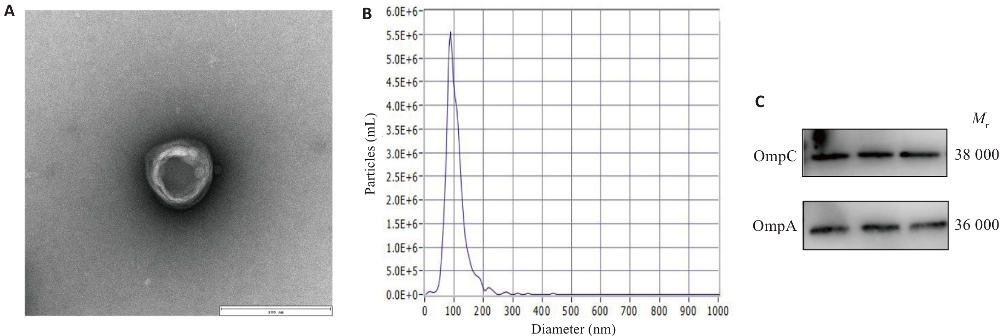
图1 E. coli-OMVs的提取与表征
Fig.1 Isolation and characterization of Escherichia coli outer membrane vesicles (E. coli-OMVs). A: Transmission electron microscopy of purified OMVs (Scale bar=200 nm). B: Size distribution of OMVs measured by NTA. C: Western blotting of OMV markers OmpA and OmpC.
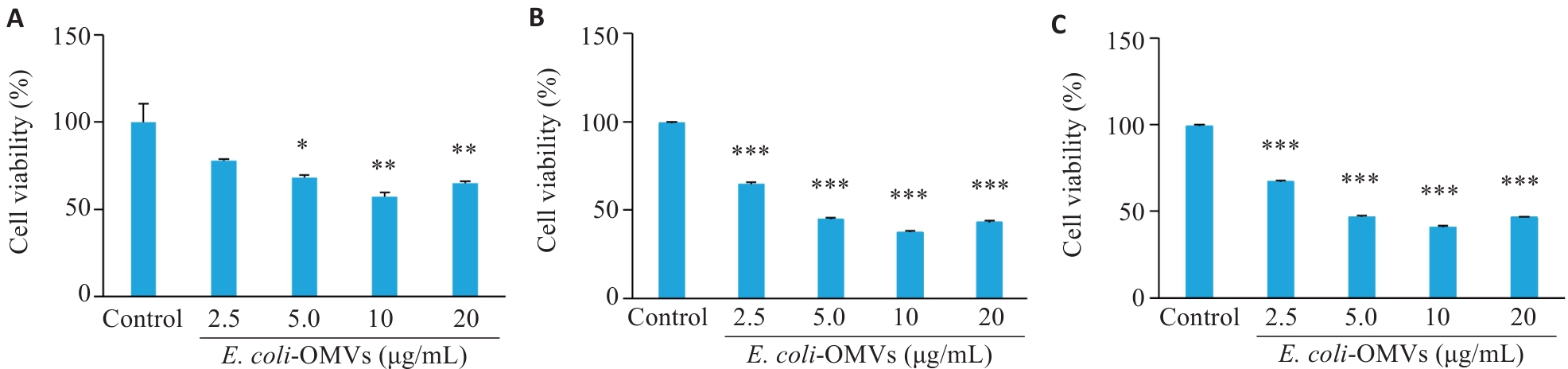
图2 CCK-8分析E. coli-OMVs对胶质瘤细胞活力的影响
Fig.2 CCK-8 analysis of the effect of E. coli-OMVs on glioma cell viability. A: Glioma cell viability after 12-h co-culture with E. coli-OMVs. B: Glioma cell viability after 24-h co-culture with E. coli-OMVs. C: Glioma cell viability after 48-h co-culture with E. coli-OMVs (n=3). *P<0.05, **P<0.01, ***P<0.001 vs control group.
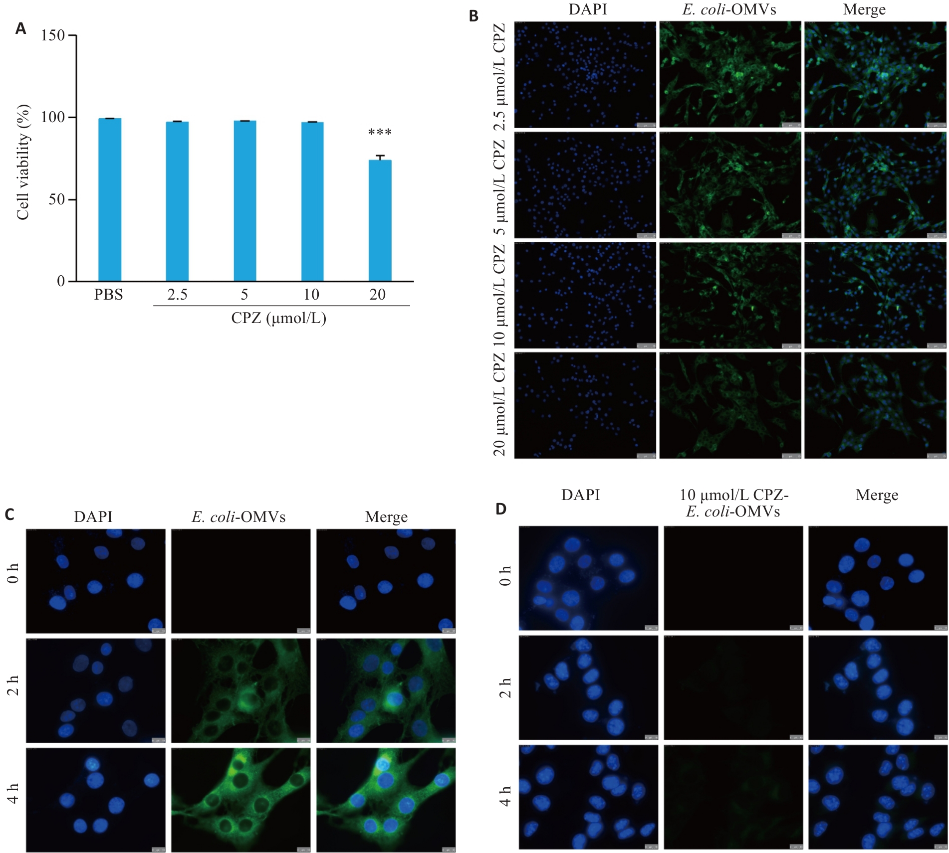
图3 CPZ对胶质瘤细胞摄取E. coli-OMVs的影响
Fig.3 Effect of CPZ on uptake of E. coli-OMVs by glioma cells. A: CCK-8 analysis of C6 cell viability after treatment with different concentrations of CPZ (n=3). B: Representative fluorescence images of C6 cells incubated with fluorescently labeled E. coli-OMVs after pretreatment with different concentrations of CPZ (Scale bar=75 μm). C: Representative fluorescence images of C6 cells incubated with fluorescently labeled E. coli-OMVs for 0 (control), 2 and 4 h (Scale bar=10 μm). D: Representative fluorescence images of C6 cells pretreated with 10 μmol/L CPZ and then incubated with fluorescently labeled E. coli-OMVs for 0, 2 and 4 h (Scale bar=10 μm). ***P<0.001 vs PBS.
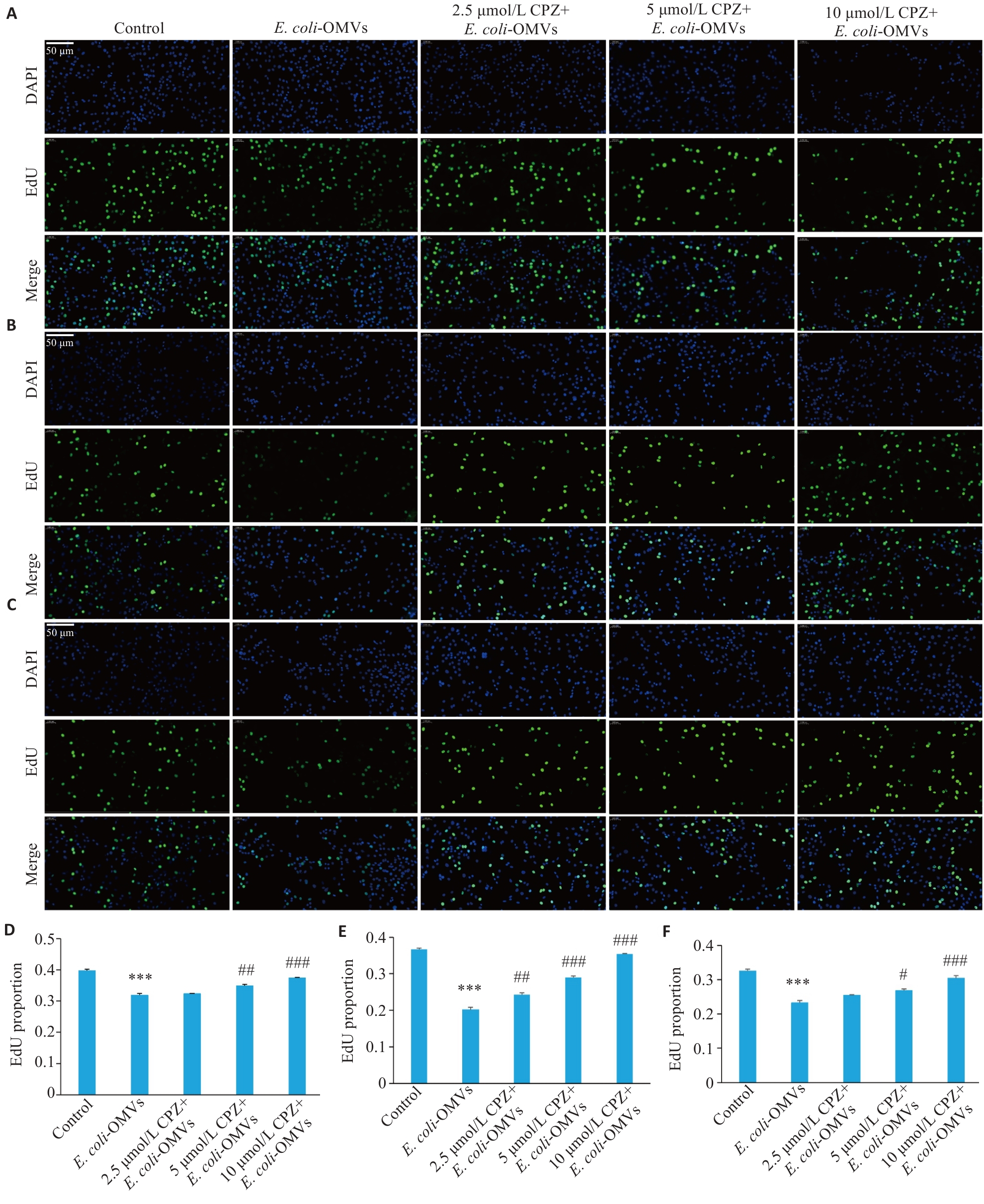
图4 CPZ对E. coli-OMVs抑制胶质瘤细胞体外增殖的影响
Fig.4 CPZ attenuates inhibitory effect of E. coli OMVs on glioma cell proliferation in vitro. A-C: Representative EdU staining images after 12, 24, and 48 h treatment (Scale bar=50 μm). D-F: Quantification of EdU-positive cells after 12, 24 and 48 h treatment (n=3). ***P<0.001 vs control. #P<0.05,##P<0.01, ###P<0.001 vs E. coli OMVs.
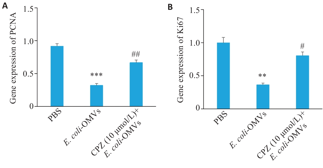
图5 CPZ对E. coli-OMVs调控胶质瘤细胞PCNA与Ki67转录水平的影响
Fig.5 Effect of CPZ on PCNA and Ki67 transcriptional levels in E. coli-OMVs-treated glioma cells. A: qRT-PCR analysis of PCNA mRNA expression levels. B: qRT-PCR analysis of Ki67 mRNA expression levels (n=3). **P<0.01, ***P<0.001 vs PBS group; #P<0.05, ##P<0.01 vs E. coli-OMVs group.
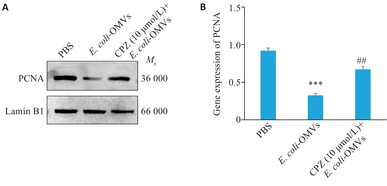
图 6 CPZ对E. coli-OMVs调控胶质瘤细胞PCNA蛋白表达的影响
Fig.6 Effect of CPZ on PCNA protein expression in E. coli-OMVs-treated glioma cells. A: Western blots showing PCNA protein levels. B: Quantitative analysis (grayscale values) of the protein bands (n=3). ***P<0.001 vs PBS group; ##P<0.01 vsE. coli-OMVs group.
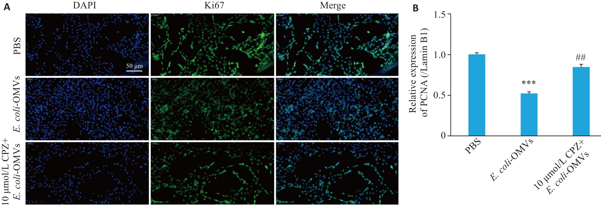
图7 CPZ对E. coli-OMVs调控胶质瘤细胞Ki67蛋白表达的影响
Fig.7 Effect of CPZ on Ki67 protein expression in E. coli-OMVs-treated glioma cells. A: Immunofluorescence images of Ki67 expression (Scale bar=50 μm). B: Quantitative analysis of Ki67-positive cells (n=3). ***P<0.001 vs PBS group; ##P<0.01 vs E. coli-OMVs group.
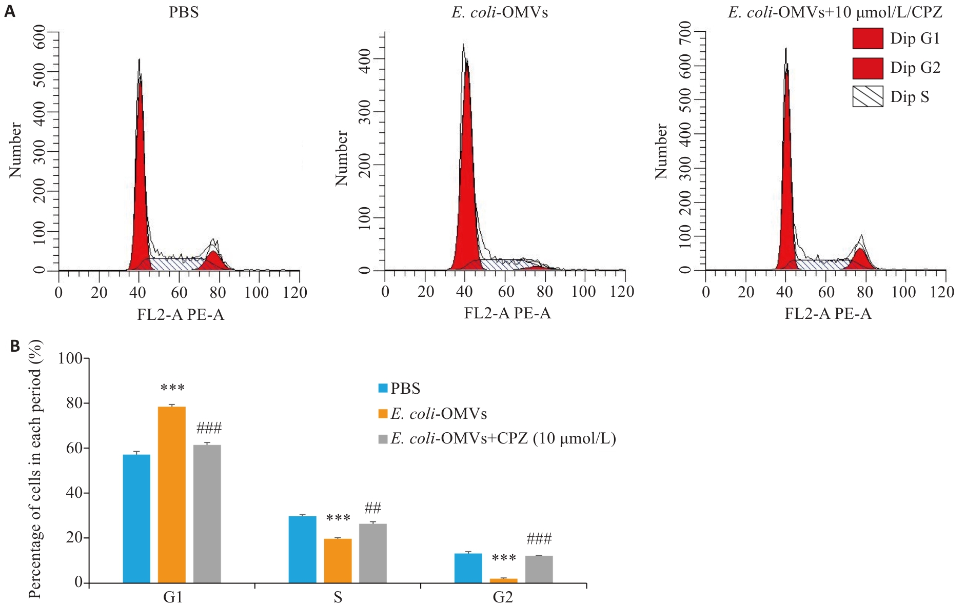
图8 CPZ 对 E. coli-OMVs 调控胶质瘤细胞周期分布的影响
Fig.8 Effect of CPZ on cell cycle distribution of E. coli-OMVs-treated glioma cells. A: Flow cytometry of C6 cell cycle distribution in PBS, E. coli-OMVs and CPZ+E. coli-OMVs groups. B: Quantitative analysis of the percentages of cells in G0/G1, S and G2/M phases (n=3). ***P<0.001 vs PBS group; ##P<0.01, ###P<0.001 vs E. coli-OMVs group.
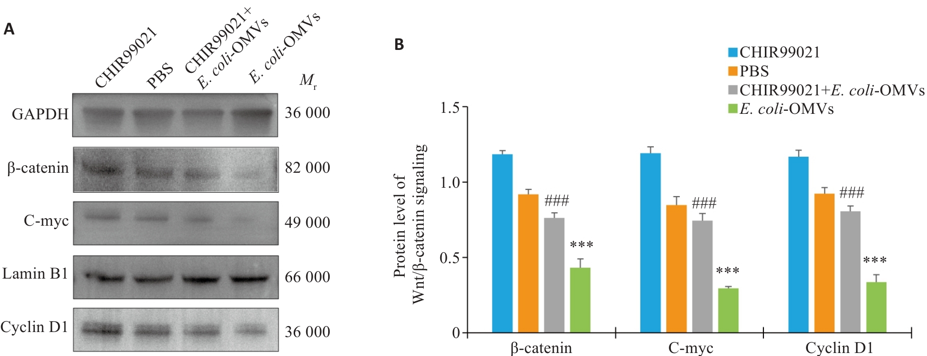
图9 E. coli-OMVs 通过调控 Wnt/β-catenin 信号通路抑制胶质瘤细胞增殖相关蛋白表达
Fig.9 E. coli-OMVs inhibit expressions of glioma cell proliferation-related proteins by regulating the Wnt/β-catenin signaling pathway. A: Western blots of β‑catenin, cyclin D1 and c-Myc proteins in PBS, OMVs, CHIR99021 and CHIR99021+OMVs groups. B: Quantitative analysis of β-catenin, cyclin D1 and c-Myc protein expression levels. (n=3). ***P<0.001 vs PBS group; ###P<0.01 vs E. coli- OMVs group.
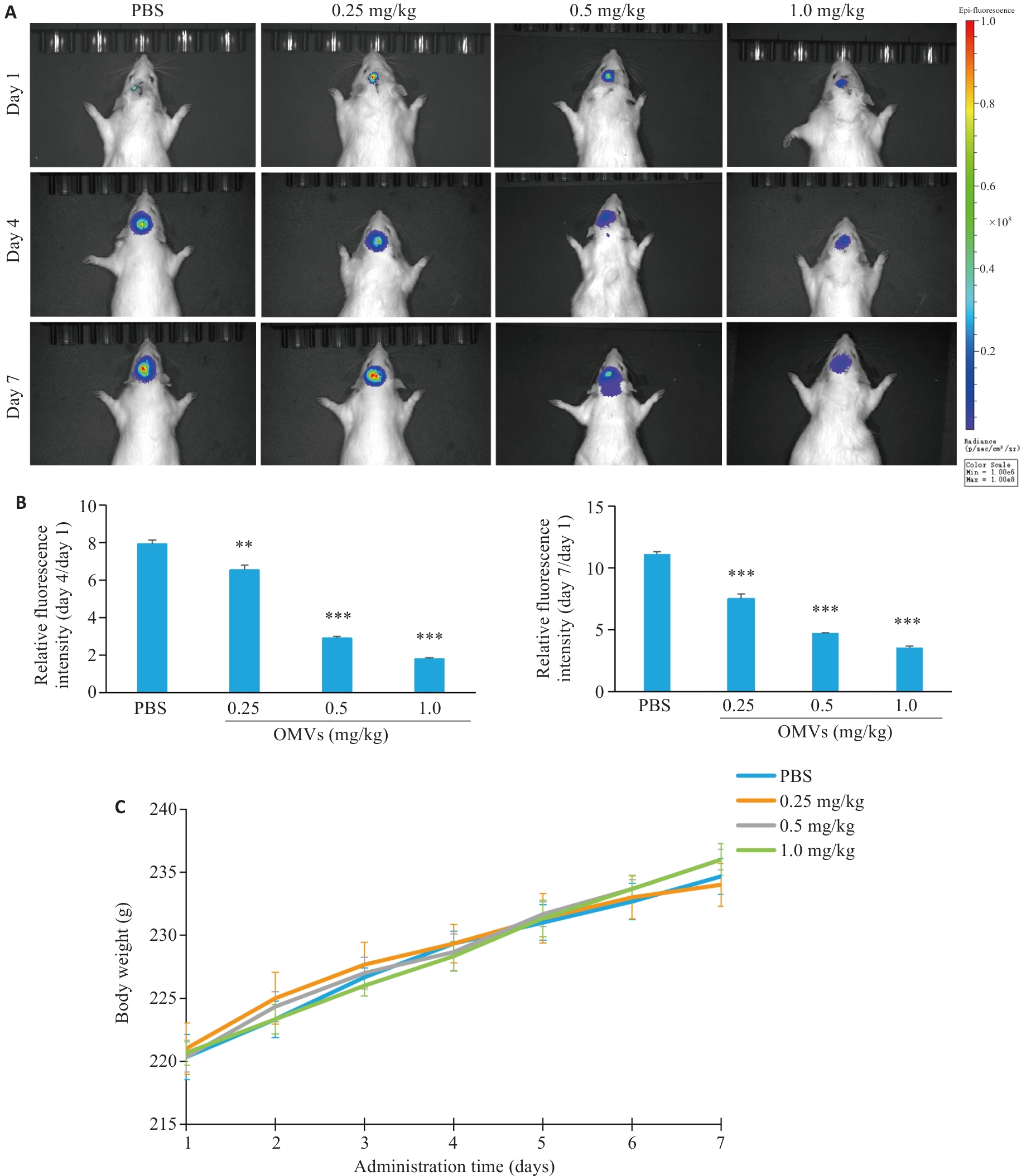
图10 E. coli-OMVs 鼻腔给药对大鼠原位胶质瘤生长的影响
Fig.10 Effect of intranasal administration of E. coli-OMVs on growth of orthotopic glioma in rats. A: In vivo bioluminescence images of orthotopic glioma-bearing rats on day 1, day 4, and day 7 after administration. B: Quantitative analysis of relative fluorescence intensity on day 4 (Day4/Day1). C: Quantitative analysis of relative fluorescence intensity on day 7 (Day7/Day1). D: Changes in body weight of rats in each group during treatment. OMVs: Outer membrane vesicles (n=3). **P<0.01, ***P<0.001 vs control group.

图11 E. coli-OMVs 鼻腔给药对大鼠原位胶质瘤组织形态的影响
Fig.11 Effect of intranasal administration of E. coli-OMVs on histopathological changes in orthotopic glioma tissues of the rats (Scale bar=20 μm).
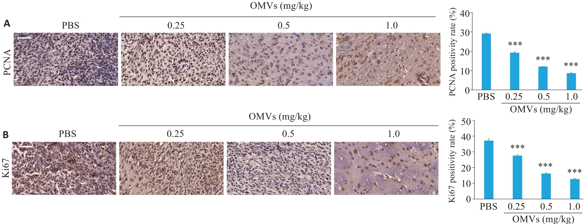
图12 E. coli-OMVs 鼻腔给药对大鼠原位胶质瘤组织中增殖相关蛋白表达的影响
Fig.12 Effect of intranasal administration of E. coli-OMVs on expressions of proliferation-related proteins in orthotopic glioma tissues of the rats (Scale bar=20 μm). A: Immunohistochemical staining and quantitative analysis of PCNA expression in brain tumor tissues in each group. B: Immunohistochemical staining and quantitative analysis of Ki67 expression in brain tumor tissues in each group (n=3). ***P<0.001 vs PBS group.
| [1] | Obrador E, Moreno-Murciano P, Oriol-Caballo M, et al. Glioblastoma therapy: past, present and future[J]. Int J Mol Sci, 2024, 25(5):2529. doi:10.3390/ijms25052529 |
| [2] | Ostrom QT, Price M, Neff C, et al. CBTRUS statistical report: primary brain and other central nervous system tumors diagnosed in the United States in 2015-2019[J]. Neuro Oncol, 2022, 24(): v1-v95. doi:10.1093/neuonc/noac202 |
| [3] | Wen PY, Kesari S. Malignant gliomas in adults[J]. N Engl J Med, 2008, 359(5): 492-507. doi:10.1056/nejmra0708126 |
| [4] | 黄秋虎, 周 建, 王子珍, 等. miR-26b-3p靶向CREB1调控神经胶质瘤细胞的增殖、迁移及侵袭[J]. 南方医科大学学报, 2024, 44(3): 578-84. doi:10.12122/j.issn.1673-4254.2024.03.20 |
| [5] | Chang C, Chavarro VS, Gerstl JVE, et al. Recurrent glioblastoma: molecular underpinnings and evolving treatment paradigms[J]. Int J Mol Sci, 2024, 25(12): 6733. doi:10.3390/ijms25126733 |
| [6] | Tang J, Karbhari N, Campian JL. Therapeutic targets in glioblastoma: molecular pathways, emerging strategies, and future directions[J]. Cells, 2025, 14(7): 494. doi:10.3390/cells14070494 |
| [7] | Schwechheimer C, Kuehn MJ. Outer-membrane vesicles from Gram-negative bacteria: biogenesis and functions[J]. Nat Rev Microbiol, 2015, 13(10): 605-19. doi:10.1038/nrmicro3525 |
| [8] | Toyofuku M, Nomura N, Eberl L. Types and origins of bacterial membrane vesicles[J]. Nat Rev Microbiol, 2019, 17(1): 13-24. doi:10.1038/s41579-018-0112-2 |
| [9] | Deng YF, Yang BY, Yang ZL, et al. Engineered E. coli OMVs carrying the membrane-binding hGC33 fragment precisely target liver cancer and effectively treat tumor[J]. Int J Nanomed, 2025, 20: 6573-90. doi:10.2147/ijn.s513508 |
| [10] | Jiang Y, Ma J, Long YQ, et al. Extracellular membrane vesicles of Escherichia coli induce apoptosis of CT26 colon carcinoma cells[J]. Microorganisms, 2024, 12(7): 1446. doi:10.3390/microorganisms12071446 |
| [11] | 王斓茜, 马官荣, 姜咏竹, 等. 大肠埃希菌外膜囊泡对乳腺癌细胞增殖及荷瘤小鼠肿瘤生长的影响[J]. 上海交通大学学报: 医学版, 2023, 43(10): 1245-54. doi:10.3969/j.issn.1674-8115.2023.10.004 |
| [12] | Scholzen T, Gerdes J. The Ki-67 protein: From the known and the unknown[J]. J Cell Phys, 2000, 182(3): 311-22. doi:10.1002/(sici)1097-4652(200003)182:3<311::aid-jcp1>3.0.co;2-9 |
| [13] | Wang X, Wang D, Yuan N, et al. The prognostic value of PCNA expression in patients with osteosarcoma: a meta-analysis of 16 studies[J]. Medicine, 2017, 96(41): e8254. doi:10.1097/md.0000000000008254 |
| [14] | Darweesh MF, Elfarouk LO, Negm MS. Evaluation of the proliferation marker Ki-67 in glioblastoma and its correlation with the histopathological findings[J]. Academic Journal of Cancer Research, 2016, 9(2): 13-8. |
| [15] | Garcia P, Mercier O, Ravent J, et al. Protocol for cell proliferation and cell death analysis of primary muscle stem cell culture using flow cytometry[J]. STAR Protoc, 2024, 5(4): 103411. doi:10.1016/j.xpro.2024.103411 |
| [16] | Salic A, Mitchison TJ. A chemical method for fast and sensitive detection of DNA synthesis in vivo [J]. Proc Natl Acad Sci USA, 2008, 105(7): 2415-20. doi:10.1073/pnas.0712168105 |
| [17] | Wang LH, Rothberg KG, Anderson RG. Mis-assembly of clathrin lattices on endosomes reveals a regulatory switch for coated pit formation[J]. J Cell Biol, 1993, 123(5): 1107-17. doi:10.1083/jcb.123.5.1107 |
| [18] | O' Donoghue EJ, Krachler AM. Mechanisms of outer membrane vesicle entry into host cells: MicroReview-OMV entry into host cells[J]. Cell Microbiol, 2016, 18(11): 1508-17. doi:10.1111/cmi.12655 |
| [19] | Kaparakis-Liaskos M, Ferrero RL. Immune modulation by bacterial outer membrane vesicles[J]. Nat Rev Immunol, 2015, 15(6): 375-87. doi:10.1038/nri3837 |
| [20] | Sales Gil R, Vagnarelli P. Ki-67: more hidden behind a 'classic proliferation marker'[J]. Trends Biochem Sci, 2018, 43(10): 747-8. doi:10.1016/j.tibs.2018.08.004 |
| [21] | Li DD, Zhang JH, Zhang K, et al. Proliferating cell nuclear antigen (PCNA) overexpression in hepatocellular carcinoma predicts poor prognosis as determined by bioinformatic analysis[J]. Chinese Medical Journal, 2020, 133(7): 848-50. doi:10.1097/cm9.0000000000001192 |
| [22] | Uxa S, Bernhart SH, Mages CF, et al. Ki-67 gene expression: characterization of the regulatory mechanism during cell cycle progression[J]. Cell Death Differ vol, 28, 12 (2021): 3357-70. |
| [23] | Lee Y, Lee JK, Ahn SH, et al. WNT signaling in glioblastoma and therapeutic opportunities[J]. Lab Investig, 2016, 96(2): 137-50. doi:10.1038/labinvest.2015.140 |
| [24] | Nager M, Bhardwaj D, Cantí C, et al. β‑catenin signalling in glioblastoma multiforme and glioma-initiating cells[J]. Chemother Res Pract,2012: 192362. doi:10.1155/2012/192362 |
| [25] | Chronopoulos A, Kalluri R. Emerging role of bacterial extracellular vesicles in cancer[J]. Oncogene, 2022,39(46):6951-60. |
| [26] | Zhao XF, Wei YS, Bu YQ, et al. Review on bacterial outer membrane vesicles: structure, vesicle formation, separation and biotechnological applications[J]. Microb Cell Fact, 2025, 24(1): 27. doi:10.1186/s12934-025-02653-9 |
| [27] | Barbieri G, Stefano A, Pietrantoni A, et al. Characterization of the small RNAs carried by outer membrane vesicles produced by hlyF-positive Shiga toxin-producing Escherichia coli [J]. Front Cell Infect Microbiol, 2025, 15: 1621341. doi:10.3389/fcimb.2025.1621341 |
| [28] | Hosseini-Giv N, Basas A, Hicks C, et al. Bacterial extracellular vesicles and their novel therapeutic applications in health and cancer[J]. Front Cell Infect Microbiol, 2022, 12: 962216. doi:10.3389/fcimb.2022.962216 |
| [29] | Liu C, Yazdani N, Moran CS, et al. Unveiling clinical applications of bacterial extracellular vesicles as natural nanomaterials in disease diagnosis and therapeutics[J]. Acta Biomater, 2024, 175: 1-20. doi:10.1016/j.actbio.2024.04.022 |
| [30] | Guo J, Huang Z, Wang Q, et al. Opportunities and challenges of bacterial extracellular vesicles in regenerative medicine[J]. J Nanobiotechnology, 2025, 23(1): 4. doi:10.1186/s12951-024-02935-1 |
| [31] | Olovo CV, Ocansey DKW, Ji Y, et al. Bacterial membrane vesicles in the pathogenesis and treatment of inflammatory bowel disease[J]. Gut Microbes, 2024,16(1):2341670. doi:10.1080/19490976.2024.2341670 |
| [32] | Sun DS, Chen P, Xi Y, et al. From trash to treasure: the role of bacterial extracellular vesicles in gut health and disease[J]. Front Immunol, 2023, 14: 1274295. doi:10.3389/fimmu.2023.1274295 |
| [33] | Mowday AM, Lieuwes NG, Biemans R, et al. Use of a luciferase-expressing orthotopic rat brain tumor model to optimize a targeted irradiation strategy for efficacy testing with temozolomide[J]. Cancers (Basel), 2020, 12(6): 1585. doi:10.3390/cancers12061585 |
| [34] | Bruinsmann FA, Vaz GR, Alves ACDS, et al. Nasal drug delivery of anticancer drugs for the treatment of glioblastoma: preclinical and clinical trials[J]. Molecules, 2019, 24(23): 4312. doi:10.3390/molecules24234312 |
| [1] | 庞一丹, 刘雅, 陈思嫒, 张荆雷, 曾今, 潘元明, 安娟. SPAG5在胃癌细胞恶性增殖中的生物学作用[J]. 南方医科大学学报, 2024, 44(8): 1497-1507. |
| [2] | 王月桂, 洪理伟, 吕国荣, 杨舒萍, 李 玲, 黄学鹏, 沈浩霖. 超声联合Ki67分析Graves病患者131I治疗后甲亢未愈或复发的相关因素[J]. 南方医科大学学报, 2022, 42(12): 1902-1906. |
| [3] | 蒋富杰, 杨海燕, 王 璐, 汪瑶台, 唐 瑜, 王狄森, 王 琦, 邹建中. 表达气体囊泡的大肠杆菌能安全地增强高强度聚焦超声的消融效果[J]. 南方医科大学学报, 2021, 41(5): 649-656. |
| [4] | 张汉运,高 杰,何肖龙,龚泽龙,万 宇,胡彤彤,李煜彬,曹 虹. 鼠李糖乳杆菌效应蛋白HM0539增强小鼠对大肠杆菌O157:H7肠道感染的抵抗力[J]. 南方医科大学学报, 2020, 40(02): 211-218. |
| [5] | 杜艳丽,韩宗利,王湘雨,万成松. 大肠杆菌O157:H7 体内多聚磷酸盐的DAPI荧光定量检测[J]. 南方医科大学学报, 2019, 39(03): 344-. |
| [6] | 王震,黄文,岑柏宏,魏媛怡,廖路敏,黎国仙,季爱民. siRNA介导的PD-L1 沉默可增强人CD8+T淋巴细胞的体外杀伤作用[J]. 南方医科大学学报, 2018, 38(07): 800-. |
| [7] | 唐波,何大维,李典,郭文浩,张丹,魏光辉. 大肠杆菌胞外囊泡对人神经母细胞瘤SK-N-SH细胞增殖、凋亡和迁移的影响[J]. 南方医科大学学报, 2018, 38(03): 334-. |
| [8] | 廖晓红,尹蔚兰,王芳,邬力祥,黄柏胜. shRNA-PAX6慢病毒载体构建及其对胶质瘤U251细胞增殖的影响[J]. 南方医科大学学报, 2017, 37(12): 1603-. |
| [9] | 李易非,史飞涛,吴承蓉,杨芳. MK、Ki67在胃癌中异常表达及其与胃癌的临床关系[J]. 南方医科大学学报, 2014, 34(10): 1534-. |
| [10] | 彭亮,潘嘉韵,罗苏,杨正慧,黄慕芳,曹虹. ppk1基因缺失对脑膜炎大肠杆菌K1株生物学功能的影响[J]. 南方医科大学学报, 2014, 34(07): 965-. |
| [11] | 屈娅荣,何肖龙,王勤,张立科,龙敏,罗军,张文炳,曹虹. 尿路致病性大肠杆菌外膜蛋白T的致病机制[J]. 南方医科大学学报, 2014, 34(02): 174-. |
| [12] | 苑召虎,胡子有,张兰兰,颜晓慧,王惠丽,吴炳义 . 槲皮素对胶质瘤U87细胞侵袭、迁移、增殖及其细胞周期的影响[J]. 南方医科大学学报, 2013, 33(02): 207-. |
| [13] | 王宏强,祝葆华,刘聪,方唯意,杨慧龄. 低氘水可选择性抑制鼻咽癌细胞增殖[J]. 南方医科大学学报, 2012, 32(10): 1394-. |
| [14] | 李岩; 鲍朗; 张会东; 李娅莎; 朱海龙;. 表达结核杆菌ESAT-6基因重组耻垢分枝杆菌的构建及其功能研究[J]. 南方医科大学学报, 2006, 26(07): 923-926. |
| [15] | 胡静1, 俞守义1, 阚飙2, 刘志华3. 缺失HPI毒力岛的EAggEC O42突变菌株的构建及鉴定[J]. 南方医科大学学报, 2005, 25(11): 1352-1356. |
| 阅读次数 | ||||||
|
全文 |
|
|||||
|
摘要 |
|
|||||
