| 1 |
Wu XM, Xu MY, Geng MY, et al. Targeting protein modifications in metabolic diseases: molecular mechanisms and targeted therapies[J]. Signal Transduct Target Ther, 2023, 8(1): 220.
|
| 2 |
Tobias DK, Merino J, Ahmad A, et al. Second international consensus report on gaps and opportunities for the clinical translation of precision diabetes medicine[J]. Nat Med, 2023, 29(10): 2438-57.
|
| 3 |
Lee YS, Olefsky J. Chronic tissue inflammation and metabolic disease[J]. Genes Dev, 2021, 35(5/6): 307-28.
|
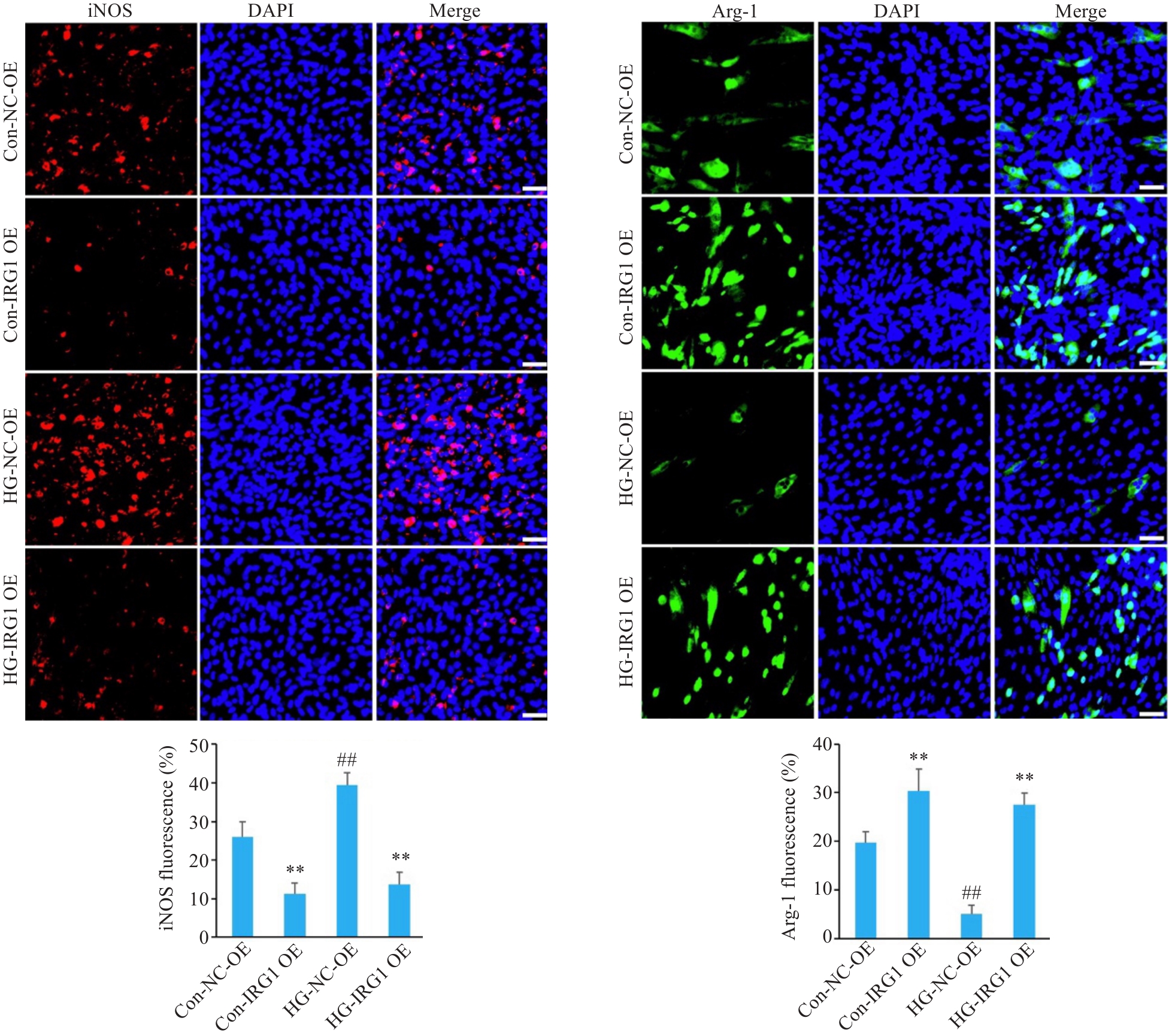
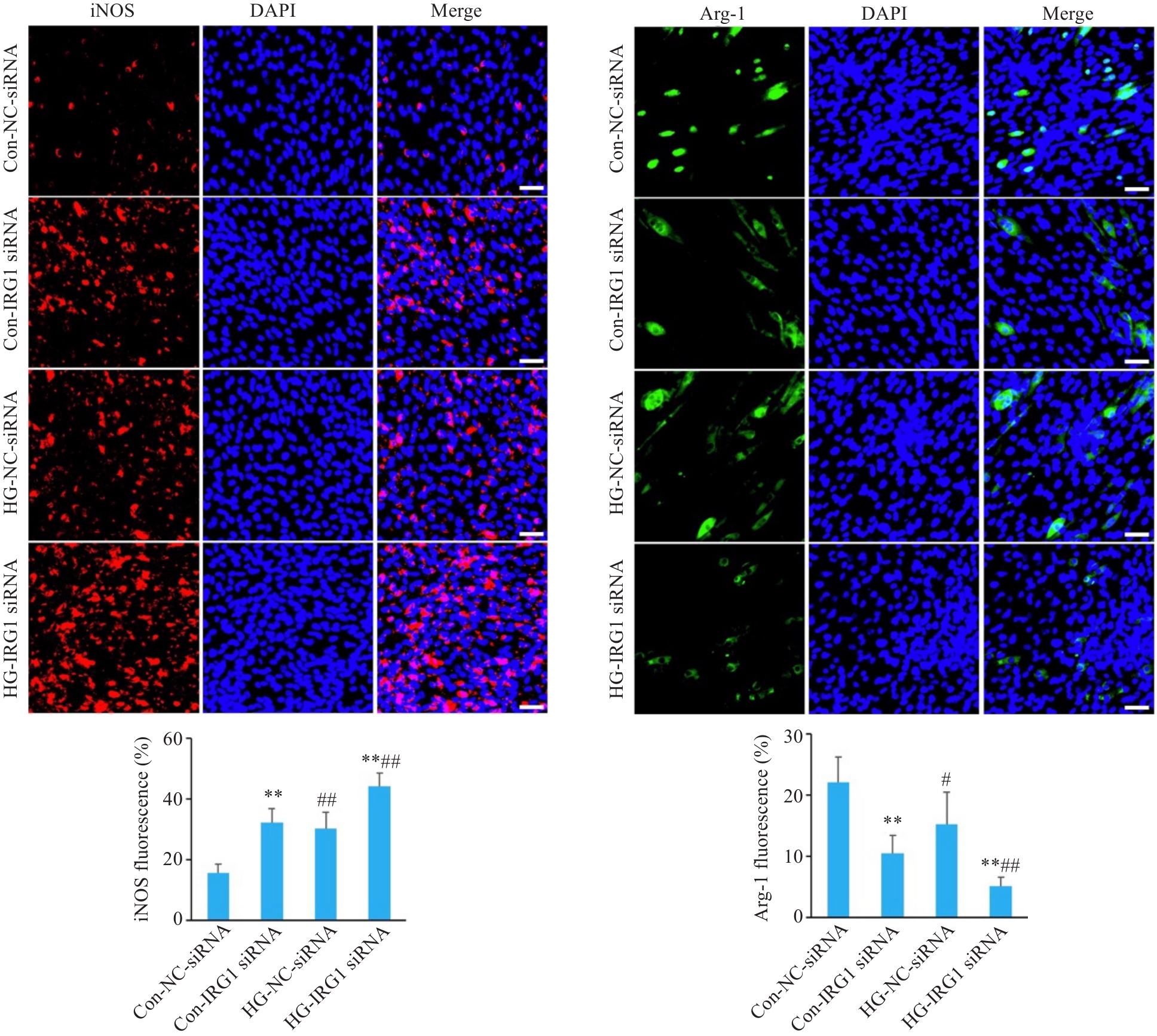
| 4 |
Franceschi C, Garagnani P, Parini P, et al. Inflammaging: a new immune-metabolic viewpoint for age-related diseases[J]. Nat Rev Endocrinol, 2018, 14(10): 576-90.
|
| 5 |
Azzu V, Vacca M, Virtue S, et al. Adipose tissue-liver cross talk in the control of whole-body metabolism: implications in nonalcoholic fatty liver disease[J]. Gastroenterology, 2020, 158(7): 1899-912.
|
| 6 |
Hasegawa Y, Okamura T, Nakajima H, et al. Metabolic outcomes and changes in innate immunity induced by diesel exhaust particles airway exposure and high-fat high-sucrose diet[J]. Life Sci, 2023, 326: 121794.
|
| 7 |
Truong TMT, Seo SH, Chung S, et al. Attenuation of hepatic fibrosis by p-Coumaric acid via modulation of NLRP3 inflammasome activation in C57BL/6 mice[J]. J Nutr Biochem, 2023, 112: 109204.
|
| 8 |
Darwish NM, Elnahas YM, AlQahtany FS. Diabetes induced renal complications by leukocyte activation of nuclear factor κ-B and its regulated genes expression[J]. Saudi J Biol Sci, 2021, 28(1): 541-9.
|
| 9 |
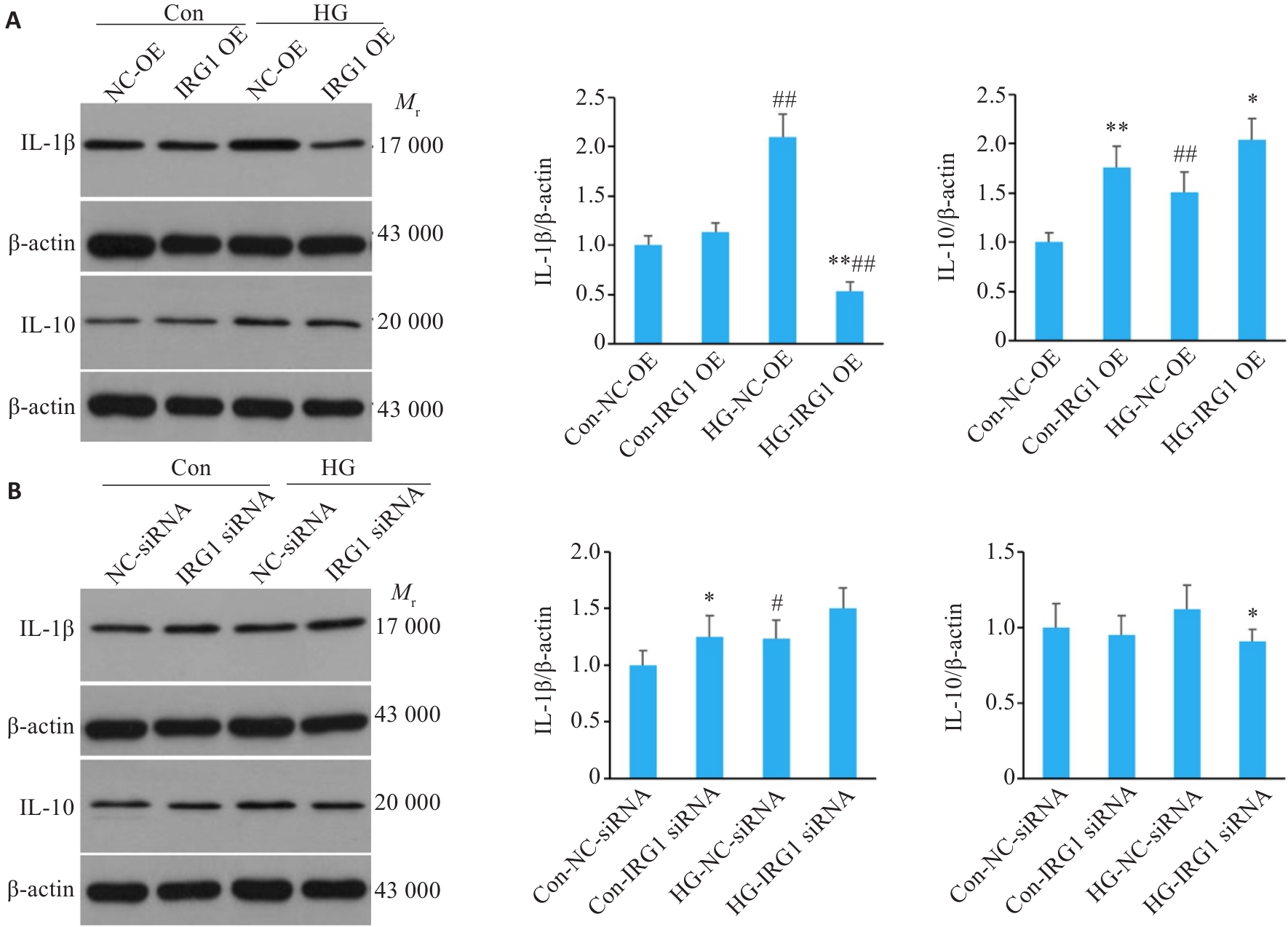
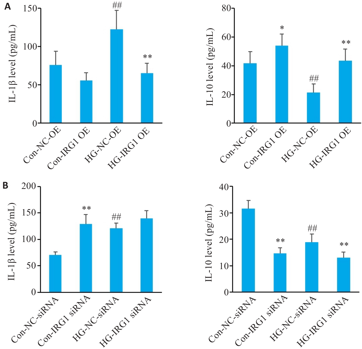
Li C, Xu MM, Wang KP, et al. Macrophage polarization and meta-inflammation[J]. Transl Res, 2018, 191: 29-44.
|
| 10 |
Li H, Meng Y, He SW, et al. Macrophages, chronic inflammation, and insulin resistance[J]. Cells, 2022, 11(19): 3001.
|
| 11 |
Wang C, Ma C, Gong LH, et al. Macrophage polarization and its role in liver disease[J]. Front Immunol, 2021, 12: 803037.
|
| 12 |
Zhang J, Muri J, Fitzgerald G, et al. Endothelial lactate controls muscle regeneration from ischemia by inducing M2-like macrophage polarization[J]. Cell Metab, 2020, 31(6): 1136-53. e7.
|
| 13 |
Cutolo M, Campitiello R, Gotelli E, et al. The role of M1/M2 macrophage polarization in rheumatoid arthritis synovitis[J]. Front Immunol, 2022, 13: 867260.
|
| 14 |
Li YG, Gong WB, Li WZ, et al. The IRG1-Itaconate axis: a regulatory hub for immunity and metabolism in macrophages[J]. Int Rev Immunol, 2023, 42(5): 364-78.
|
| 15 |
Chen LL, Morcelle C, Cheng ZL, et al. Itaconate inhibits TET DNA dioxygenases to dampen inflammatory responses[J]. Nat Cell Biol, 2022, 24(3): 353-63.
|
| 16 |
Lei I, Huang W, Noly PE, et al. Metabolic reprogramming by immune-responsive gene 1 up-regulation improves donor heart pres-ervation and function[J]. Sci Transl Med, 2023, 15(682): eade3782.
|
| 17 |
Sendra M, Saco A, Rey-Campos M, et al. Immune-responsive gene 1 (IRG1) and dimethyl itaconate are involved in the mussel immune response[J]. Fish Shellfish Immunol, 2020, 106: 645-55.
|
| 18 |
罗 维, 艾 磊, 周 越.小鼠巨噬细胞RAW264.7细胞电穿孔转染条件的建立和验证[J].中国实验动物学报, 2020, 28(2): 193-200.
|
| 19 |
Johansson C, Kirsebom FCM. Neutrophils in respiratory viral infections[J]. Mucosal Immunol, 2021, 14(4): 815-27.
|
| 20 |
夏崇建, 黄俊杰, 张 煜, 等.膜联蛋白A1的N末端肽Ac2-26对高糖刺激的小鼠巨噬细胞极化的影响[J].中国病理生理杂志, 2022, 38(11): 1998-2004.
|
| 21 |
Luo W, Zhou Y, Tang Q, et al. Downhill running and caloric restriction attenuate insulin resistance associated skeletal muscle atrophy via the promotion of M2-like macrophages through TRIB3-AKT pathway[J]. Free Radic Biol Med, 2024, 210: 271-85.
|
| 22 |
Kieler M, Hofmann M, Schabbauer G. More than just protein building blocks: how amino acids and related metabolic pathways fuel macrophage polarization[J]. FEBS J, 2021, 288(12): 3694-714.
|
| 23 |
Luo W, Zhou Y, Tang Q, et al. Modulation of TRIB3 and macrophage phenotype to attenuate insulin resistance after downhill running in mice[J]. Front Physiol, 2021, 12: 637432.
|
| 24 |
罗 维, 艾 磊, 王博发, 等. 慢性高糖作用下小鼠骨骼肌成肌细胞对巨噬细胞极化的影响[J]. 免疫学杂志, 2020, 36(11): 935-42.
|
| 25 |
Xia YY, He F, Wu XY, et al. GABA transporter sustains IL-1β production in macrophages[J]. Sci Adv, 2021, 7(15): eabe9274.
|
| 26 |
Eddie Ip WKE, Hoshi N, Shouval DS, et al. Anti-inflammatory effect of IL-10 mediated by metabolic reprogramming of macrophages[J]. Science, 2017, 356(6337): 513-9.
|
| 27 |
徐龙飞, 韩 晶, 杨 喆, 等. LRG1抑制小鼠肝巨噬细胞活化从而改善代谢相关脂肪性肝病: 基于增强TGF-β1信号通路[J]. 南方医科大学学报, 2023, 43(7): 1164-71.
|
| 28 |
Li YK, Zhang P, Wang CC, et al. Immune responsive gene 1 (IRG1) promotes endotoxin tolerance by increasing A20 expression in macrophages through reactive oxygen species[J]. J Biol Chem, 2013, 288(23): 16225-34.
|
| 29 |
Domínguez-Andrés J, Novakovic B, Li Y, et al. The itaconate pathway is a central regulatory node linking innate immune toler-ance and trained immunity[J]. Cell Metab, 2019, 29(1): 211-20. e5.
|
| 30 |
Mills EL, Kelly B, Logan A, et al. Succinate dehydrogenase supports metabolic repurposing of mitochondria to drive inflammatory macrophages[J]. Cell, 2016, 167(2): 457-70. e13.
|
 ), 王宇航1, 刘延松1, 王媛媛1, 艾磊2(