
南方医科大学学报 ›› 2026, Vol. 46 ›› Issue (10): 2536-2547.doi: 10.12122/j.issn.1673-4254.2026.10.21
• • 上一篇
刘燕1( ), 张洪齐2()
收稿日期:2026-01-29
出版日期:2026-10-20
发布日期:2026-10-26
通讯作者:
张洪齐
E-mail:yanliucqmu@163.com;zhanghongqicqmu@163.com
作者简介:刘 燕,在读硕士研究生,E-mail: yanliucqmu@163.com
基金资助:
Yan LIU1(), Hongqi ZHANG2()
Received:2026-01-29
Online:2026-10-20
Published:2026-10-26
Contact:
Hongqi ZHANG
E-mail:yanliucqmu@163.com;zhanghongqicqmu@163.com
摘要:
目的 探究紫丁香苷(SYR)对脓毒症小鼠心肌的保护作用及其机制。 方法 使用C57小鼠构建脓毒症模型,分为假手术组、假手术+SYR组、脓毒症(CLP)组、CLP+SYR组、CLP+SYR+3-TYP组(SIRT3抑制剂)。采用超声检测心功能,HE染色评估心肌损伤,ELISA检测血清cTnt、LDH、MDA、SOD、GSH-Px水平;免疫组化和荧光检测活性氧水平以及p-DRP1、MFN1、NOX4表达;Western blotting分析SIRT3/NOX2/p-DRP1等通路蛋白;原代小鼠心肌细胞(MNCMs)使用LPS处理以构建脓毒症模型,分为对照组、LPS组、LPS+SYR组、LPS+SYR+3-TYP组、LPS+Honokiol组。免疫荧光检测线粒体膜电位变化。Western blotting分析SIRT3/NOX2/p-DRP1通路蛋白表达水平。 结果 与假手术组相比,CLP组心功能下降,心肌组织损伤增加,血清cTnt, LDH含量增加(P<0.01),氧化应激标志蛋白和线粒体损伤蛋白升高(P<0.01)。与CLP组相比,CLP+SYR组心脏组织结构完整,心功能恢复,线粒体损伤减轻,氧化应激减少。与CLP+SYR组相比,CLP+SYR+3-TYP组的MDA和cTnt升高(P<0.01),SOD下降,活性氧水平增加,氧化应激标志蛋白和线粒体损伤蛋白升高(P<0.01)。在NMCMs中,LPS处理导致线粒体膜电位降低,AC-SOD2, NOX2和p-DRP1蛋白升高(P<0.01),MFN1水平下降。LPS+SYR处理可保护细胞免受LPS诱导的氧化应激和线粒体损伤,但这一作用被3-TYP抑制,此外,使用小干扰RNA抑制SIRT3蛋白后,SYR的保护作用消失。 结论 SYR通过激活SIRT3的表达,可有效减轻脓毒症诱导的心肌损伤,改善心功能,抑制氧化应激并减轻线粒体损伤。
刘燕, 张洪齐. 紫丁香苷通过激活SIRT3改善脓毒症小鼠心肌线粒体动力学和减轻氧化应激损伤[J]. 南方医科大学学报, 2026, 46(10): 2536-2547.
Yan LIU, Hongqi ZHANG. Syringin improves myocardial mitochondrial dynamics and alleviates oxidative stress injury in septic mice by activating SIRT3[J]. Journal of Southern Medical University, 2026, 46(10): 2536-2547.
| Gene | Forward primer | Reverse primer |
|---|---|---|
| NOX2 | TGCCAACTTCCTCAGCTACAATA | GTGTTCACTTGCAATGGTCTTGA |
| NRF2 | TGGCAGAGACATTCCCATTTGTA | CTTGCTCCATGTCCTGCTCTATG |
| NOX4 | CCTCCTGGCTGCATTAGTCTTAA | AAAGGCACAAAGGTCCAGAAATC |
| GAPDH | GGCATTGTGGAAGGGCTCAT | AGATCCACGACGGACACATT |
表1 引物序列
Tab.1 Primer sequences for RT-qPCR
| Gene | Forward primer | Reverse primer |
|---|---|---|
| NOX2 | TGCCAACTTCCTCAGCTACAATA | GTGTTCACTTGCAATGGTCTTGA |
| NRF2 | TGGCAGAGACATTCCCATTTGTA | CTTGCTCCATGTCCTGCTCTATG |
| NOX4 | CCTCCTGGCTGCATTAGTCTTAA | AAAGGCACAAAGGTCCAGAAATC |
| GAPDH | GGCATTGTGGAAGGGCTCAT | AGATCCACGACGGACACATT |
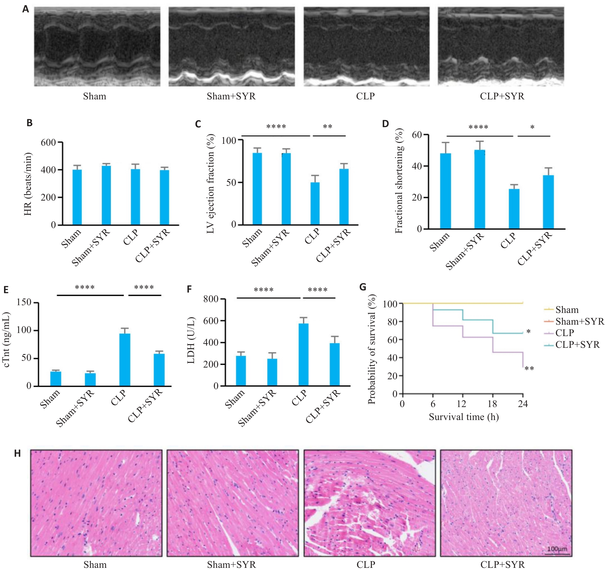
图1 SYR减轻脓毒症小鼠心肌损伤
Fig.1 Syringin (SYR) alleviates myocardial injury in mice with sepsis. A: Representative left ventricular M-mode echocardiograms of the mice in different groups. B: Statistical analysis of heart rate (n=6). C: Statistical analysis of LVEF (n=6). D: Statistical analysis of LVFS (n=6). E: Serum cTnt levels in the mice. F: LDH activity in mouse serum. G: Survival rates of the mice with or without SYR therapy 24 h after cecal ligation and puncture (CLP) (n=20). Time (h) refers to the survival hours after sepsis. H: HE staining (Scale bar=100 μm). *P<0.05, **P<0.01, ****P<0.0001.
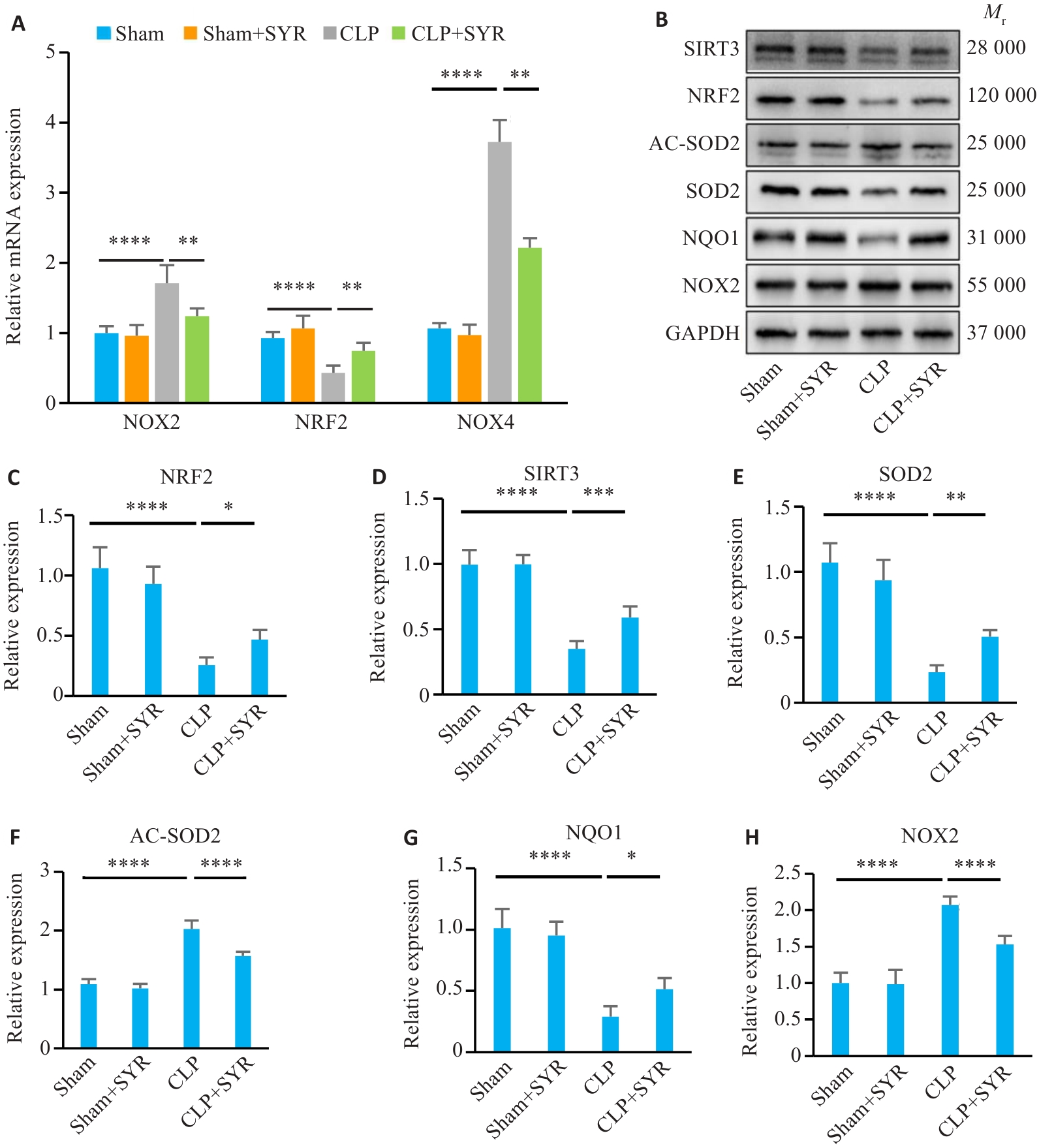
图2 SYR治疗减轻脓毒症小鼠氧化应激相关基因和蛋白表达
Fig.2 SYR treatment alleviates sepsis-induced aberrant expressions of oxidative stress-related genes and proteins in mice. A: NOX2, NRF2 and NOX4 mRNA levels in mouse myocardial tissue. B: Target proteins examined by Western blotting. C: Statistical analysis of NRF2/GAPDH (n=6). D: Statistical analysis of SIRT3/GAPDH (n=6). E: Statistical analysis of SOD2/GAPDH (n=6). F: Statistical analysis of AC-SOD2/GAPDH (n=6). G: Statistical analysis of NQO1/GAPDH (n=6). H: Statistical analysis of NOX2/GAPDH (n=6). Comparisons among multiple groups were made using one-way ANOVA. *P<0.05, **P<0.01, ***P<0.001, ****P<0.0001.
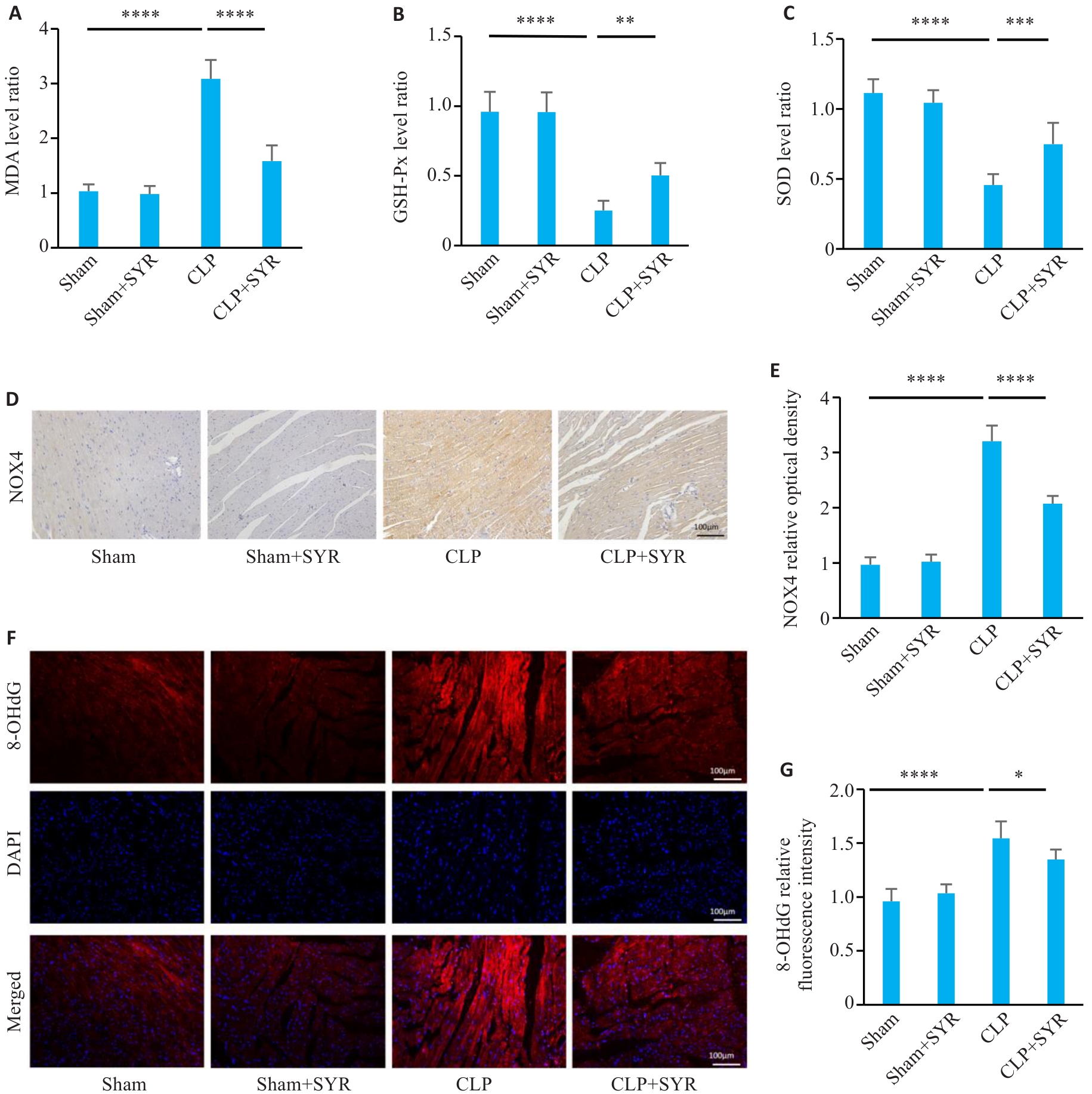
图3 SYR治疗减轻脓毒症小鼠血清和组织氧化应激水平
Fig.3 SYR treatment reduces the levels of oxidative stress in the serum and tissues of septic mice. A: MDA content in mouse serum (n=6). B: GSH-Px content in mouse serum (n=6). C: SOD content in mouse serum (n=6). D: Expression and location of NOX4 determined by immunohistochemistry (Scale bar=100 μm). E: Statistical analysis of NOX4 area (n=6). F: Expression and location of 8-OHdG determined by immunofluorescence staining (Scale bar=100 μm). G: Statistical analysis of fluorescence intensity (n=6). *P<0.05, **P<0.01, ***P<0.001, ****P<0.0001.
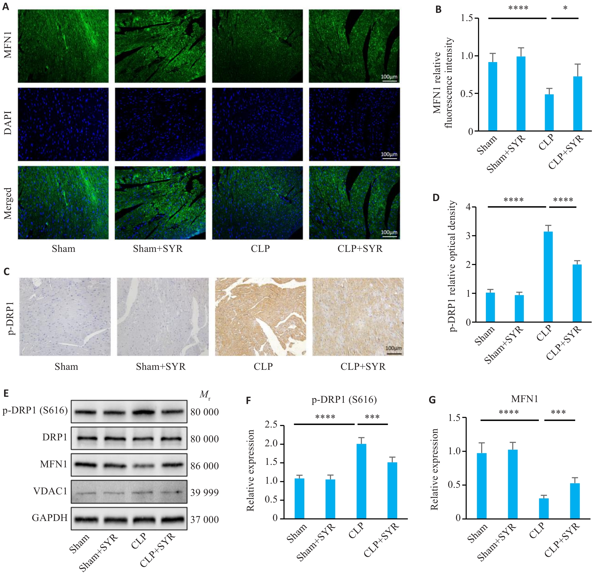
图4 SYR治疗减轻脓毒症小鼠线粒体损伤
Fig.4 SYR treatment alleviates mitochondrial damage in septic mice. A: Expression and location of MFN1 determined by immunofluorescence staining (Scale bar=100 μm). B: Statistical analysis of fluorescence intensity (n=6). C: Expression and location of p-DRP1 were determined by immunohistochemistry (Scale bar=100 μm). D: Statistical analysis of p-DRP1 area (n=6). E: Target proteins examined by Western blotting. F: Statistical analysis of p-DRP1(S616)/DRP1 (n=6). G: Statistical analysis of MFN1/GAPDH (n=6). *P<0.05, ***P<0.001, ****P<0.0001.
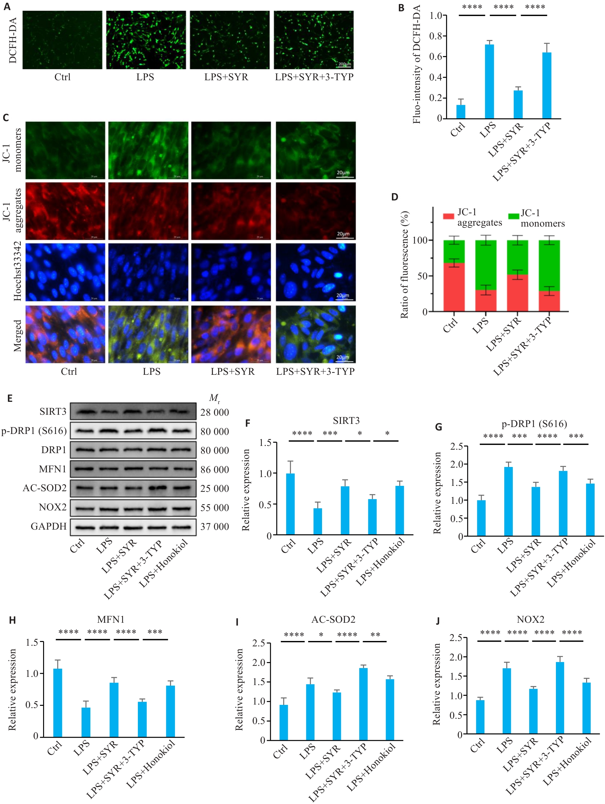
图5 SYR通过激活SIRT3减轻新生小鼠心肌细胞氧化应激和线粒体损伤
Fig.5 SYR attenuates oxidative stress and mitochondrial damage in primary culture of neonatal mouse cardiomyocytes (NMCMs) by activating SIRT3. A: Expression of DCFH-DA determined by immunofluorescence staining (Scale bar=200 μm). B: Statistical analysis of fluorescence intensity (n=6). C: JC-1 staining for measuring the mitochondrial membrane potential in NMCMs showing depolarization (green) and hyperpolarization (red) of the mitochondria. D: Statistical analysis of fluorescence intensity (n=6). E: Target proteins examined by Western blotting. F: Statistical analysis of SIRT3/GAPDH (n=6). G: Statistical analysis of p-DRP1/DRP1 (n=6). H: Statistical analysis of MFN1/GAPDH (n=6). I: Statistical analysis of AC-SOD2/GAPDH (n=6). J: Statistical analysis of NOX2/GAPDH (n=6). *P<0.05, **P<0.01, ***P<0.001, ****P<0.0001.
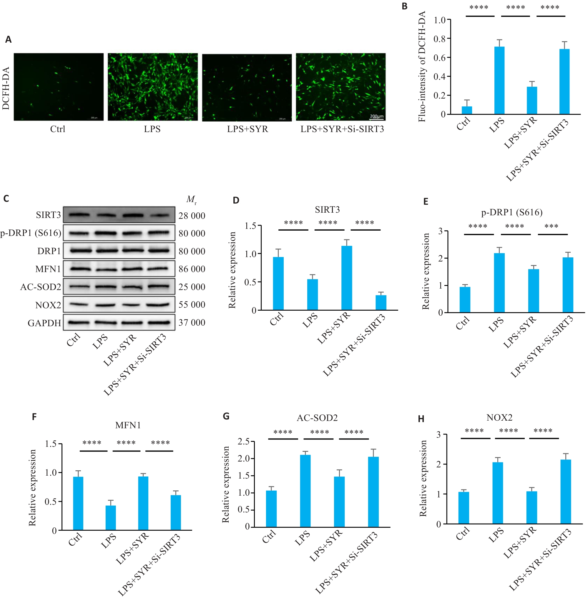
图6 抑制SIRT3可消除SYR对新生小鼠心肌细胞氧化应激抑制和线粒体的保护作用
Fig.6 Inhibiting SIRT3 eliminates the inhibitory effects of SYR on oxidative stress in neonatal mouse cardiomyocytes and its protective effect on mitochondria. A: Expression of DCFH-DA determined by immunofluorescence staining (Scale bar=200 μm). B: Statistical analysis of fluorescence intensity (n=6). C: Target proteins examined by Western blotting. D: Statistical analysis of SIRT3/GAPDH (n=6). E: Statistical analysis of p-DRP1/DRP1 (n=6). F: Statistical analysis of MFN1/GAPDH (n=6). G: Statistical analysis of AC-SOD2/GAPDH (n=6). H: Statistical analysis of NOX2/GAPDH (n=6). ***P<0.001, ****P<0.0001.
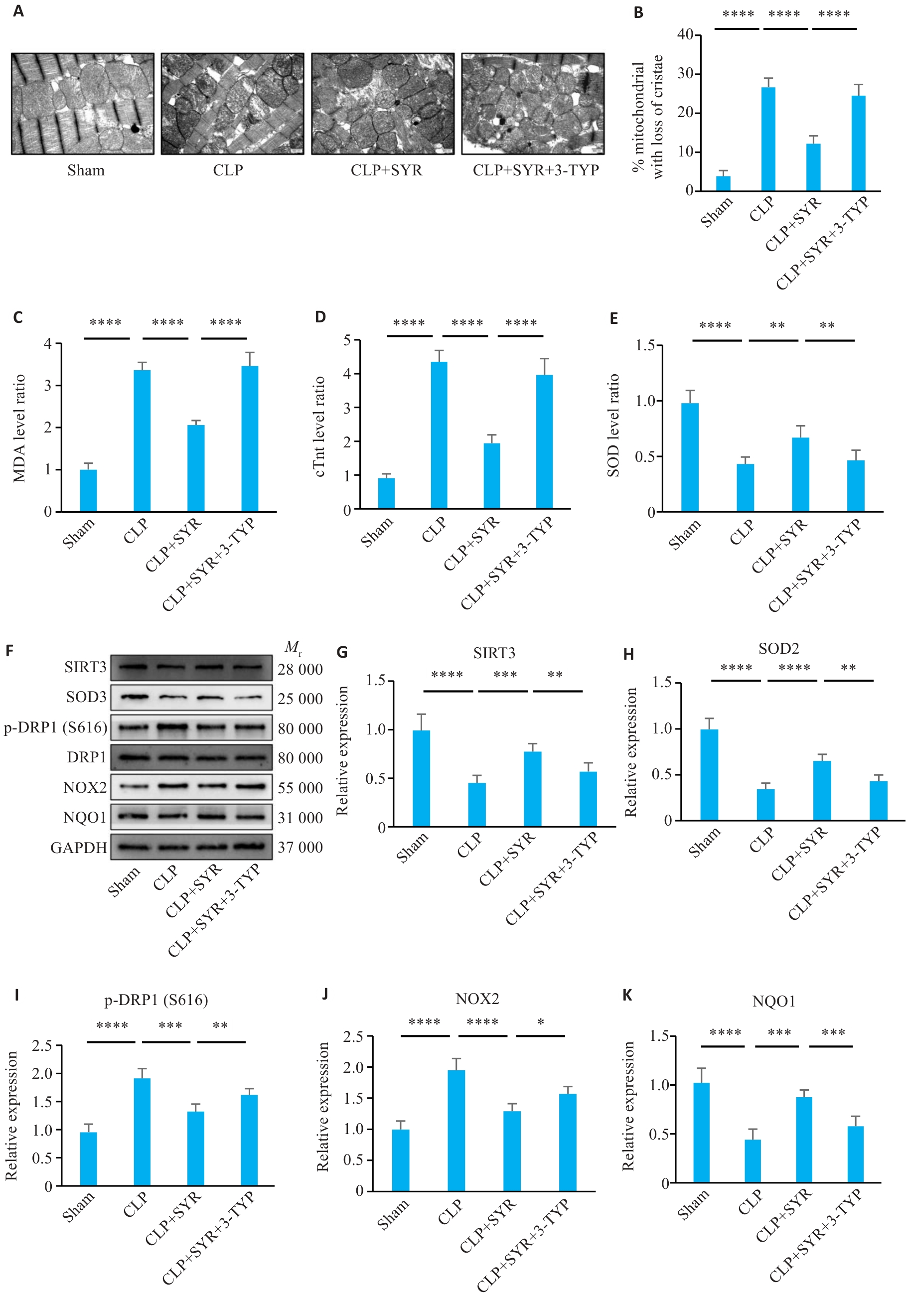
图7 SYR通过激活SIRT3从而在脓毒症小鼠心肌中发挥保护作用
Fig.7 SYR produces protective effect on the myocardium in septic mice by activating SIRT3. A: Representative electron microscopy images showing damaged mitochondrial with loss of cristae in mouse cardiomyocytes (Scale bar=500 nm). B: Quantification of damaged mitochondria in mouse cardiomyocytes (n=6). C: MDA content in mouse serum (n=6). D: cTnt content in mouse (n=6). E: SOD content in mouse serum (n=6). F: Target proteins examined by Western blotting. G: Statistical analysis of SIRT3/GAPDH (n=6). H: Statistical analysis of SOD2/GAPDH (n=6). I: Statistical analysis of p-DRP1/DRP1 (n=6). J: Statistical analysis of NOX2/GAPDH (n=6). K: Statistical analysis of NQO1/GAPDH (n=6). *P<0.05, **P<0.01, ***P<0.001, ****P<0.0001.
| [1] | Sato R, Sanfilippo F, Lanspa M, et al. Sepsis-induced cardio-myopathy[J]. Chest, 2025, 168(6): 1383-94. doi:10.1016/j.chest.2025.08.013 |
| [2] | Pamporis K, Karakasis P, Pantelidaki A, et al. Sepsis-induced cardiomyopathy and cardiac arrhythmias: pathophysiology and implications for novel therapeutic approaches[J]. Biomedicines, 2025, 13(11): 2643. doi:10.3390/biomedicines13112643 |
| [3] | Hasegawa D, Ishisaka Y, Maeda T, et al. Prevalence and prognosis of sepsis-induced cardiomyopathy: a systematic review and meta-analysis[J]. J Intensive Care Med, 2023, 38(9): 797-808. doi:10.1177/08850666231180526 |
| [4] | Shvilkina T, Shapiro N. Sepsis-Induced myocardial dysfunction: heterogeneity of functional effects and clinical significance[J]. Front Cardiovasc Med, 2023, 10: 1200441. doi:10.3389/fcvm.2023.1200441 |
| [5] | L' Heureux M, Sternberg M, Brath L, et al. Sepsis-induced cardiomyopathy: a comprehensive review[J]. Curr Cardiol Rep, 2020, 22(5): 35. doi:10.1007/s11886-020-01277-2 |
| [6] | Marques E, Kramer R, Ryan DG. Multifaceted mitochondria in innate immunity[J]. npj Metab Health Dis, 2024, 2: 6. doi:10.1038/s44324-024-00008-3 |
| [7] | Kuroshima T, Kawaguchi S, Okada M. Current perspectives of mitochondria in sepsis-induced cardiomyopathy[J]. Int J Mol Sci, 2024, 25(9): 4710. doi:10.3390/ijms25094710 |
| [8] | Nedel W, Strogulski NR, Kopczynski A, et al. Assessment of mitochondrial function and its prognostic role in sepsis: a literature review[J]. Intensive Care Med Exp, 2024, 12(1): 107. doi:10.1186/s40635-024-00694-9 |
| [9] | Chen W, Zhao ZZ, Geng ZG, et al. Advances in mitochondria-nucleus crosstalk in septic cardiomyopathy[J]. Cell Biol Toxicol, 2025, 41(1): 136. doi:10.1007/s10565-025-10090-y |
| [10] | West AP, Shadel GS. Mitochondrial DNA in innate immune responses and inflammatory pathology[J]. Nat Rev Immunol, 2017, 17(6): 363-75. doi:10.1038/nri.2017.21 |
| [11] | Yao X, Carlson D, Sun YX, et al. Mitochondrial ROS induces cardiac inflammation via a pathway through mtDNA damage in a pneumonia-related sepsis model[J]. PLoS One, 2015, 10(10): e0139416. doi:10.1371/journal.pone.0139416 |
| [12] | Hu DX, Sheeja Prabhakaran H, Zhang YY, et al. Mitochondrial dysfunction in sepsis: mechanisms and therapeutic perspectives[J]. Crit Care, 2024, 28(1): 292. doi:10.1186/s13054-024-05069-w |
| [13] | 董永杰, 董 静, 岳 峰, 等. 紫丁香苷调节PD-1/PD-L1信号通路对胃癌细胞增殖、凋亡和免疫逃逸的影响 [J]. 中国老年学杂志, 2025, 45(17): 4304-8. |
| [14] | 任兰鑫, 左振魁, 王 琳, 等. 基于JAK2/STAT3信号通路探究紫丁香苷对溃疡性结肠炎肠上皮细胞凋亡的影响 [J]. 中药材, 2025, 48(6): 1528-33. |
| [15] | Niu HS, Liu IM, Cheng JT, et al. Hypoglycemic effect of syringin from Eleutherococcus senticosus in streptozotocin-induced diabetic rats[J]. Planta Med, 2008, 74(2): 109-13. doi:10.1055/s-2008-1034275 |
| [16] | Zhao D, Liu KT, Wang J, et al. Syringin exerts anti-inflammatory and antioxidant effects by regulating SIRT1 signaling in rat and cell models of acute myocardial infarction[J]. Immunity Inflam & Disease, 2023, 11(2): e775. doi:10.1002/iid3.775 |
| [17] | Yao JP, Li Y, Jin YP, et al. Synergistic cardioptotection by tilianin and syringin in diabetic cardiomyopathy involves interaction of TLR4/NF‑κB/NLRP3 and PGC1a/SIRT3 pathways[J]. Int Immunopharmacol, 2021, 96: 107728. doi:10.1016/j.intimp.2021.107728 |
| [18] | Xu ZW, Huang FJ, Wu ZM, et al. Syringin attenuates Alzheimer's disease-associated neuroinflammation by inhibiting NLRP3 inflammasome activation[J]. Phytomedicine, 2025, 148: 157454. doi:10.1016/j.phymed.2025.157454 |
| [19] | Wang WL, Feng XY, Guo X, et al. Syringin attenuates fibrogenesis and inflammation through SIRT3-regulated P2X7 receptor signaling pathway in hepatic fibrosis[J]. Phytomedicine, 2025, 148: 157461. doi:10.1016/j.phymed.2025.157461 |
| [20] | Peng YC, Xu J, Wei LY, et al. Melatonin alleviates sepsis-induced acute lung injury by inhibiting necroptosis via reducing circulating mtDNA release[J]. Mol Med, 2025, 31(1): 176. doi:10.1186/s10020-025-01228-z |
| [21] | Guo ML, Zhuang HJ, Su YM, et al. SIRT3 alleviates imiquimod-induced psoriatic dermatitis through deacetylation of XBP1s and modulation of TLR7/8 inducing IL-23 production in macrophages[J]. Front Immunol, 2023, 14: 1128543. doi:10.3389/fimmu.2023.1128543 |
| [22] | 彭煜策, 姜 毅, 马 丹, 等. 异牡荆素通过促进SIRT3表达减轻糖尿病小鼠的心肌氧化应激损伤 [J]. 南方医科大学学报,2026,46(2): 316-24. |
| [23] | Marconi GD, Della Rocca Y, Fonticoli L, et al. The beneficial effect of carvacrol in HL-1 cardiomyocytes treated with LPS-G: anti-inflammatory pathway investigations[J]. Antioxidants, 2022, 11(2): 386. doi:10.3390/antiox11020386 |
| [24] | Huang pian-pian, Fu J, Liu li-hua, et al. Honokiol antagonizes doxorubicin-induced cardiomyocyte senescence by inhibiting TXNIP-mediated NLRP3 inflammasome activation[J]. Int J Mol Med, 2019,45(1): 186-94. |
| [25] | Yu WL, Lyu JS, Jia LL, et al. Dexmedetomidine ameliorates hippocampus injury and cognitive dysfunction induced by hepatic ischemia/reperfusion by activating SIRT3-mediated mitophagy and inhibiting activation of the NLRP3 inflammasome in young rats[J]. Oxid Med Cell Longev, 2020, 2020: 7385458. doi:10.1155/2020/7385458 |
| [26] | Aissaoui N, Boissier F, Chew M, et al. Sepsis-induced cardiomyopathy[J]. Eur Heart J, 2025, 46(34): 3339-53. doi:10.1093/eurheartj/ehaf340 |
| [27] | Yu XT, Gao J, Zhang CX. Sepsis-induced cardiac dysfunction: mitochondria and energy metabolism[J]. Intensive Care Med Exp, 2025, 13(1): 20. doi:10.1186/s40635-025-00728-w |
| [28] | Haileselassie B, Mukherjee R, Joshi AU, et al. Drp1/Fis1 interaction mediates mitochondrial dysfunction in septic cardiomyopathy[J]. J Mol Cell Cardiol, 2019, 130: 160-9. doi:10.1016/j.yjmcc.2019.04.006 |
| [29] | Liu AB, Wang S, Shen Y, et al. The role of mitochondrial dysfunction and inflammatory response in the pathogenesis of sepsis-induced myocardial injury: a mechanistic study[J]. J Inflamm Res, 2025, 18: 15207-35. doi:10.2147/jir.s552730 |
| [30] | Bertozzi G, Ferrara M, Di Fazio A, et al. Oxidative stress in sepsis: a focus on cardiac pathology[J]. Int J Mol Sci, 2024, 25(5): 2912. doi:10.3390/ijms25052912 |
| [31] | Wan CL, Sun SY, Du YQ, et al. A study on the mechanism of action of Syringin in the treatment of type 2 diabetes by integrating metabolomics, network pharmacology, molecular docking technology and experimental validation[J]. Int Immunopharmacol, 2025, 167: 115716. doi:10.1016/j.intimp.2025.115716 |
| [32] | Cai XM, Wu YN, Liu FX, et al. Syringin alleviates ROS-induced acute lung injury by activating SIRT1/STAT6 signaling pathway to inhibit ferroptosis[J]. Tissue Cell, 2025, 93: 102698. doi:10.1016/j.tice.2024.102698 |
| [1] | 徐璐, 潘缘瑾, 汪雨辰, 陈振宇, 蒋伟, 於江泉, 郑瑞强. 血必净注射液通过上调磷脂转运蛋白调控巨噬细胞JNK/c-JUN通路改善脓毒症相关急性呼吸窘迫综合征[J]. 南方医科大学学报, 2026, 46(9): 2035-2045. |
| [2] | 陈元欢, 岳斌, 武权生, 毛海燕, 吉秀家, 褚翠君, 陈琳, 张雨知, 李冰霞, 张小花, 冯鹏. CaMKKβ/AMPK/HIF-1α信号通路通过调控线粒体自噬诱导子宫内膜异位症血管生成[J]. 南方医科大学学报, 2026, 46(9): 2046-2055. |
| [3] | 李晓敏, 徐洁, 高彩月, 朱季军, 王玉, 李冬冬, 王晓燕. circUSP25编码的线粒体蛋白促进结肠癌细胞增殖及迁移[J]. 南方医科大学学报, 2026, 46(9): 2123-2129. |
| [4] | 于梦洁, 赵安博, 郝艺璇, 王奕人, 刘月娴, 叶红伟, 高琴. 敲除脂质运载蛋白2通过抑制NF-κB通路改善脓毒症急性肺损伤小鼠血管内皮屏障功能[J]. 南方医科大学学报, 2026, 46(8): 1779-1789. |
| [5] | 乔通, 张龙涛, 章雨, 黄菊, 李晴晴, 耿志军, 胡建国, 李静. 异香草酸激活PPARγ通路改善线粒体功能缓解葡聚糖硫酸钠诱导的小鼠溃疡性结肠炎[J]. 南方医科大学学报, 2026, 46(7): 1487-1497. |
| [6] | 尹维帅, 李飞, 曹奕, 杨琪琪, 邓则天, 曹世杰, 孙永旗, 毕昕阳, 梁正鹏, 伍思嘉, 王保国. 化瘀通络灸联合神经干细胞移植对血管性痴呆大鼠Drp1/Mfn蛋白表达及线粒体动力学的影响[J]. 南方医科大学学报, 2026, 46(7): 1520-1532. |
| [7] | 李淼, 于桂莹, 王金城, 谢抗, 欧阳雨林, 王能, 温蕊嘉, 王伟, 李春. 金银花通过调控STAT3/PGC-1α轴减轻氧化应激改善缺血后心力衰竭[J]. 南方医科大学学报, 2026, 46(7): 1563-1575. |
| [8] | 郭晓喻, 吴家羽, 韩冰, 杨婷, 殷欢欢, 熊津怡, 谢汝佳. C/EBP环磷酸腺苷反应元件结合转录因子同源蛋白通过抑制内质网自噬并增强内质网-线粒体互作促进肝细胞焦亡[J]. 南方医科大学学报, 2026, 46(7): 1648-1659. |
| [9] | 王炎炎, 宋志会, 郭流漓, 肖扬, 陈瑞, 王怡. 通脉养心丸通过激活ERK信号通路增强线粒体功能减轻大鼠心肌缺血再灌注损伤[J]. 南方医科大学学报, 2026, 46(6): 1203-1215. |
| [10] | 李瑾, 贺一杰, 毕芳芳, 陈浚鑫, 范桢颜, 张宇, 陈恩宇, 肖鸿文, 吴云, 田华, 周宇宏. 靶向抑制miR-503可通过上调Apelin表达减轻小鼠心肌梗死损伤[J]. 南方医科大学学报, 2026, 46(6): 1235-1243. |
| [11] | 尹林, 章雨, 张可妮, 乔通, 张龙涛, 黄菊, 李静, 胡建国, 耿志军. 1,3-二咖啡酰奎宁酸通过抑制PI3K/Akt通路减轻氧化应激缓解葡聚糖硫酸钠诱导的小鼠结肠炎[J]. 南方医科大学学报, 2026, 46(6): 1244-1255. |
| [12] | 王梓渝, 伍静雯, 王涵冰, 徐艳, 姜春玲, 周莉. 红细胞分布宽度/红细胞压积与脓毒症患者28 d全因死亡率的关系[J]. 南方医科大学学报, 2026, 46(6): 1256-1266. |
| [13] | 于静, 储继军, 梁程程. 慢性铁过载通过铁代谢紊乱引发氧化应激与内分泌失调诱导小鼠卵巢储备功能减退[J]. 南方医科大学学报, 2026, 46(6): 1313-1322. |
| [14] | 宋明朗, 苏海, 夏洪兴, 陈诗雨, 邵丽丽, 蒋志滨. 痛泻要方通过调控SREBP-1/SCD1通路改善5-HT诱导的BRL 3A细胞氧化应激损伤[J]. 南方医科大学学报, 2026, 46(6): 1385-1394. |
| [15] | 祝金超, 邵宁宁, 黄泽彧, 刘燕青, 孟祥艳, 刘子泉, 孙予杰, 董津睿, 樊毫军. 薤白超细粉通过抑制铁死亡并调控NF-κB/STAT3信号轴缓解脂多糖诱导的小鼠急性肺损伤[J]. 南方医科大学学报, 2026, 46(5): 1006-1017. |
| 阅读次数 | ||||||
|
全文 |
|
|||||
|
摘要 |
|
|||||
