南方医科大学学报 ›› 2026, Vol. 46 ›› Issue (2): 345-352.doi: 10.12122/j.issn.1673-4254.2026.02.12
• 英文单位2和3反了 • 上一篇
魏金燕1( ), 郑海锐1, 赵云腾1, 于言1, 徐莹莹2, 李刚1,3()
收稿日期:2025-06-26
出版日期:2026-02-20
发布日期:2026-03-10
通讯作者:
李刚
E-mail:2320925662@qq.com;lg@smu.edu.cn
作者简介:魏金燕,在读硕士研究生,E-mail: 2320925662@qq.com
基金资助:
Jinyan WEI1(), Hairui ZHENG1, Yunteng ZHAO1, Yan YU1, Yingying XU2, Gang LI1,3()
Received:2025-06-26
Online:2026-02-20
Published:2026-03-10
Contact:
Gang LI
E-mail:2320925662@qq.com;lg@smu.edu.cn
摘要:
目的 开发一种经济、简便的人鼻粘膜类器官培养方法,用于建立EB病毒(EBV)体外感染模型。 方法 从手术获得的鼻息肉组织,经清洗、剪切、消化、过滤后得到细胞团,在无基质胶的条件下经动态悬浮培养扩增为未分化的鼻粘膜类器官,再通过14 d的促分化处理以形成分化的鼻粘膜类器官,通过免疫荧光、免疫组化鉴定类器官主要细胞组成。加入EBV体外感染鼻粘膜类器官,通过RT-qPCR检测病毒特异性基因EBNA1、BALF5的表达,并通过EBNA1免疫荧光染色确认病毒感染。 结果 动态悬浮培养法可在无基质胶的培养体系中,成功培养出由基底细胞、粘液细胞、纤毛细胞构成的鼻粘膜类器官。分化的鼻粘膜类器官高表达EBV相关受体EphA2、NRP1、NMHCⅡ-A。未分化和分化的鼻粘膜类器官均可被EBV感染,类器官中病毒复制随病毒暴露载量的增加而增加,分化类器官可能更有利于病毒复制(P<0.001)。 结论 无基质胶的动态悬浮培养法是一种经济、简便的鼻粘膜类器官建模方法,人鼻粘膜类器官能够作为EBV感染模型,为上皮EBV感染相关研究提供一个良好的平台。
魏金燕, 郑海锐, 赵云腾, 于言, 徐莹莹, 李刚. 人鼻粘膜类器官EB病毒感染模型的建立[J]. 南方医科大学学报, 2026, 46(2): 345-352.
Jinyan WEI, Hairui ZHENG, Yunteng ZHAO, Yan YU, Yingying XU, Gang LI. Establishment of an Epstein-Barr virus infection model using human nasal organoids[J]. Journal of Southern Medical University, 2026, 46(2): 345-352.
| Sample number | Source | Reason for surgery |
|---|---|---|
| ZZH-9580 | Nasal polyps | CRSwNP |
| LHX-4635 | Nasal polyps | CRSwNP |
| XWJ-4024 | Nasal polyps | CRSwNP |
| XCY-8640 | Nasal polyps | CRSwNP |
| LJL-3202 | Nasal polyps | CRSwNP |
| LHB-4635 | Nasal polyps | CRSwNP |
| TRJ-8166 | Nasal polyps | CRSwNP |
表1 鼻粘膜样本信息
Tab.1 Information of the nasal mucosa samples
| Sample number | Source | Reason for surgery |
|---|---|---|
| ZZH-9580 | Nasal polyps | CRSwNP |
| LHX-4635 | Nasal polyps | CRSwNP |
| XWJ-4024 | Nasal polyps | CRSwNP |
| XCY-8640 | Nasal polyps | CRSwNP |
| LJL-3202 | Nasal polyps | CRSwNP |
| LHB-4635 | Nasal polyps | CRSwNP |
| TRJ-8166 | Nasal polyps | CRSwNP |
| Antibody | Brand | Cat No. | dilution ratio |
|---|---|---|---|
| Anti-Cytokeratin 5 antibody | Abcam | ab75869 | 1:100 |
| Anti-Cytokeratin 14 antibody | Abcam | ab119695 | 1:100 |
| Anti-p63 antibody | Abcam | ab124762 | 1:100 |
| Anti-MUC5AC antibody | Abcam | ab198294 | 1:200 |
| Anti-beta Ⅳ tubulin antibody | Abcam | ab179509 | 1:200 |
| Anti-FoxJ1 antibody | Abcam | ab235445 | 1:2000 |
| COL4A2 Polyclonal antibody | Proteintech | 55131-1-AP | 1:300 |
| MYH9 Monoclonal antibody | Proteintech | 60233-1-IG | 1:200 |
| EPHA2 Monoclonal antibody | Proteintech | 66736-1-IG | 1:200 |
| Neuropilin 1/CD304 Recombinant antibody | Proteintech | 84429-5-RR | 1:300 |
| Anti-EBNA1 antibody | Abcam | ab316860 | 1:2000 |
| Goat anti-Rabbit IgG (H+L) Highly Cross-Adsorbed Secondary Antibody, Alexa Fluor™ 488 | Invitrogen | A11034 | 1:500 |
| Goat anti-Mouse IgG (H+L) Highly Cross-Adsorbed Secondary Antibody, Alexa Fluor™ 488 | Invitrogen | A11029 | 1:500 |
| HRP-conjugated Goat Anti-Rabbit/mouse IgG for IHC (ready to use) | Proteintech | PR30009 | No dilution needed |
表2 免疫荧光和免疫组化抗体信息
Tab.2 Antibodies for immunofluorescence assay and immunohistochemistry
| Antibody | Brand | Cat No. | dilution ratio |
|---|---|---|---|
| Anti-Cytokeratin 5 antibody | Abcam | ab75869 | 1:100 |
| Anti-Cytokeratin 14 antibody | Abcam | ab119695 | 1:100 |
| Anti-p63 antibody | Abcam | ab124762 | 1:100 |
| Anti-MUC5AC antibody | Abcam | ab198294 | 1:200 |
| Anti-beta Ⅳ tubulin antibody | Abcam | ab179509 | 1:200 |
| Anti-FoxJ1 antibody | Abcam | ab235445 | 1:2000 |
| COL4A2 Polyclonal antibody | Proteintech | 55131-1-AP | 1:300 |
| MYH9 Monoclonal antibody | Proteintech | 60233-1-IG | 1:200 |
| EPHA2 Monoclonal antibody | Proteintech | 66736-1-IG | 1:200 |
| Neuropilin 1/CD304 Recombinant antibody | Proteintech | 84429-5-RR | 1:300 |
| Anti-EBNA1 antibody | Abcam | ab316860 | 1:2000 |
| Goat anti-Rabbit IgG (H+L) Highly Cross-Adsorbed Secondary Antibody, Alexa Fluor™ 488 | Invitrogen | A11034 | 1:500 |
| Goat anti-Mouse IgG (H+L) Highly Cross-Adsorbed Secondary Antibody, Alexa Fluor™ 488 | Invitrogen | A11029 | 1:500 |
| HRP-conjugated Goat Anti-Rabbit/mouse IgG for IHC (ready to use) | Proteintech | PR30009 | No dilution needed |
| Gene | Foward (5'→3') | Reverse (5'→3') |
|---|---|---|
| GAPDH | GGAGCGAGATCCCTCCAAAAT | GGCTGTTGTCATACTTCTCATGG |
| EPHA2 | CCCGATGAGATCACCGTCAG | GGCACCGATATCCTGGAAGG |
| NMHC-ⅡA | GAGCAAATGGGCCTGCT | TGTTGTCGGGCATGGA |
| NRP1 | ACCCAAGTGAAAAATGCGAATG | CCTCCAAATCGAAGTGAGGGTT |
| EBNA1 | GGTCGTGGACGTGGAGAAAA | GGTGGAGACCCGGATGATG |
| BALF5 | GAGCGATCTTGGCAATCTCT | TGGTCATGGATCTGCTAAACC |
表3 RT-qPCR引物序列
Tab.3 Primer sequence for RT-qPCR
| Gene | Foward (5'→3') | Reverse (5'→3') |
|---|---|---|
| GAPDH | GGAGCGAGATCCCTCCAAAAT | GGCTGTTGTCATACTTCTCATGG |
| EPHA2 | CCCGATGAGATCACCGTCAG | GGCACCGATATCCTGGAAGG |
| NMHC-ⅡA | GAGCAAATGGGCCTGCT | TGTTGTCGGGCATGGA |
| NRP1 | ACCCAAGTGAAAAATGCGAATG | CCTCCAAATCGAAGTGAGGGTT |
| EBNA1 | GGTCGTGGACGTGGAGAAAA | GGTGGAGACCCGGATGATG |
| BALF5 | GAGCGATCTTGGCAATCTCT | TGGTCATGGATCTGCTAAACC |
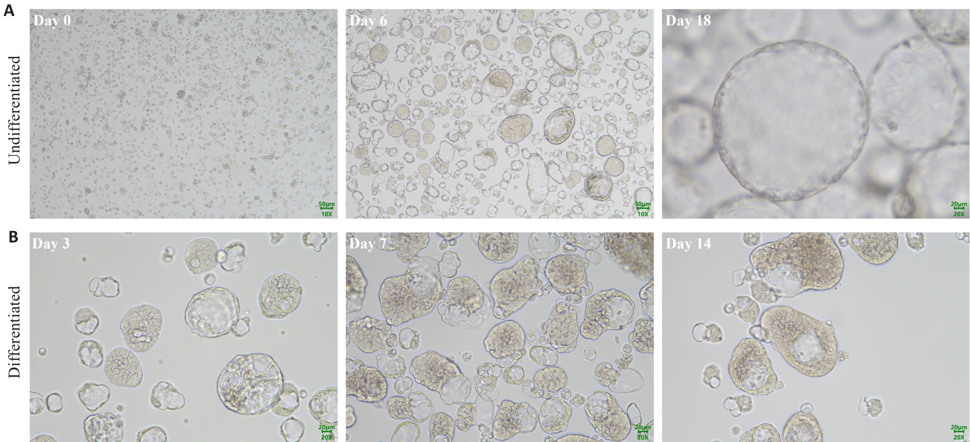
图1 鼻粘膜类器官培养光镜图
Fig.1 Phase-contrast microscopy of nasal organoids in culture. A: Representative images at different time points during the expansion of nasal organoids. B: Representative images of the nasal organoids on days 3, 7, and 14 during differentiation induction.
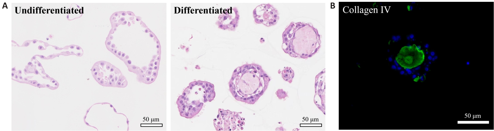
图2 鼻粘膜类器官HE染色结果及基底膜标志物染色
Fig.2 HE staining of the nasal organoids and immunofluorescence staining for the basal lamina marker collagen IV. A: HE staining of undifferentiated and differentiated nasal organoids. The differentiated nasal organoids exhibit a higher density of cilia on their apical surface. B: Immunofluorescence staining for collagen IV in differentiated nasal organoids.
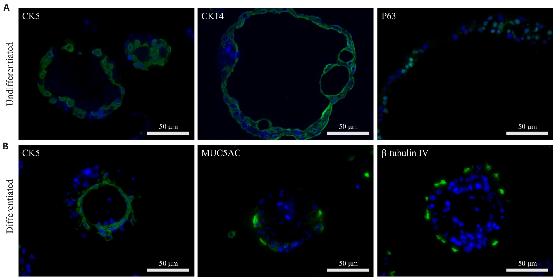
图3 未分化和分化的鼻粘膜类器官组成细胞免疫荧光
Fig.3 Immunofluorescence analysis of cellular composition of undifferentiated and differentiated nasal organoids. A: Representative images of undifferentiated organoids showing positive expressions of epithelial basal stem cell markers CK5, CK14, and p63. B: Representative images of differentiated organoids consisting of basal cells (CK5), goblet cells (MUC5AC), and ciliated cells (β-tubulin IV).
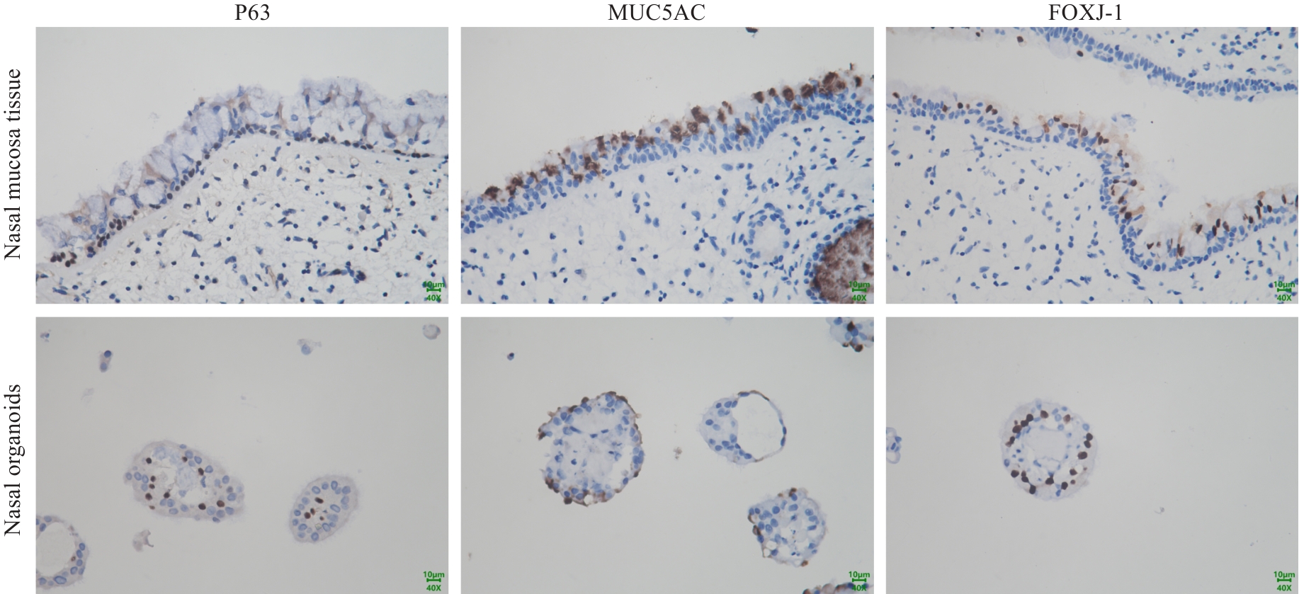
图 4 鼻粘膜组织和类器官组成细胞免疫组化
Fig.4 Immunohistochemical analysis of cellular composition of nasal mucosal tissue and organoids. Representative images of nasal mucosal tissue are shown in the upper panel and differentiated nasal organoids in the lower panel. The organoids exhibit comparable expression patterns to nasal mucosal tissue for P63 (basal cells), MUC5AC (goblet cells), and FOXJ-1 (ciliated cells).
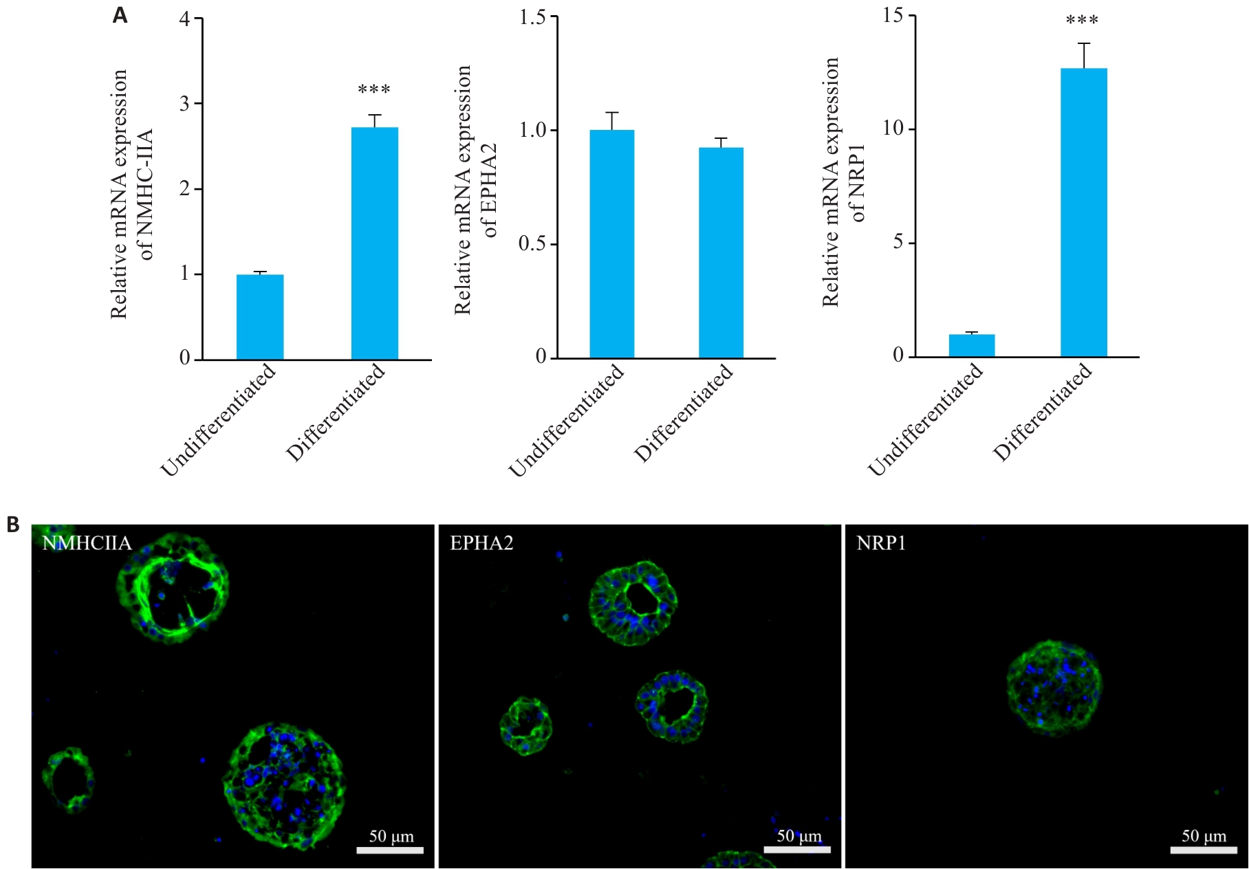
图5 分化的鼻粘膜类器官高表达EBV感染受体
Fig.5 Differentiated nasal organoids exhibit high expressions of Epstein-Barr virus (EBV) receptors. A: Relative mRNA expressions of EBV receptors NMHC-IIA, EPHA2 and NRP1 in undifferentiated and differentiated nasal organoids. ***P<0.001 vs undifferentiated group. B: Immunofluorescence staining for EBV receptors NMHC-IIA, EPHA2 and NRP1 in differentiated nasal organoids (n=3).
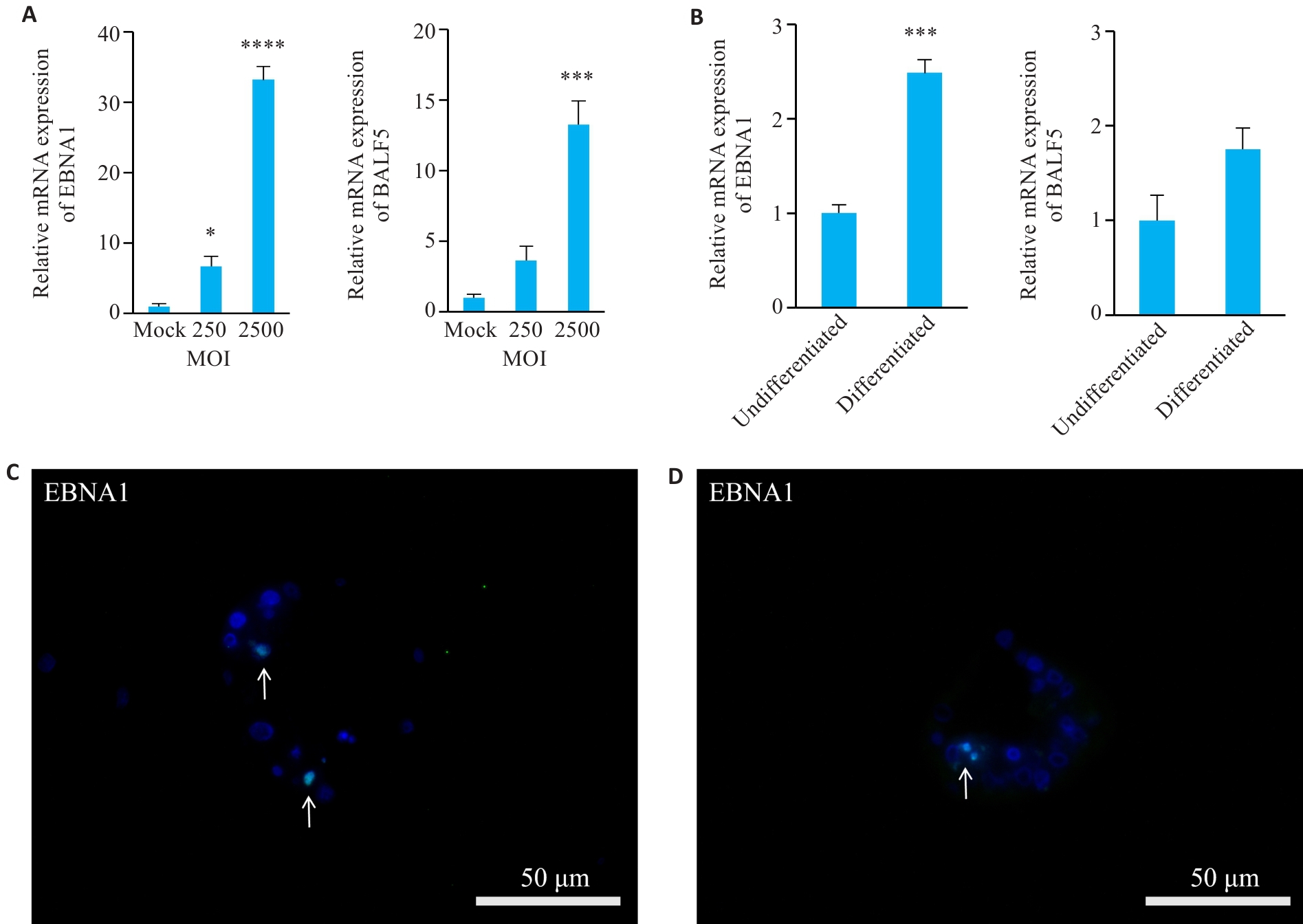
图6 鼻粘膜类器官EBV感染模型的建立
Fig.6 Establishment of EBV-infected nasal organoids. A, B: Relative mRNA expression levels of EBV-specific genes EBNA1 and BALF5 in differentiated organoids harvested at 72 h post-EBV infection at MOI=250 and 2500 (A; *P<0.05, ***P<0.001, ****P<0.0001 vs Mock group) and in both undifferentiated and differentiated organoids harvested 72 h after EBV infection at MOI=2500 (B; ***P<0.001 vs undifferentiated group). C, D: Immunofluorescence staining for EBNA1 undifferentiated (C) and differentiated (D) organoids with EBV infection at MOI=2500 (n=3).
| [1] | Alsaadawe M, Radman BA, Long JY, et al. Epstein Barr virus: a cellular hijacker in cancer[J]. Biochim Biophys Acta BBA Rev Cancer, 2024, 1879(6): 189218. doi:10.1016/j.bbcan.2024.189218 |
| [2] | Damania B, Kenney SC, Raab-Traub N. Epstein-Barr virus: Biology and clinical disease[J]. Cell, 2022, 185(20): 3652-70. doi:10.1016/j.cell.2022.08.026 |
| [3] | Schindele A, Holm A, Kraft S, et al. Cross-evaluating Epstein-Barr virus, human Papilloma virus, human cytomegalovirus and human adenovirus in nasal polyps and turbinate mucosa[J]. Acta Otolaryngol, 2025, 145(2): 164-7. doi:10.1080/00016489.2024.2445025 |
| [4] | Zaravinos A, Bizakis J, Spandidos DA. Prevalence of human Papilloma virus and human herpes virus types 1-7 in human nasal polyposis[J]. J Med Virol, 2009, 81(9): 1613-9. doi:10.1002/jmv.21534 |
| [5] | 谭学君, 姚文昊, 陈晓平, 等.三峡库区重庆万州段鼻息肉与HPV及EBV阳性表达的相关性研究[J].中国中西医结合耳鼻咽喉科杂志,2015, 23(1): 7-11. |
| [6] | Jia Z, Zhang D, Zhu L, et al. Animal models of human herpesvirus infection[J]. Animal Model Exp Med, 2025, 8(4): 615-28. doi:10.1002/ame2.12575 |
| [7] | Huang PY, Zeng TT, Li MQ, et al. Proteomic analysis of a nasopharyngeal carcinoma cell line and a nasopharyngeal epithelial cell line[J]. Tumori, 2015, 101(6): 676-83. doi:10.5301/tj.5000345 |
| [8] | Li HM, Man C, Jin Y, et al. Molecular and cytogenetic changes involved in the immortalization of nasopharyngeal epithelial cells by telomerase[J]. Int J Cancer, 2006, 119(7): 1567-76. doi:10.1002/ijc.22032 |
| [9] | Song LB, Zeng MS, Liao WT, et al. Bmi-1 is a novel molecular marker of nasopharyngeal carcinoma progression and immortalizes primary human nasopharyngeal epithelial cells[J]. Cancer Res, 2006, 66(12): 6225-32. doi:10.1158/0008-5472.can-06-0094 |
| [10] | Collett S, Torresi J, Silveira LE, et al. Investigating virus–host cell interactions: Comparative binding forces between hepatitis C virus-like particles and host cell receptors in 2D and 3D cell culture models[J]. J Colloid Interface Sci, 2021, 592: 371-84. doi:10.1016/j.jcis.2021.02.067 |
| [11] | Yan HHN, Chan AS, Lai FP, et al. Organoid cultures for cancer modeling[J]. Cell Stem Cell, 2023, 30(7): 917-37. doi:10.1016/j.stem.2023.05.012 |
| [12] | Zhu S, Chen D, Yang X, et al. Organoid models to study human infectious diseases[J]. Cell Prolif, 2025: e70004. doi:10.1111/cpr.70004 |
| [13] | 汪珂,于言,韩日,等.分化可控的人类鼻粘膜类器官模型的建立[J].南方医科大学学报,2022,42(06):868-77. |
| [14] | 于言,曹浚垣, 刘 蓉, 等.人鼻粘膜类器官冠状病毒感染模型可用于抗病毒药物的筛选和评价[J].南方医科大学学报,2024,44(11):2227-34. |
| [15] | Kaur S, Kaur I, Rawal P, et al. Non-matrigel scaffolds for organoid cultures[J]. Cancer Lett, 2021, 504: 58-66. doi:10.1016/j.canlet.2021.01.025 |
| [16] | Co JY, Margalef-Català M, Monack DM, et al. Controlling the polarity of human gastrointestinal organoids to investigate epithelial biology and infectious diseases[J]. Nat Protoc, 2021, 16(11): 5171-92. doi:10.1038/s41596-021-00607-0 |
| [17] | Chiu MC, Li C, Liu X, et al. Human nasal organoids model SARS-CoV-2 upper respiratory infection and recapitulate the differential infectivity of emerging variants[J]. mBio, 2022, 13(4): e0194422. doi:10.1128/mbio.01944-22 |
| [18] | Xia TL, Li XY, Wang XP, et al. N(6)‑methyladenosine-binding protein YTHDF1 suppresses EBV replication and promotes EBV RNA decay[J]. EMBO Rep, 2021, 22(4): e50128. doi:10.15252/embr.202050128 |
| [19] | Zhong LY, Xie C, Zhang LL, et al. Research landmarks on the 60th anniversary of Epstein-Barr virus[J]. Sci China Life Sci, 2025, 68(2): 354-80. doi:10.1007/s11427-024-2766-0 |
| [20] | 谭德重, 冯 源, 胡 月, 等. 病毒感染与鼻息肉关系的Meta分析[J].临床耳鼻咽喉头颈外科杂志,2018, 32(12): 910-6. |
| [21] | Wong KCW, Hui EP, Lo KW, et al. Nasopharyngeal carcinoma: an evolving paradigm[J]. Nat Rev Clin Oncol, 2021, 18(11): 679-95. doi:10.1038/s41571-021-00524-x |
| [22] | Tsang CM, Zhang G, Seto E, et al. Epstein-Barr virus infection in immortalized nasopharyngeal epithelial cells: regulation of infection and phenotypic characterization[J]. Int J Cancer, 2010, 127(7): 1570-83. doi:10.1002/ijc.25173 |
| [23] | Wang HB, Zhang H, Zhang JP, et al. Neuropilin 1 is an entry factor that promotes EBV infection of nasopharyngeal epithelial cells[J]. Nat Commun, 2015, 6: 6240. doi:10.3410/f.725353089.793504525 |
| [24] | Kozlowski MT, Crook CJ, Ku HT. Towards organoid culture without Matrigel [J]. Commun Biol, 2021, 4(1): 1387. doi:10.1038/s42003-021-02910-8 |
| [25] | Marchini A, Gelain F. Synthetic scaffolds for 3D cell cultures and organoids: applications in regenerative medicine[J]. Crit Rev Biotechnol, 2022, 42(3): 468-86. doi:10.1080/07388551.2021.1932716 |
| [26] | Capeling MM, Huang S, Childs CJ, et al. Suspension culture promotes serosal mesothelial development in human intestinal organoids[J]. Cell Rep, 2022, 38(7): 110379. doi:10.1016/j.celrep.2022.110379 |
| [27] | Kumar SV, Er PX, Lawlor KT, et al. Kidney micro-organoids in suspension culture as a scalable source of human pluripotent stem cell-derived kidney cells[J]. Development, 2019, 146(5): dev172361. doi:10.1242/dev.172361 |
| [28] | Wu H, Wang J, Liu S, et al. Large-scale production of expandable hepatoblast organoids and polarised hepatocyte organoids from hESCs under 3D static and dynamic suspension conditions[J]. Cell Prolif, 2025, 58(7): e70001. doi:10.1111/cpr.70001 |
| [29] | Stroulios G, Brown T, Moreni G, et al. Apical-out airway organoids as a platform for studying viral infections and screening for antiviral drugs[J]. Sci Rep, 2022, 12(1): 7673. doi:10.1038/s41598-022-11700-z |
| [30] | Zhao KY, Du YX, Cao HM, et al. The biological macromolecules constructed Matrigel for cultured organoids in biomedical and tissue engineering[J]. Colloids Surf B Biointerfaces, 2025, 247: 114435. doi:10.1016/j.colsurfb.2024.114435 |
| [31] | Bu GL, Xie C, Kang YF, et al. How EBV infects: the tropism and underlying molecular mechanism for viral infection[J]. Viruses, 2022, 14(11): 2372. doi:10.3390/v14112372 |
| [32] | Hayman IR, Temple RM, Burgess CK, et al. New insight into Epstein-Barr virus infection using models of stratified epithelium[J]. PLoS Pathog, 2023, 19(1): e1011040. doi:10.1371/journal.ppat.1011040 |
| [33] | Nawandar DM, Wang A, Makielski K, et al. Differentiation-dependent KLF4 expression promotes lytic Epstein-Barr virus infection in epithelial cells[J]. PLoS Pathog, 2015, 11(10): e1005195. doi:10.1371/journal.ppat.1005195 |
| [1] | 龚雪, 樊雍扬, 罗开元, 燕翼, 李忠豪. 利用人诱导多能干细胞构建的心脏类器官在心脏疾病建模及药物评价中的应用价值[J]. 南方医科大学学报, 2025, 45(11): 2444-2455. |
| [2] | 于言, 曹浚垣, 刘蓉, 周旻旻, 魏金燕, 郑海锐, 王薇, 李刚. 人鼻粘膜类器官冠状病毒感染模型可用于抗病毒药物的筛选和评价[J]. 南方医科大学学报, 2024, 44(11): 2227-2234. |
| [3] | 汪 珂, 于 言, 韩 日, 王显文, 赵云腾, 唐浩程, 李 刚. 分化可控的人类鼻粘膜类器官模型的建立[J]. 南方医科大学学报, 2022, 42(6): 868-877. |
| [4] | 楼俪泓,曾悦,周慧,陆颖影,王兴鹏. 脱氧胆酸对C57BL/6小鼠回肠类器官生长的影响[J]. 南方医科大学学报, 2017, 37(01): 6-. |
| [5] | 乌维秋1, 黄盛光2, 马宗1, 张永泉1, 史剑波3. 四黄灌洗液对慢性鼻窦炎和鼻息肉术后粘膜形态和功能转归的影响[J]. 南方医科大学学报, 2005, 25(04): 424-427. |
| [6] | 方唯意1, 郑文岭4, 马文丽3, 刘腾飞1, 王爽2, 谢卫兵2, 李虹1, 任彩萍1, 姚开泰1,2. EBV病毒全基因组cDNA探针的设计与制备[J]. 南方医科大学学报, 2005, 25(03): 246-250. |
| 阅读次数 | ||||||
|
全文 |
|
|||||
|
摘要 |
|
|||||