Journal of Southern Medical University ›› 2026, Vol. 46 ›› Issue (5): 1167-1174.doi: 10.12122/j.issn.1673-4254.2026.05.21
Previous Articles Next Articles
Dual-wavelength photoacoustic microscopy for evaluation of microcirculatory dysfunction and spatial heterogeneity in mice with liver fibrosis
Qiansong XIA1,2( ), Yang QIU1,2, Honghui LI2(), Liming NIE1,2()
- 1.School of Biomedical Engineering, Southern Medical University, Guangzhou 510515, China
2.Medical Research Institute, Guangdong Provincial People's Hospital (Guangdong Academy of Medical Sciences), Southern Medical University, Guangzhou 510080, China
-
Received:2026-02-23Online:2026-05-20Published:2026-05-27 -
Contact:Honghui LI, Liming NIE E-mail:3188010198@i.smu.edu.cn;lihonghui@gdph.org.cn;nieliming@gdph.org.cn -
Supported by:National Natural Science Foundation of China(82402340)

Cite this article
Qiansong XIA, Yang QIU, Honghui LI, Liming NIE. Dual-wavelength photoacoustic microscopy for evaluation of microcirculatory dysfunction and spatial heterogeneity in mice with liver fibrosis[J]. Journal of Southern Medical University, 2026, 46(5): 1167-1174.
share this article
Add to citation manager EndNote|Ris|BibTeX
URL: https://www.j-smu.com/EN/10.12122/j.issn.1673-4254.2026.05.21
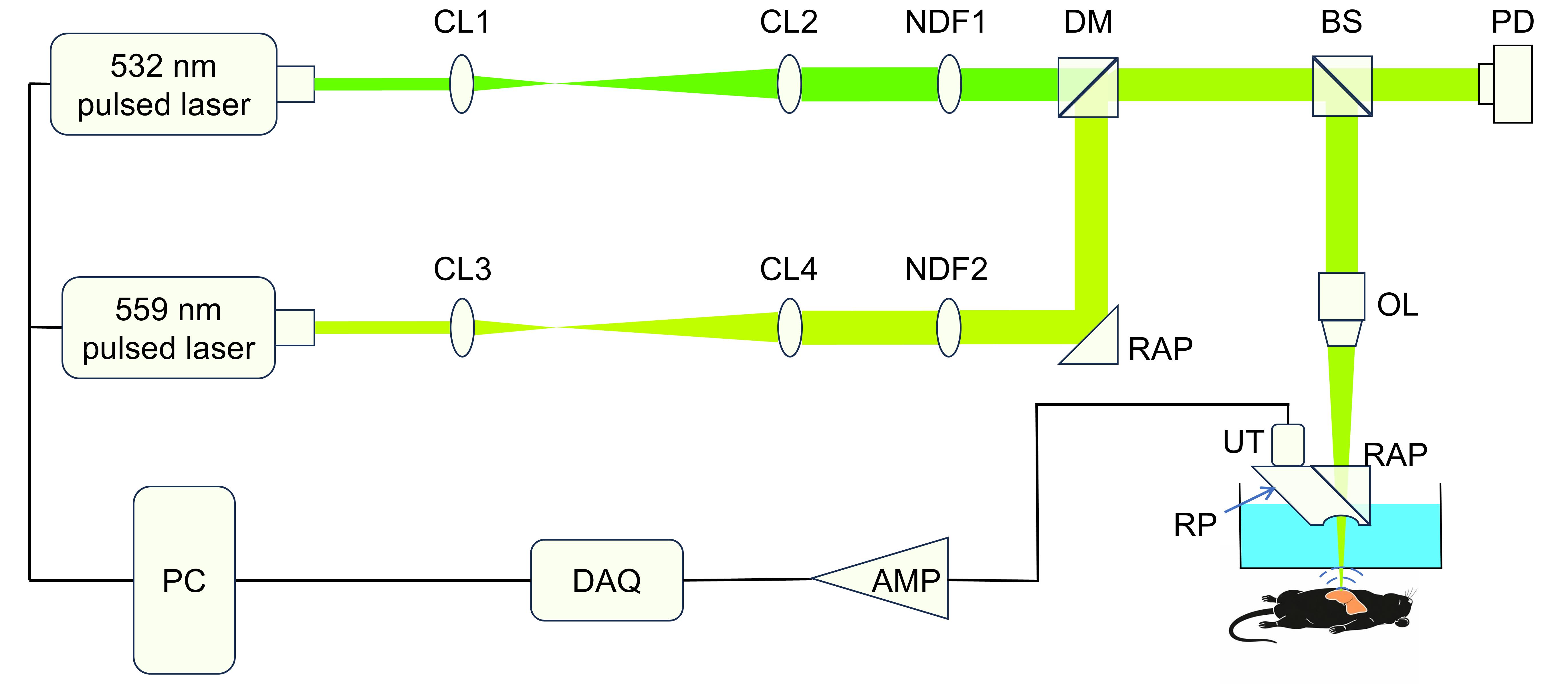
Fig.1 Schematic diagram of the dual-wavelength photoacoustic microscopic imaging system. CL: Convex lens; NDF: Neutral density filter; DM: Dichroic mirror; BS: Beam splitter; PD: Photodiode; OL: Objective lens; RAP: Right-angle prism; RP: Rhomboid prism; UT: Ultrasonic transducer; AMP: Amplifier; DAQ: Data acquisition unit; PC: Personal computer.
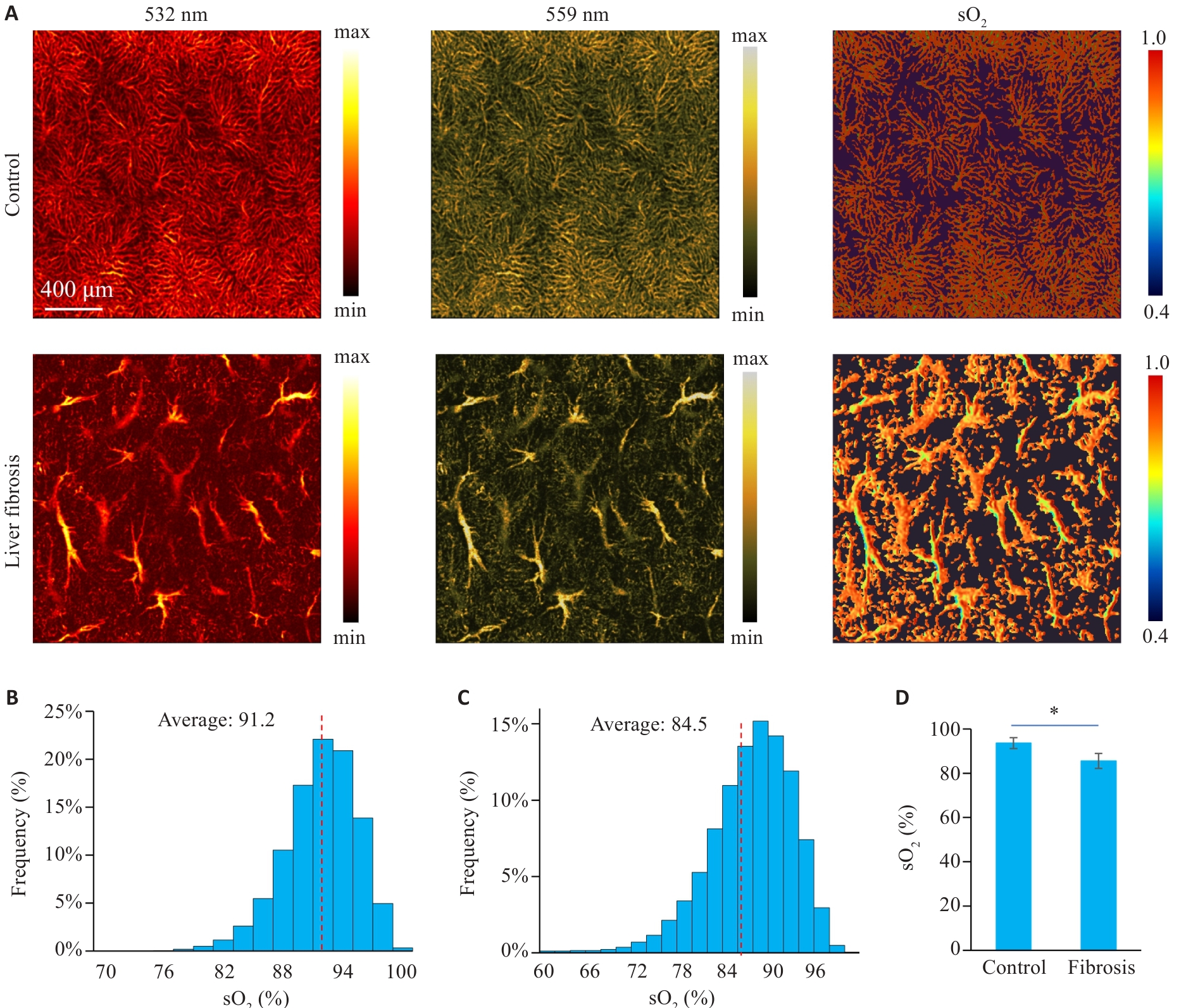
Fig.2 Dual-wavelength imaging of livers from normal and liver fibrosis mice. A: Images of liver vessels and blood oxygen saturation at 532 nm and 559 nm in normal and liver fibrosis mice. B, C: Histogram of blood oxygen saturation distribution in hepatic vessels of normal and liver fibrosis mice, respectively. Red dashed line indicates the average blood oxygen saturation value measured. The average sO2 values within the displayed representative image are shown in the histograms. D: Comparison of intrahepatic microvascular oxygen saturation between normal and liver fibrosis groups. *P<0.05. Data are presented as Mean±SD, n=3.
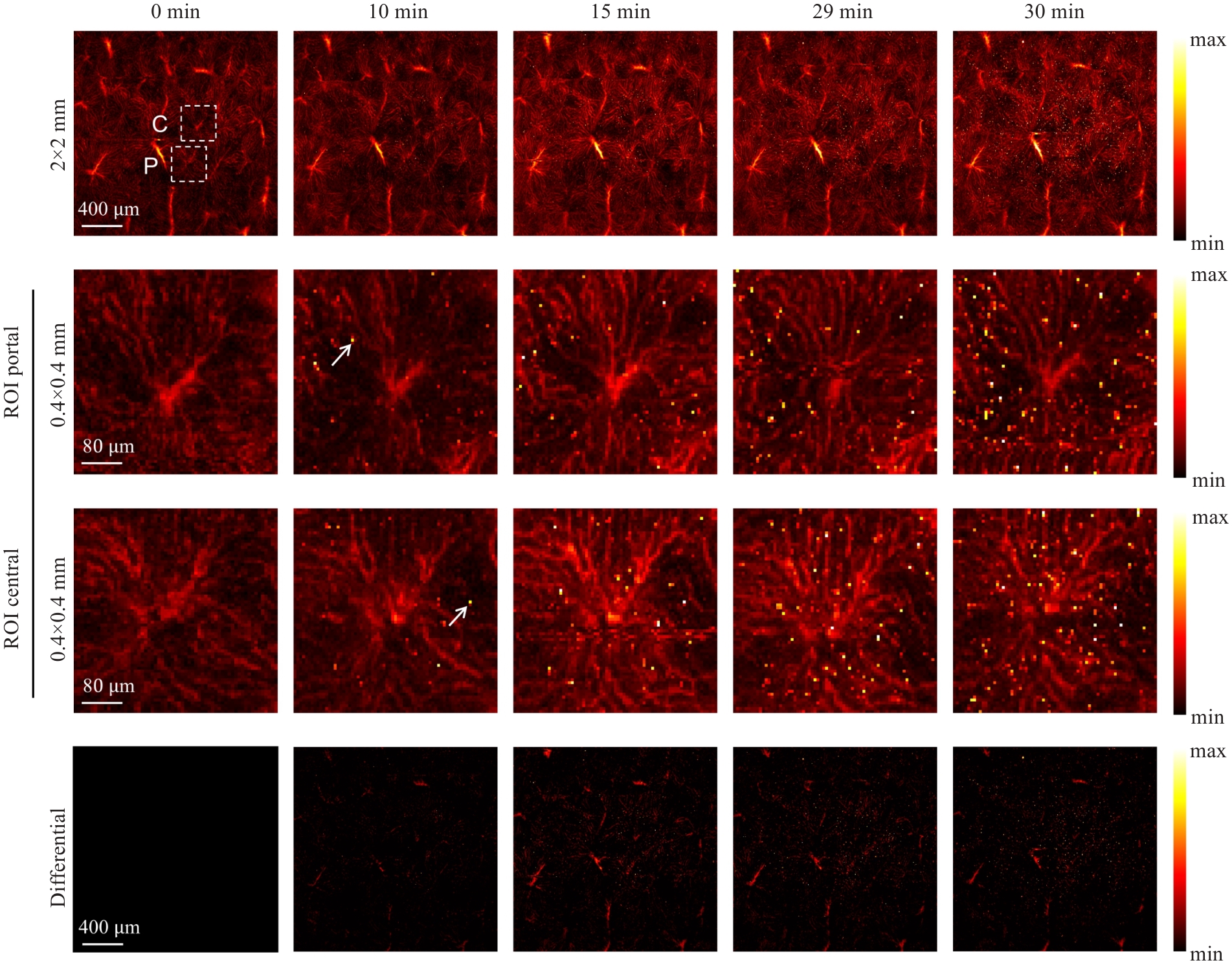
Fig.3 Detection of delayed Evens Blue exudation in normal mice liver.
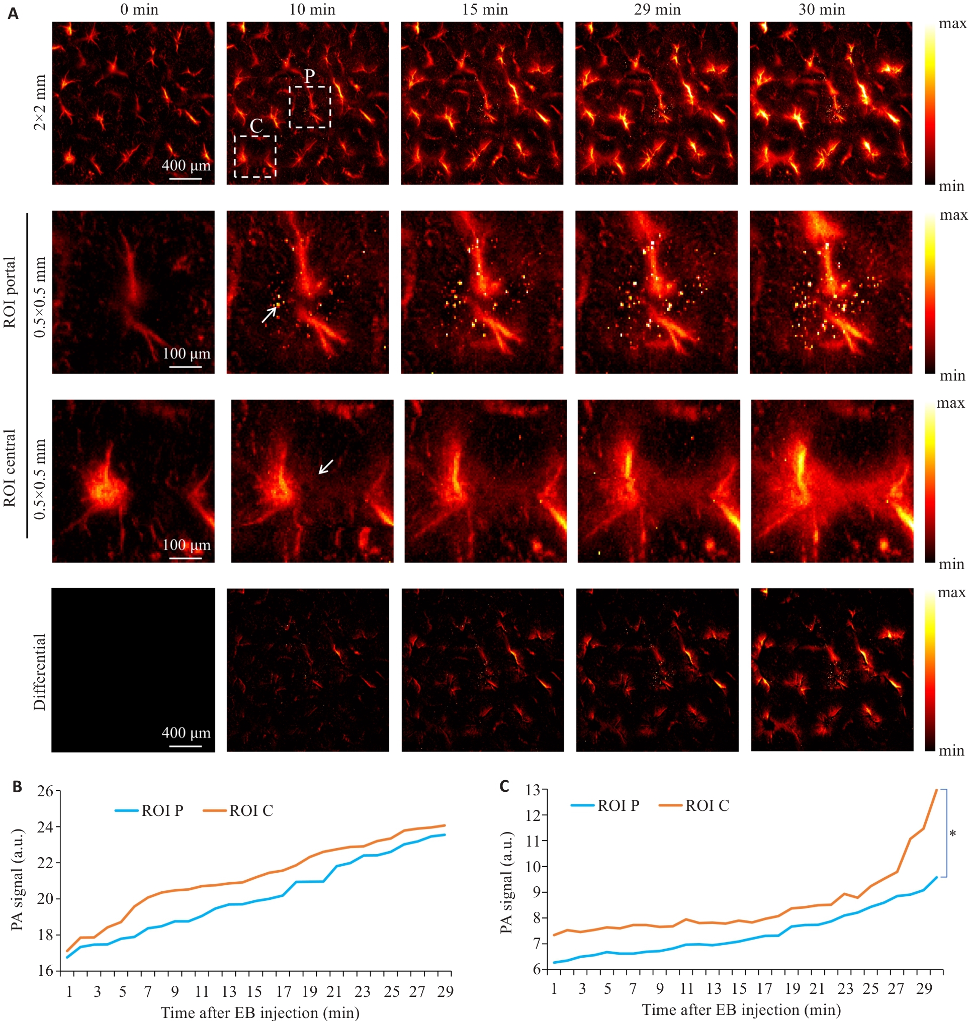
Fig.4 Delayed detection of Evans Blue extravasation in the liver of mice with liver fibrosis. A: Evans Blue extravasation within 30 min post-injection. B, C: Changes in Evans Blue exudation in the portal and central regions of the liver lobules over time in normal mice and the mice with liver fibrosis, respectively. *P<0.05.
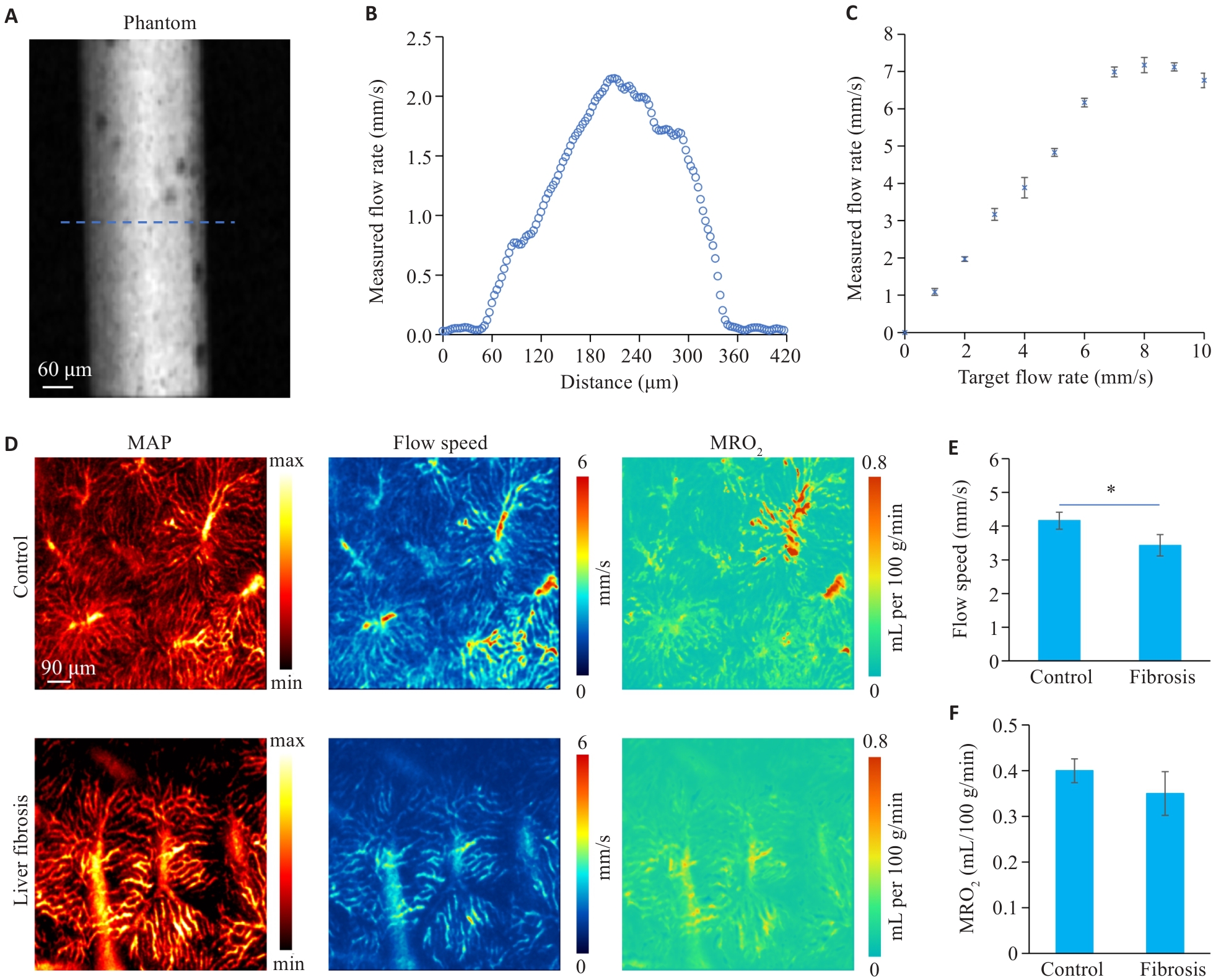
Fig.5 Blood flow and oxygen metabolism imaging of the livers of normal mice and mice with liver fibrosis. A: Blood flow measurement phantom image. B: Profile of blood flow speed across the dashed line in (A). C: Graph showing the relationship between the set flow rate and measured flow rate. D: Images of vascular structures, blood flow, and oxygen metabolism in the livers of normal mice and mice with liver fibrosis. E: Comparison of intrahepatic microvascular blood flow speed between the normal and liver fibrosis groups. F: Comparison of intrahepatic microvascular metabolic rate of oxygen between the normal and liver fibrosis groups. Data are presented as Mean±SD, n=3. *P<0.05.
| [1] | Bataller R, Brenner DA. Liver fibrosis[J]. J Clin Invest, 2005, 115(2): 209-18. doi:10.1172/JCI200524282 |
| [2] | Vollmar B, Menger MD. The hepatic microcirculation: mechanistic contributions and therapeutic targets in liver injury and repair[J]. Physiol Rev, 2009, 89(4): 1269-339. doi:10.1152/physrev.00027.2008 |
| [3] | Liu P, Liang WL, Huang RT, et al. Hepatic microcirculatory disturbance in liver diseases: intervention with traditional Chinese medicine[J]. Front Pharmacol, 2024, 15: 1399598. doi:10.3389/fphar.2024.1399598 |
| [4] | Qiu Y, Li HH, Yu K, et al. Collagen fibers quantification for liver fibrosis assessment using linear dichroism photoacoustic microscopy[J]. Photoacoustics, 2025, 42: 100694. doi:10.1016/j.pacs.2025.100694 |
| [5] | Bedossa P, Dargère D, Paradis V. Sampling variability of liver fibrosis in chronic hepatitis C[J]. Hepatology, 2003, 38(6): 1449-57. doi:10.1016/j.hep.2003.09.022 |
| [6] | Reiter R, Shahryari M, Tzschätzsch H, et al. Spatial heterogeneity of hepatic fibrosis in primary sclerosing cholangitis vs. viral hepatitis assessed by MR elastography[J]. Sci Rep, 2021, 11: 9820. doi:10.1038/s41598-021-89372-4 |
| [7] | Angelini G, Panunzi S, Castagneto-Gissey L, et al. Accurate liquid biopsy for the diagnosis of non-alcoholic steatohepatitis and liver fibrosis[J]. Gut, 2023, 72(2): 392-403. doi:10.1136/gutjnl-2022-327498 |
| [8] | Wang TT, Chen JL, Nie LM, et al. Photoacoustic microscopy for multiscale biological system visualization and clinical translation[J]. Adv Sci, 2026, 13(9): e21173. doi:10.1002/advs.202521173 |
| [9] | Mathew RC, Bourque JM, Salerno M, et al. Cardiovascular imaging techniques to assess microvascular dysfunction[J]. JACC Cardiovasc Imaging, 2020, 13(7): 1577-90. doi:10.1016/j.jcmg.2019.09.006 |
| [10] | Dewey M, Siebes M, Kachelrieß M, et al. Clinical quantitative cardiac imaging for the assessment of myocardial ischaemia[J]. Nat Rev Cardiol, 2020, 17(7): 427-50. doi:10.1038/s41569-020-0341-8 |
| [11] | Chen HJ, Tong X, Lang LX, et al. Quantification of tumor vascular permeability and blood volume by positron emission tomography[J]. Theranostics, 2017, 7(9): 2363-76. doi:10.7150/thno.19898 |
| [12] | Mahesh M, Ansari AJ, Mettler FA Jr. Patient exposure from radiologic and nuclear medicine procedures in the United States and worldwide: 2009–2018[J]. Radiology, 2023, 307: e221263. doi:10.1148/radiol.239013 |
| [13] | Li HH, Sun MY, Zhu BW, et al. Multidimensional dynamic optical imaging unveils anesthetic-driven hemispheric lateralization in blood-brain barrier homeostasis[J]. Sci Adv, 2025, 11(49): eady6275. doi:10.1126/sciadv.ady6275 |
| [14] | Qin W, Li HH, Chen JL, et al. Amphiphilic hemicyanine molecular probes crossing the blood-brain barrier for intracranial optical imaging of glioblastoma[J]. Sci Adv, 2025, 11(3): eadq5816. doi:10.1126/sciadv.adq5816 |
| [15] | Bi RZ, Dinish US, Goh CC, et al. In vivo label-free functional photoacoustic monitoring of ischemic reperfusion[J]. J Biophotonics, 2019, 12(7): e201800454. doi:10.1002/jbio.201800454 |
| [16] | Zhou Y, Liang JY, Maslov KI, et al. Calibration-free in vivo transverse blood flowmetry based on cross correlation of slow time profiles from photoacoustic microscopy[J]. Opt Lett, 2013, 38(19): 3882-5. doi:10.1364/ol.38.003882 |
| [17] | Yao JJ, Wang LV. Transverse flow imaging based on photoacoustic Doppler bandwidth broadening[J]. J Biomed Opt, 2010, 15(2): 1. doi:10.1117/1.3339953 |
| [18] | Chen SL, Xie ZX, Carson PL, et al. In vivo flow speed measurement of capillaries by photoacoustic correlation spectroscopy[J]. Opt Lett, 2011, 36(20): 4017-9. doi:10.1364/OL.36.004017 |
| [19] | Wang TX, Sun ND, Cao R, et al. Multiparametric photoacoustic microscopy of the mouse brain with 300-kHz A-line rate[J]. Neurophotonics, 2016, 3(4): 045006. doi:10.1117/1.nph.3.4.045006 |
| [20] | Liu C, Liang YZ, Wang LD. Single-shot photoacoustic microscopy of hemoglobin concentration, oxygen saturation, and blood flow in sub-microseconds[J]. Photoacoustics, 2019, 17: 100156. doi:10.1016/j.pacs.2019.100156 |
| [21] | Li WY, Lv J, Li HH, et al. Quantification of vascular remodeling and sinusoidal capillarization to assess liver fibrosis with photoacoustic imaging[J]. Radiology, 2025, 314: e241275. doi:10.1148/radiol.241275 |
| [22] | Zhou Q, Huang GJ, Chen JL, et al. Multiparameter assessment of hepatobiliary excretion dysfunction in cholestasis via dynamical photoacoustic and NIR-II fluorescence imaging[J]. Photon Res, 2025, 13(9): 2679. doi:10.1364/prj.564269 |
| [23] | Zhou Q, Liu J, Li HH, et al. Optical imaging unveils morphological heterogeneity at the hepatic lobule level during metabolic dysfunction-associated fatty liver disease progression[J]. Biomed Opt Express, 2025, 16(10): 4040. doi:10.1364/boe.573860 |
| [24] | Fujimoto Y, Urashima T, Shimura D, et al. Low cardiac output leads hepatic fibrosis in right heart failure model rats[J]. PLoS One, 2016, 11(2): e0148666. doi:10.1371/journal.pone.0148666 |
| [25] | He QY, He W, Dong H, et al. Role of liver sinusoidal endothelial cell in metabolic dysfunction-associated fatty liver disease[J]. Cell Commun Signal, 2024, 22(1): 346. doi:10.1186/s12964-024-01720-9 |
| [26] | Friedman SL. Mechanisms of hepatic fibrogenesis[J]. Gastroenterology, 2008, 134(6): 1655-69. doi:10.1053/J.GASTRO.2008.03.003 |
| [27] | Cai JY, Hu M, Chen ZY, et al. The roles and mechanisms of hypoxia in liver fibrosis[J]. J Transl Med, 2021, 19(1): 186. doi:10.1186/s12967-021-02854-x |
| [28] | Gan C, Yaqoob U, Lu JW, et al. Liver sinusoidal endothelial cells contribute to portal hypertension through collagen type IV-driven sinusoidal remodeling[J]. JCI Insight, 2024, 9(11): e174775. doi:10.1172/jci.insight.174775 |
| [29] | Van Beers BE, Leconte I, Materne R, et al. Hepatic perfusion parameters in chronic liver disease: dynamic CT measurements correlated with disease severity[J]. Am J Roentgenol, 2001, 176(3): 667-73. doi:10.2214/ajr.176.3.1760667 |
| [1] | Tingting YANG, Li ZHAO. A stable mouse model of chronic liver fibrosis induced by vitamin A deficiency and intraperitoneal CCl4 injection [J]. Journal of Southern Medical University, 2025, 45(7): 1527-1534. |
| [2] | Jiawen YU, Yi ZHOU, Chunmei QIAN, Lan MU, Renye QUE. Effects of liver fibrosis induced by iron overload on M2 polarization of macrophages in mice [J]. Journal of Southern Medical University, 2025, 45(4): 684-691. |
| [3] | Daiping HUA, Qiaoyu XUAN, Lanting SUN, Qingsheng YU, Qin WANG, Tao WANG, Qiyan MA, Wenming YANG, Han WANG. LncRNA Meg3 expression level is negatively correlated with liver fibrosis severity in patients with Wilson disease [J]. Journal of Southern Medical University, 2025, 45(11): 2365-2374. |
| [4] | Yuming ZHANG, Shicheng XIA, Linlin ZHANG, Mengxi CHEN, Xiaojing LIU, Qin GAO, Hongwei YE. Protective effect of Lonicerae japonicae flos extract against doxorubicin-induced liver injury in mice [J]. Journal of Southern Medical University, 2024, 44(8): 1571-1581. |
| [5] | CAO Jiafan, SUN Yue, DING Xin, LI Shengwen, CHEN Bo, LAN Tian. Arbutin ameliorates liver fibrosis in mice by inhibiting macrophage recruitment and regulating the Akt/NF-κB and Smad signaling pathways [J]. Journal of Southern Medical University, 2024, 44(4): 652-659. |
| [6] | XU Xiaohui, FENG Jinmei, LUO Ying, HE Xinyu, ZANG Jinbao, HUANG Daochao. Adeno-associated virus-mediated hepatocyte-specific NDUFA13 overexpression protects against CCl4-induced liver fibrosis in mice by inhibiting hepatic NLRP3 activation [J]. Journal of Southern Medical University, 2024, 44(2): 201-209. |
| [7] | Wen ZHAO, Hejing RUAN, Siyuan WANG, Yuzhe CHENG, Miao LEI, Jiufa ZHAO, Chuanmiao LIU. Inhibiting Yes-associated protein alleviates CCl4 liver fibrosis in mice by reducing epithelial mesenchymal transition [J]. Journal of Southern Medical University, 2024, 44(10): 1839-1849. |
| [8] | LIN Jiayi, LOU Anni, LI Xu. Lipopolysaccharide stimulates macrophages to secrete exosomes containing miR-155-5p to promote activation and migration of hepatic stellate cells [J]. Journal of Southern Medical University, 2023, 43(6): 994-1001. |
| [9] | YANG Xuejia, LI Yujie, WU Dengqiang, MA Yili, ZHOU Sufang. Screening and identification of key genes ATP1B3 and ENAH in the progression of hepatocellular carcinoma: based on data mining and clinical validation [J]. Journal of Southern Medical University, 2022, 42(6): 815-823. |
| [10] | DENG Ya, WANG Chunyan, FU Yiming, LI Zhongbin, JI Dong. A high relapse risk of chronic drug-induced liver injury is correlated with a greater severity of liver fibrosis [J]. Journal of Southern Medical University, 2022, 42(11): 1655-1661. |
| [11] | ZHAO Chenling, DONG Ting, SUN Lunyan, HU Huibing, WANG Qiong, TIAN Liwei, JIANG Zhangsheng. Establishment and validation of a predictive nomogram for liver fibrosis in patients with Wilson disease and abnormal lipid metabolism [J]. Journal of Southern Medical University, 2022, 42(11): 1720-1725. |
| [12] | ZHAO Zhibin, DONG Hui, LI Binghang, SHEN Bo, GUO Yuecheng, GU Tianyi, QU Ying, CAI Xiaobo, LU Lungen. Hydroxynitone suppresses hepatic stellate cell activation by inhibiting TGF-β1 phosphorylation to alleviate CCl4-induced liver fibrosis in rats [J]. Journal of Southern Medical University, 2022, 42(10): 1511-1516. |
| [13] | ZHANG Chunyan, YAN Yuxin, GAO Xiaoyang, MA Yuehong. Therapeutic mechanism of the Mongolian medicine Qiwei Qinggan Powder against liver fibrosis based on UHPLC-TOF-MS combined with network pharmacological methods [J]. Journal of Southern Medical University, 2021, 41(8): 1131-1141. |
| [14] | . Baoganning formula alleviates liver fibrosis in mice by inhibiting hepatic IDO1 expression and promoting phenotypic maturation of dendritic cells [J]. Journal of Southern Medical University, 2021, 41(7): 1002-1011. |
| [15] | . Inhibitory effect of Xinhui citrus fermentation liquor on liver fibrosis in mice [J]. Journal of Southern Medical University, 2021, 41(4): 588-592. |
| Viewed | ||||||
|
Full text |
|
|||||
|
Abstract |
|
|||||