Journal of Southern Medical University ›› 2024, Vol. 44 ›› Issue (5): 950-959.doi: 10.12122/j.issn.1673-4254.2024.05.17
• Techniques and Methods • Previous Articles Next Articles
Chen WANG1,2( ), Mingqiang MENG1,2, Mingqiang LI2, Yongbo WANG1,2, Dong ZENG1,2, Zhaoying BIAN1,2, Jianhua MA1,2()
Received:2023-10-31
Online:2024-05-20
Published:2024-06-06
Contact:
Jianhua MA
E-mail:wangchen9909@outlook.com;jhma@smu.edu.cn
Supported by:Chen WANG, Mingqiang MENG, Mingqiang LI, Yongbo WANG, Dong ZENG, Zhaoying BIAN, Jianhua MA. Reconstruction from CT truncated data based on dual-domain transformer coupled feature learning[J]. Journal of Southern Medical University, 2024, 44(5): 950-959.
Add to citation manager EndNote|Ris|BibTeX
URL: https://www.j-smu.com/EN/10.12122/j.issn.1673-4254.2024.05.17
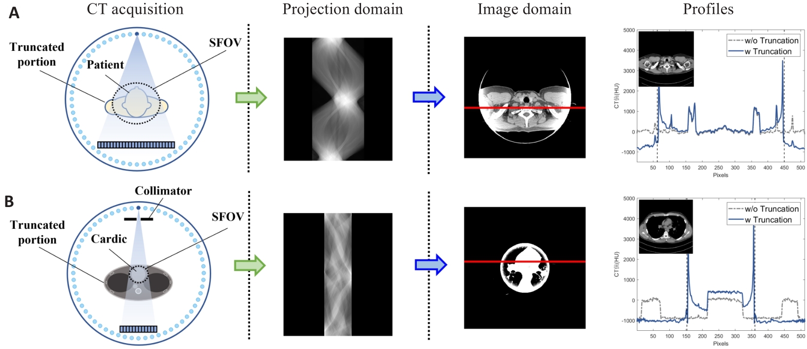
Fig.1 Illustration of truncation artifacts of the projection data collected under two truncation conditions, the reconstructed images and profiles comparison with the non-truncated image at the position marked by red solid line (the vertical dashed lines represent the truncation boundaries). A: Partial truncation at the patient's shoulder. B: Cardiac interior scanning.
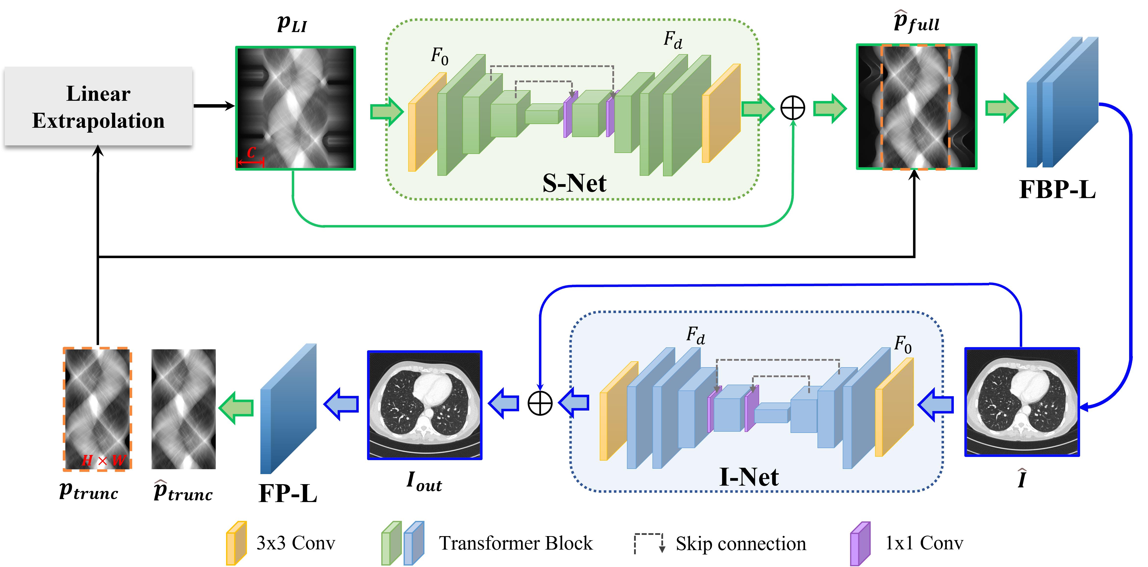
Fig.2 Overall architecture of the proposed DDTrans.
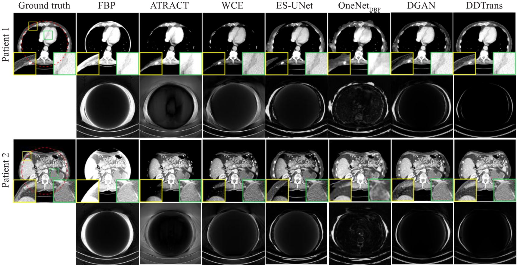
Fig.3 Comparison of truncation artifacts correction results with different methods for partial truncation data. The display window is [-200, 200]HU. The second and fourth rows are absolute residual images of different algorithm results with respect to the ground truth.
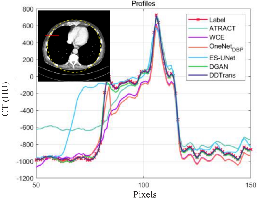
Fig.4 Comparison of the profiles at red solid line locations for partial truncation data.
| Methods | RMSE | SSIM | PSNR | |||
|---|---|---|---|---|---|---|
| Inside FOV | Outside FOV | Inside FOV | Outside FOV | Inside FOV | Outside FOV | |
| FBP | 189.0240 | 269.5267 | 0.9134 | 0.5618 | 18.9133 | 4.3666 |
| ATRACT | 62.8714 | 234.9390 | 0.9290 | 0.6436 | 29.4219 | 7.0502 |
| WCE | 26.1827 | 152.8316 | 0.9628 | 0.7531 | 38.0734 | 9.5631 |
| ES-UNet | 40.6024 | 155.7068 | 0.9458 | 0.8858 | 33.2551 | 9.2289 |
| OneNetDBP | 61.3758 | 158.5035 | 0.8821 | 0.8530 | 27.1814 | 9.2492 |
| DGAN | 22.0072 | 138.6643 | 0.9837 | 0.8917 | 41.8713 | 10.3352 |
| DDTrans | 5.4097 | 116.9005 | 0.9991 | 0.9250 | 52.4910 | 12.0010 |
Tab.1 Comparison of quantitative results of different methods for partial truncation data
| Methods | RMSE | SSIM | PSNR | |||
|---|---|---|---|---|---|---|
| Inside FOV | Outside FOV | Inside FOV | Outside FOV | Inside FOV | Outside FOV | |
| FBP | 189.0240 | 269.5267 | 0.9134 | 0.5618 | 18.9133 | 4.3666 |
| ATRACT | 62.8714 | 234.9390 | 0.9290 | 0.6436 | 29.4219 | 7.0502 |
| WCE | 26.1827 | 152.8316 | 0.9628 | 0.7531 | 38.0734 | 9.5631 |
| ES-UNet | 40.6024 | 155.7068 | 0.9458 | 0.8858 | 33.2551 | 9.2289 |
| OneNetDBP | 61.3758 | 158.5035 | 0.8821 | 0.8530 | 27.1814 | 9.2492 |
| DGAN | 22.0072 | 138.6643 | 0.9837 | 0.8917 | 41.8713 | 10.3352 |
| DDTrans | 5.4097 | 116.9005 | 0.9991 | 0.9250 | 52.4910 | 12.0010 |
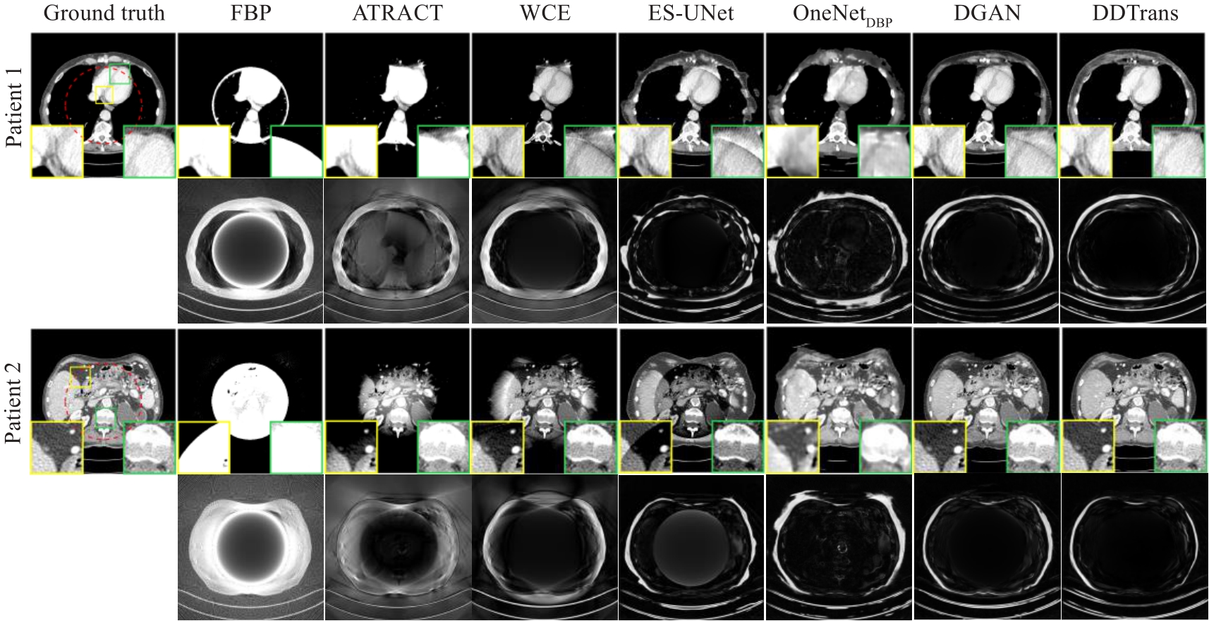
Fig.5 Comparison of truncation artifacts correction results with different methods for interior scanning. The display window is [-200, 200]HU. The second and fourth rows are absolute residual images of different algorithm results with respect to the ground truth.
| Methods | RMSE | SSIM | PSNR | |||
|---|---|---|---|---|---|---|
| Inside FOV | Outside FOV | Inside FOV | Outside FOV | Inside FOV | Outside FOV | |
| FBP | 488.4375 | 556.6830 | 0.6466 | 0.2275 | 7.5358 | 7.0234 |
| ATRACT | 62.3716 | 287.6859 | 0.8911 | 0.5542 | 25.8800 | 13.1452 |
| WCE | 37.0367 | 225.5691 | 0.8819 | 0.5915 | 32.9671 | 14.9137 |
| ES-UNet | 54.7751 | 226.6859 | 0.8906 | 0.8062 | 29.2464 | 14.8481 |
| OneNetDBP | 48.7271 | 219.1173 | 0.8279 | 0.7781 | 26.1069 | 15.2767 |
| DGAN | 29.6017 | 201.8265 | 0.9537 | 0.7769 | 33.4705 | 15.4285 |
| DDTrans | 8.0043 | 174.9918 | 0.9836 | 0.8317 | 42.9410 | 17.0266 |
Tab.2 Comparison of quantitative results of different methods for interior scanning data
| Methods | RMSE | SSIM | PSNR | |||
|---|---|---|---|---|---|---|
| Inside FOV | Outside FOV | Inside FOV | Outside FOV | Inside FOV | Outside FOV | |
| FBP | 488.4375 | 556.6830 | 0.6466 | 0.2275 | 7.5358 | 7.0234 |
| ATRACT | 62.3716 | 287.6859 | 0.8911 | 0.5542 | 25.8800 | 13.1452 |
| WCE | 37.0367 | 225.5691 | 0.8819 | 0.5915 | 32.9671 | 14.9137 |
| ES-UNet | 54.7751 | 226.6859 | 0.8906 | 0.8062 | 29.2464 | 14.8481 |
| OneNetDBP | 48.7271 | 219.1173 | 0.8279 | 0.7781 | 26.1069 | 15.2767 |
| DGAN | 29.6017 | 201.8265 | 0.9537 | 0.7769 | 33.4705 | 15.4285 |
| DDTrans | 8.0043 | 174.9918 | 0.9836 | 0.8317 | 42.9410 | 17.0266 |
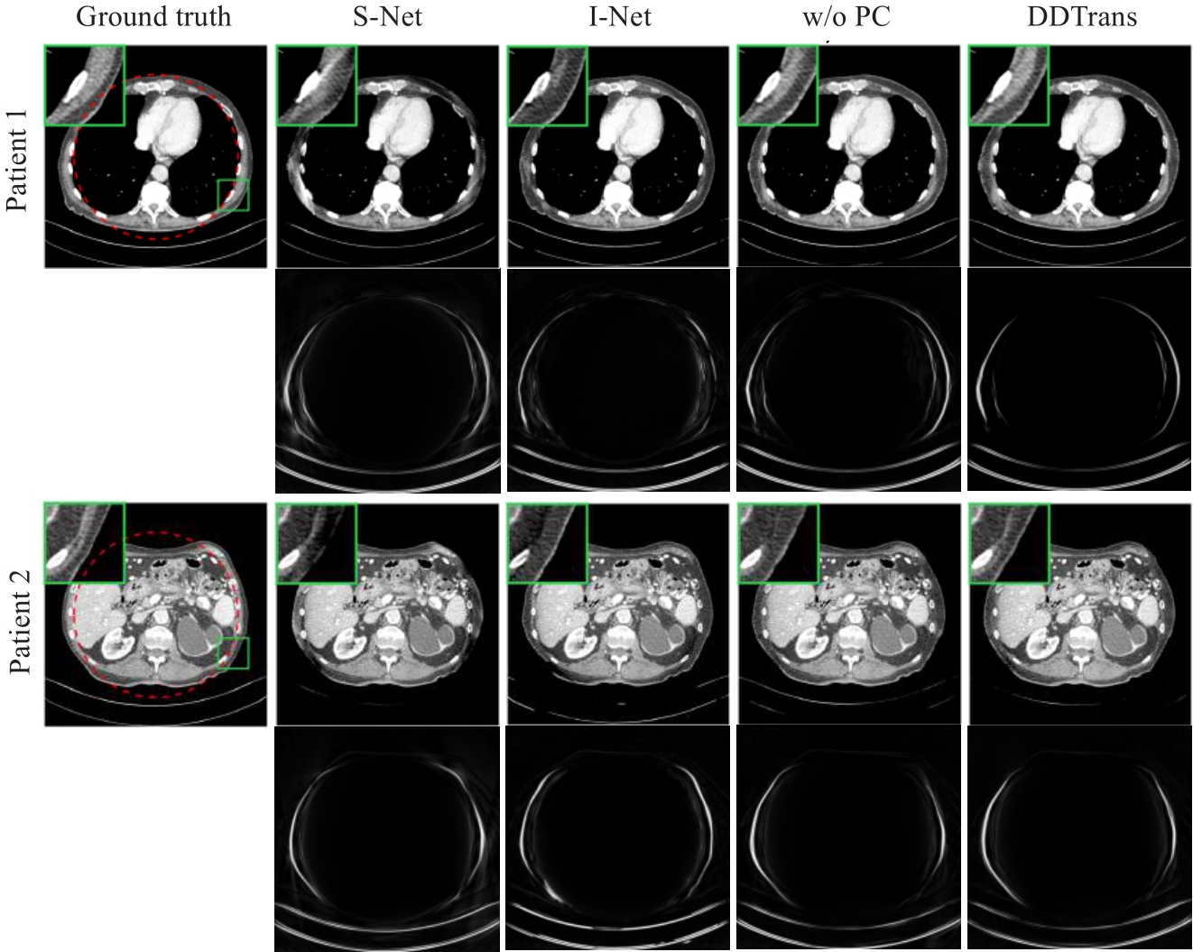
Fig.6 Ablation experiment results of single projection domain network (S-Net), single image domain network (I-Net), dual domain network without projection consistency constraint layer (w/o PC) and DDTrans. The display window is [-200, 200]HU.
| Methods | RMSE | SSIM | PSNR | |||
|---|---|---|---|---|---|---|
| Inside FOV | Outside FOV | Inside FOV | Outside FOV | Inside FOV | Outside FOV | |
| S-Net | 6.5405 | 120.0671 | 0.9982 | 0.8898 | 49.0927 | 11.5285 |
| I-Net | 11.3366 | 130.1608 | 0.9965 | 0.9124 | 41.9385 | 10.7919 |
| w/o PC | 6.5004 | 117.1258 | 0.9989 | 0.9232 | 50.4997 | 11.7845 |
| DDTrans | 5.4079 | 116.9005 | 0.9991 | 0.9250 | 52.4910 | 12.0010 |
Tab.3 Quantitative comparison of ablation experimental results
| Methods | RMSE | SSIM | PSNR | |||
|---|---|---|---|---|---|---|
| Inside FOV | Outside FOV | Inside FOV | Outside FOV | Inside FOV | Outside FOV | |
| S-Net | 6.5405 | 120.0671 | 0.9982 | 0.8898 | 49.0927 | 11.5285 |
| I-Net | 11.3366 | 130.1608 | 0.9965 | 0.9124 | 41.9385 | 10.7919 |
| w/o PC | 6.5004 | 117.1258 | 0.9989 | 0.9232 | 50.4997 | 11.7845 |
| DDTrans | 5.4079 | 116.9005 | 0.9991 | 0.9250 | 52.4910 | 12.0010 |
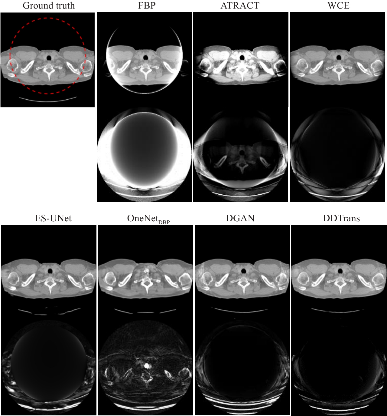
Fig.7 Comparison of the generalization capabilities of different algorithms on shoulder data. The head data display window is [-500, 500]HU. The second and fourth rows are absolute residual images of different algorithm results with respect to the ground truth.
| Methods | RMSE | SSIM | PSNR | |||
|---|---|---|---|---|---|---|
| Inside FOV | Outside FOV | Inside FOV | Outside FOV | Inside FOV | Outside FOV | |
| FBP | 341.9528 | 460.3208 | 0.6594 | 0.5833 | 14.7162 | 9.5040 |
| ATRACT | 100.3245 | 261.8279 | 0.8597 | 0.7811 | 24.1792 | 13.2714 |
| WCE | 22.6755 | 187.2308 | 0.9611 | 0.8212 | 41.6179 | 16.1640 |
| ES-UNet | 36.4977 | 149.5005 | 0.9148 | 0.8647 | 40.7829 | 16.2713 |
| OneNetDBP | 108.2811 | 159.9267 | 0.8676 | 0.8098 | 23.5470 | 15.7454 |
| DGAN | 19.9203 | 174.9319 | 0.9800 | 0.8205 | 41.3990 | 15.4051 |
| DDTrans | 16.9099 | 144.0732 | 0.9897 | 0.8891 | 42.4004 | 16.7163 |
Tab.4 Quantitative comparison of generalization capabilities of different methods.
| Methods | RMSE | SSIM | PSNR | |||
|---|---|---|---|---|---|---|
| Inside FOV | Outside FOV | Inside FOV | Outside FOV | Inside FOV | Outside FOV | |
| FBP | 341.9528 | 460.3208 | 0.6594 | 0.5833 | 14.7162 | 9.5040 |
| ATRACT | 100.3245 | 261.8279 | 0.8597 | 0.7811 | 24.1792 | 13.2714 |
| WCE | 22.6755 | 187.2308 | 0.9611 | 0.8212 | 41.6179 | 16.1640 |
| ES-UNet | 36.4977 | 149.5005 | 0.9148 | 0.8647 | 40.7829 | 16.2713 |
| OneNetDBP | 108.2811 | 159.9267 | 0.8676 | 0.8098 | 23.5470 | 15.7454 |
| DGAN | 19.9203 | 174.9319 | 0.9800 | 0.8205 | 41.3990 | 15.4051 |
| DDTrans | 16.9099 | 144.0732 | 0.9897 | 0.8891 | 42.4004 | 16.7163 |
| Methods | FBP | ATRACT | WCE | ES-UNet | OneNetDBP | DGAN | DDTrans |
|---|---|---|---|---|---|---|---|
| Time-consumption (s) | 85.7 | 111.3 | 93.9 | 63.5 | 353.5 | 114.1 | 40.6 |
Tab.5 Comparison of time consumption of different methods for reconstructing 526 images
| Methods | FBP | ATRACT | WCE | ES-UNet | OneNetDBP | DGAN | DDTrans |
|---|---|---|---|---|---|---|---|
| Time-consumption (s) | 85.7 | 111.3 | 93.9 | 63.5 | 353.5 | 114.1 | 40.6 |
| 1 | 曾更生. 医学图像重建[M]. 北京: 高等教育出版社, 2010. |
| 2 | Fan FX, Kreher B, Keil H, et al. Fiducial marker recovery and detection from severely truncated data in navigation-assisted spine surgery[J]. Med Phys, 2022, 49(5): 2914-30. DOI: 10.1002/mp.15617 |
| 3 | Wang G, Yu HY, De Man B. An outlook on X-ray CT research and development[J]. Med Phys, 2008, 35(3): 1051-64. DOI: 10.1118/1.2836950 |
| 4 | Kudo H, Suzuki T, Rashed EA. Image reconstruction for sparse-view CT and interior CT-introduction to compressed sensing and differentiated backprojection[J]. Quant Imaging Med Surg, 2013, 3(3): 147-61. DOI: 10.3978/j.issn.2223-4292.2013.06.01 |
| 5 | Ohnesorge B, Flohr T, Schwarz K, et al. Efficient correction for CT image artifacts caused by objects extending outside the scan field of view[J]. Med Phys, 2000, 27(1): 39-46. DOI: 10.1118/1.598855 |
| 6 | Hsieh J, Chao E, Thibault J, et al. A novel reconstruction algorithm to extend the CT scan field-of-view[J]. Med Phys, 2004, 31(9): 2385-91. DOI: 10.1118/1.1776673 |
| 7 | Sourbelle K, Kachelriess M, Kalender WA. Reconstruction from truncated projections in CT using adaptive detruncation[J]. Eur Radiol, 2005, 15(5): 1008-14. DOI: 10.1007/s00330-004-2621-9 |
| 8 | Noo F, Clackdoyle R, Pack JD. A two-step Hilbert transform method for 2D image reconstruction[J]. Phys Med Biol, 2004, 49(17): 3903-23. DOI: 10.1088/0031-9155/49/17/006 |
| 9 | Kudo H, Courdurier M, Noo F, et al. Tiny a priori knowledge solves the interior problem in computed tomography[J]. Phys Med Biol, 2008, 53(9): 2207-31. DOI: 10.1088/0031-9155/53/9/001 |
| 10 | Courdurier M, Noo F, Defrise M, et al. Solving the interior problem of computed tomography using a priori knowledge[J]. Inverse Probl, 2008, 24(6): 065001. DOI: 10.1088/0266-5611/24/6/065001 |
| 11 | Coussat A, Rit S, Clackdoyle R, et al. Region-of-interest CT reconstruction using object extent and singular value decomposition[J]. IEEE Trans Radiat Plasma Med Sci, 2022, 6(5): 537-51. DOI: 10.1109/trpms.2021.3091288 |
| 12 | Xia Y, Hofmann H, Dennerlein F, et al. Towards clinical application of a Laplace operator-based region of interest reconstruction algorithm in C-arm CT[J]. IEEE Trans Med Imaging, 2014, 33(3): 593-606. DOI: 10.1109/tmi.2013.2291622 |
| 13 | Han WM, Yu HY, Wang G. A general total variation minimization theorem for compressed sensing based interior tomography[J]. J Biomed Imag, 2009, 2009: 21. DOI: 10.1155/2009/125871 |
| 14 | Yang JS, Yu HY, Jiang M, et al. High-order total variation minimization for interior tomography[J]. Inverse Probl, 2010, 26(3): 035013. DOI: 10.1088/0266-5611/26/3/035013 |
| 15 | Yu HY, Wang G. Compressed sensing based interior tomography[J]. Phys Med Biol, 2009, 54(9): 2791-805. DOI: 10.1088/0031-9155/54/9/014 |
| 16 | Han Y, Ye JC. One network to solve all ROIs: deep learning CT for any ROI using differentiated backprojection[J]. Med Phys, 2019, 46(12): e855-e872. DOI: 10.1002/mp.13631 |
| 17 | Li YS, Li K, Zhang CZ, et al. Learning to reconstruct computed tomography images directly from sinogram data under A variety of data acquisition conditions[J]. IEEE Trans Med Imaging, 2019, 38(10): 2469-81. DOI: 10.1109/tmi.2019.2910760 |
| 18 | Fournié É, Baer-Beck M, Stierstorfer K. CT field of view extension using combined channels extension and deep learning methods[J]. arXiv preprint, arXiv:1908.09529, 2019, |
| 19 | Huang YX, Preuhs A, Manhart M, et al. Data extrapolation from learned prior images for truncation correction in computed tomography[J]. IEEE Trans Med Imaging, 2021, 40(11): 3042-53. DOI: 10.1109/tmi.2021.3072568 |
| 20 | Shamshad F, Khan S, Zamir SW, et al. Transformers in medical imaging: a survey[J]. Med Image Anal, 2023, 88: 102802. DOI: 10.1016/j.media.2023.102802 |
| 21 | Guo Y, Chen J, Wang JD, et al. Closed-loop matters: dual regression networks for single image super-resolution[C]//2020 IEEE/CVF Conference on Computer Vision and Pattern Recognition (CVPR). June 13-19, 2020. Seattle, WA, USA. IEEE, 2020: 5047-16. DOI: 10.1109/cvpr42600.2020.00545 |
| 22 | Ronchetti M. TorchRadon: fast differentiable routines for computed tomography[J]. arXiv preprint arXiv:2009.14788, 2020 |
| 23 | Zamir SW, Arora A, Khan S, et al. Restormer: efficient transformer for high-resolution image restoration[C]//2022 IEEE/CVF Conference on Computer Vision and Pattern Recognition (CVPR). June 18-24, 2022. New Orleans, LA, USA. IEEE, 2022: 5278-39. DOI: 10.1109/cvpr52688.2022.00564 |
| 24 | AAPM. Low dose CT grand challenge[EB/OL]. , 2017. |
| 25 | Moen TR, Chen BY, Holmes DR 3rd, et al. Low-dose CT image and projection dataset[J]. Med Phys, 2021, 48(2): 902-11. DOI: 10.1002/mp.14594 |
| 26 | Paszke A, Gross S, Chintala S, et al. Automatic differentiation in PyTorch[C]. Proceedings of the 31st Conference on Neural Information Processing Systems. Long Beach, USA, 2017. |
| 27 | Cheung JP, Shugard E, Mistry N, et al. Evaluating the impact of extended field-of-view CT reconstructions on CT values and dosimetric accuracy for radiation therapy[J]. Med Phys, 2019, 46(2): 892-901. DOI: 10.1002/mp.13299 |
| 28 | Fonseca GP, Baer-Beck M, Fournie E, et al. Evaluation of novel AI-based extended field-of-view CT reconstructions[J]. Med Phys, 2021, 48(7): 3583-94. DOI: 10.1002/mp.14937 |
| 29 | Wallace N, Schaffer NE, Freedman BA, et al. Computer-assisted navigation in complex cervical spine surgery: tips and tricks[J]. J Spine Surg, 2020, 6(1): 136-44. DOI: 10.21037/jss.2019.11.13 |
| [1] | LONG Kaixing, WENG Danyi, GENG Jian, LU Yanmeng, ZHOU Zhitao, CAO Lei. Automatic classification of immune-mediated glomerular diseases based on multi-modal multi-instance learning [J]. Journal of Southern Medical University, 2024, 44(3): 585-593. |
| [2] | XIAO Hui, FANG Weiyang, LIN Mingjun, ZHOU Zhenzhong, FEI Hongwen, CHEN Chaomin. A multiscale carotid plaque detection method based on two-stage analysis [J]. Journal of Southern Medical University, 2024, 44(2): 387-396. |
| [3] | HUANG Pinyu, ZHONG Liming, ZHENG Kaiyi, CHEN Zeli, XIAO Ruolin, QUAN Xianyue, YANG Wei. Multi-phase CT synthesis-assisted segmentation of abdominal organs [J]. Journal of Southern Medical University, 2024, 44(1): 83-92. |
| [4] | MI Jia, ZHOU Yujia, FENG Qianjin. A 3D/2D registration method based on reconstruction of orthogonal-view Xray images [J]. Journal of Southern Medical University, 2023, 43(9): 1636-1643. |
| [5] | CHU Zhiqin, QU Yaoming, ZHONG Tao, LIANG Shujun, WEN Zhibo, ZHANG Yu. A Dual-Aware deep learning framework for identification of glioma isocitrate dehydrogenase genotype using magnetic resonance amide proton transfer modalities [J]. Journal of Southern Medical University, 2023, 43(8): 1379-1387. |
| [6] | YU Jiahong, ZHANG Kunpeng, JIN Shuang, SU Zhe, XU Xiaotong, ZHANG Hua. Sinogram interpolation combined with unsupervised image-to-image translation network for CT metal artifact correction [J]. Journal of Southern Medical University, 2023, 43(7): 1214-1223. |
| [7] | ZHOU Hao, ZENG Dong, BIAN Zhaoying, MA Jianhua. A semi-supervised network-based tissue-aware contrast enhancement method for CT images [J]. Journal of Southern Medical University, 2023, 43(6): 985-993. |
| [8] | TENG Lin, WANG Bin, FENG Qianjin. Deep learning-based dose prediction in radiotherapy planning for head and neck cancer [J]. Journal of Southern Medical University, 2023, 43(6): 1010-1016. |
| [9] | WU Xueyang, ZHANG Yu, ZHANG Hua, ZHONG Tao. Whole-brain parcellation for macaque brain magnetic resonance images based on attention mechanism and multi-modality feature fusion [J]. Journal of Southern Medical University, 2023, 43(12): 2118-2125. |
| [10] | HE Fawei, WANG Yongbo, TAO Xi, ZHU Manman, HONG Zixuan, BIAN Zhaoying, MA Jianhua. Low-dose helical CT projection data restoration using noise estimation [J]. Journal of Southern Medical University, 2022, 42(6): 849-859. |
| [11] | WANG Lei, WANG Yongbo, BIAN Zhaoying, MA Jianhua, HUANG Jing. A nonlocal spectral similarity-induced material decomposition method for noise reduction of dual-energy CT images [J]. Journal of Southern Medical University, 2022, 42(5): 724-732. |
| [12] | FU Shuai, LI Mingqiang, BIAN Zhaoying, MA Jianhua. Performance of low-dose CT image reconstruction for detecting intracerebral hemorrhage: selection of dose, algorithms and their combinations [J]. Journal of Southern Medical University, 2022, 42(2): 223-231. |
| [13] | ZHANG Kunpeng, YU Jiahong, JIN Shuang, SU Zhe, XU Xiaotong, ZHANG Hua. Prediction of respiratory motion based on sequential embedding combined with relational embedding [J]. Journal of Southern Medical University, 2022, 42(12): 1858-1866. |
| [14] | SI Wenbin, FENG Yanqiu. A multi-channel input convolutional neural network for artifact reduction in quantitative susceptibility mapping [J]. Journal of Southern Medical University, 2022, 42(12): 1799-1806. |
| [15] | HUANG Jinhong, ZHOU Genjiao, YU Zefeng, HU Wenyu. Deep parallel MRI reconstruction based on a complex-valued loss function [J]. Journal of Southern Medical University, 2022, 42(12): 1755-1764. |
| Viewed | ||||||
|
Full text |
|
|||||
|
Abstract |
|
|||||