| [1] |
Wang R, Wang N, Han Y, et al. Dulaglutide alleviates LPS-induced injury in cardiomyocytes[J]. ACS Omega, 2021, 6(12): 8271-8. doi:10.1021/acsomega.0c06326
|
| [2] |
Du J, Zhou Y. Propofol reduces lipopolysaccharide-induced cardiomyocyte injury in sepsis by activating SIRT1-mediated autophagy[J]. Exp Ther Med, 2023, 25(4): 187. doi:10.3892/etm.2023.11886
|
| [3] |
Breglio AM, May LA, Barzik M, et al. Exosomes mediate sensory hair cell protection in the inner ear[J]. J Clin Invest, 2020, 130(5): 2657-72. doi:10.1172/jci128867
|
| [4] |
Gao L, Mei S, Zhang S, et al. Cardio-renal exosomes in myocardial infarction serum regulate proangiogenic paracrine signaling in adipose mesenchymal stem cells[J]. Theranostics, 2020, 10(3): 1060-73. doi:10.7150/thno.37678
|
| [5] |
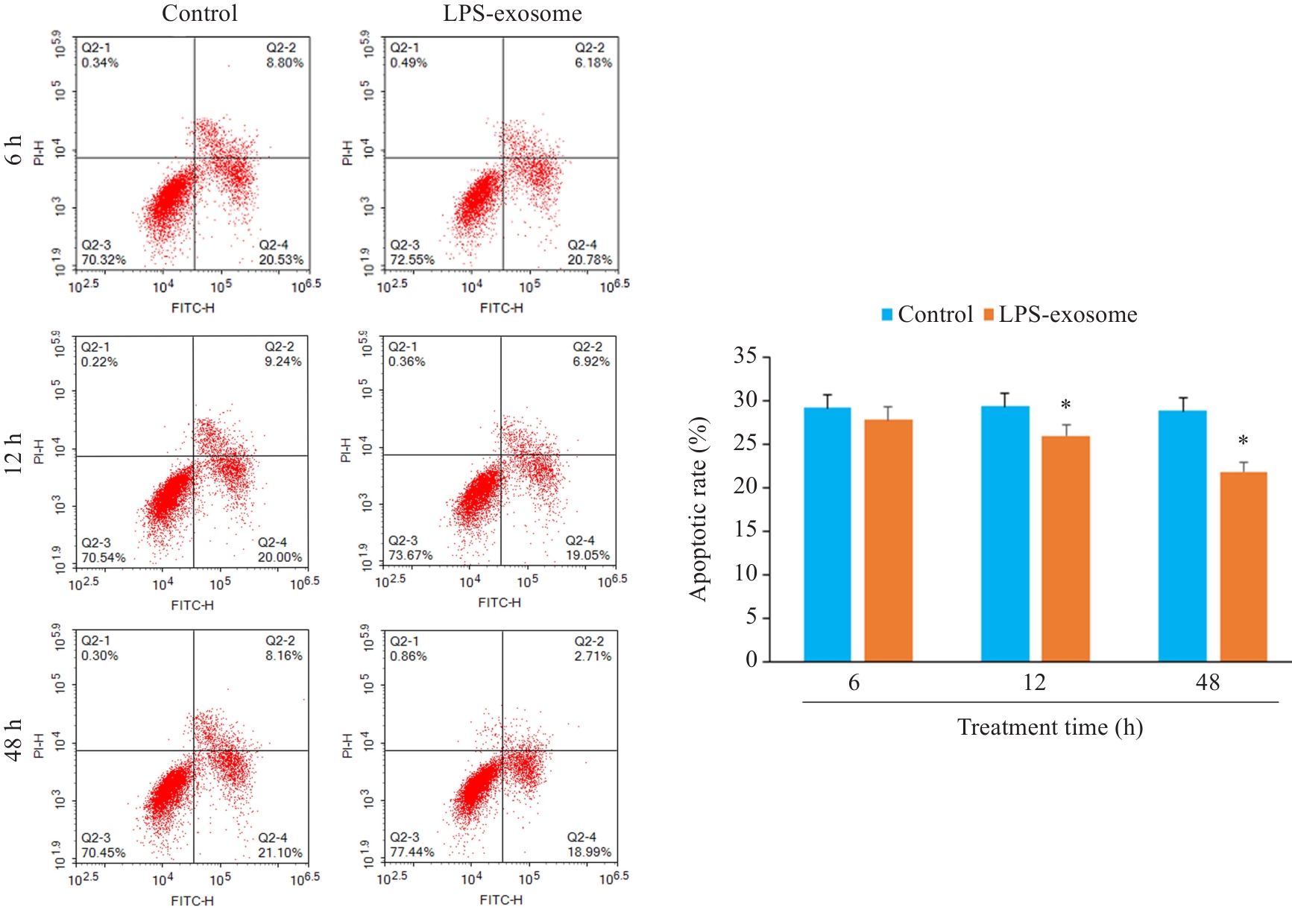
Cao Y, Wang Y, Xiao L, et al. Endothelial-derived exosomes induced by lipopolysaccharide alleviate rat cardiomyocytes injury and apoptosis[J]. Am J Transl Res, 2021, 13(3): 1432-44.
|
| [6] |
Zhang Z, Xu Y, Cao C, et al. Exosomes as a messager to regulate the crosstalk between macrophages and cardiomyocytes under hypoxia conditions[J]. J Cell Mol Med, 2022, 26(5): 1486-500. doi:10.1111/jcmm.17162
|
| [7] |
Henning RJ. Cardiovascular exosomes and microRNAs in cardiovascular physiology and pathophysiology[J]. J Cardiovasc Transl Res, 2021, 14(2): 195-212. doi:10.1007/s12265-020-10040-5
|
| [8] |
Jiang C, Liu X, Wang M, et al. High blood miR-802 is associated with poor prognosis in HCC patients by regulating DNA damage response 1 (REDD1)-mediated function of T cells[J]. Oncol Res, 2019, 27(9): 1025-34. doi:10.3727/096504018x15456687424096
|
| [9] |
Montgomery RL, Hullinger TG, Semus HM, et al. Therapeutic inhibition of miR-208a improves cardiac function and survival during heart failure[J]. Circulation, 2011, 124(14): 1537-47. doi:10.1161/circulationaha.111.030932
|
| [10] |
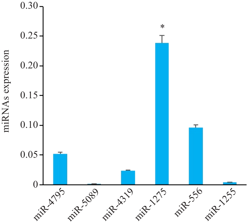
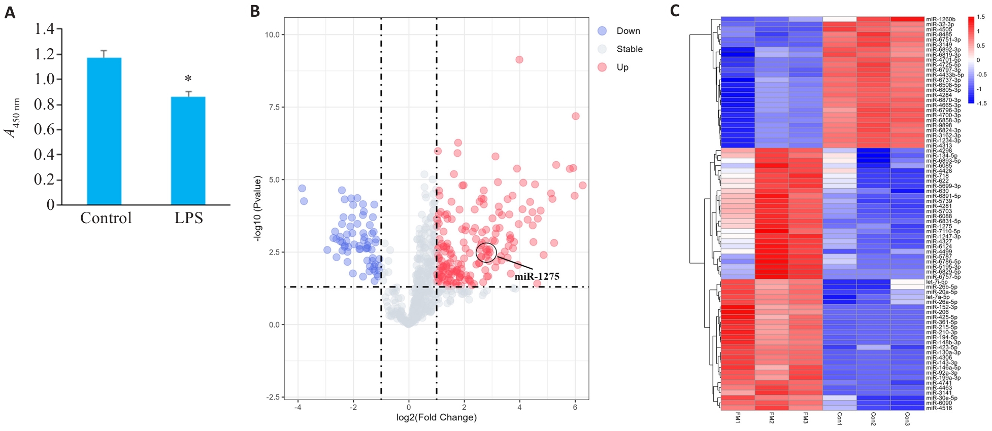
Zeng Z, Ma H, Chen J, et al. Knockdown of miR-1275 protects against cardiomyocytes injury through promoting neuromedin U type 1 receptor[J]. Cell Cycle, 2020, 19(24): 3639-49. doi:10.1080/15384101.2020.1860310
|
| [11] |
Zou Z, Dai R, Deng N, et al. Exosomal miR-1275 secreted by prostate cancer cells modulates osteoblast proliferation and activity by targeting the SIRT2/RUNX2 cascade[J]. Cell Transplant, 2021, 30: 9636897211052977. doi:10.1177/09636897211052977
|
| [12] |
陈 杰. 肝癌外泌体源性miR-1275调控肿瘤免疫的机制研究[D].桂林医学院, 2024.
|
| [13] |
Li JW, Qu H, Wang Y. Aib1 deficiency exacerbates inflammatory responses in acute myocardial infarction mice[J]. J Mol Med, 2022, 100(8): 1181-90. doi:10.1007/s00109-022-02231-1
|
| [14] |
Kim S, Lee JD, Kim BK, et al. Association between left ventricular systolic dysfunction and mortality in patients with septic shock[J]. J Korean Med Sci, 2020, 35(4): e24. doi:10.3346/jkms.2020.35.e24
|
| [15] |
Xiao J, Pan Y, Li XH, et al. Cardiac progenitor cell-derived exosomes prevent cardiomyocytes apoptosis through exosomal miR-21 by targeting PDCD4[J]. Cell Death Dis, 2016, 7(6): e2277. doi:10.1038/cddis.2016.181
|
| [16] |
Kalluri R, LeBleu VS.The biology, function, and biomedical applications of exosomes[J]. Science, 2020, 367(6478): eaau6977. doi:10.1126/science.aau6977
|
| [17] |
He X, Liu S, Zhang Z, et al. M1 macrophage-derived exosomes inhibit cardiomyocyte proliferation through delivering miR-155[J]. BMC Cardiovasc Disord, 2024, 24(1): 365. doi:10.1186/s12872-024-03893-0
|
| [18] |
Mayourian J, Ceholski DK, Gorski PA, et al. Exosomal microRNA-21-5p mediates mesenchymal stem cell paracrine effects on human cardiac tissue contractility[J]. Circ Res, 2018, 122(7): 933-44. doi:10.1161/circresaha.118.312420
|
| [19] |
Bayoumi AS, Park KM, Wang YC, et al. A carvedilol-responsive microRNA, miR-125b-5p protects the heart from acute myocardial infarction by repressing pro-apoptotic bak1 and klf13 in cardiomyocytes[J]. J Mol Cell Cardiol, 2018, 114: 72-82. doi:10.1016/j.yjmcc.2017.11.003
|
| [20] |
Gupta S, Knowlton AA. HSP60 trafficking in adult cardiac myocytes: role of the exosomal pathway[J]. Am J Physiol Heart Circ Physiol, 2007, 292(6): H3052-6. doi:10.1152/ajpheart.01355.2006
|
| [21] |
Tong QH, Hu HY, Chai H, et al. Dysregulation of the miR-1275/HK2 axis contributes to the progression of hypoxia/reoxygenation-induced myocardial injury[J]. Arch Med Res, 2021, 52(5): 461-70. doi:10.1016/j.arcmed.2021.01.006
|
| [22] |
林芸芸, 黄 珊, 宋艳玲, 等. 阿托伐他汀诱导EPC-MVs增多对STEMI患者心肌细胞的保护作用研究[J]. 中国药房, 2019,30(10):1396-402.
|
| [23] |
Gong Z, Wen M, Zhang W, et al. Plasma exosomes induce inflammatory immune response in patients with acute myocardial infarction[J]. Arch Physiol Biochem, 2023, 129(5): 1168-76. doi:10.1080/13813455.2021.1912102
|
| [24] |
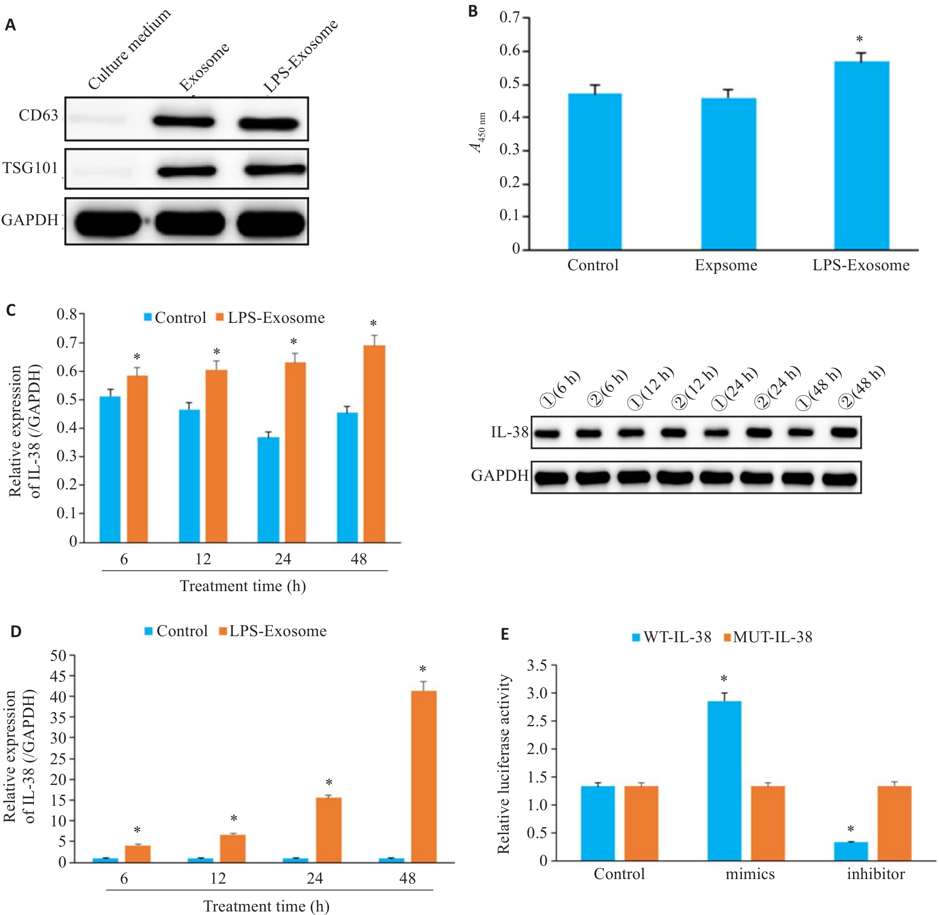
Li Z, Ding Y, Peng Y, et al. Effects of IL-38 on macrophages and myocardial ischemic injury[J]. Front Immunol, 2022, 13: 894002. doi:10.3389/fimmu.2022.894002
|
| [25] |
刘 越.IL-38对脓毒症心肌损伤的作用及机制研究[D].南昌大学,2022.DOI:10.27232/d.cnki.gnchu.2022.000118 .
|
| [26] |
Song J, Chen X, Wang M, et al. Cardiac endothelial cell-derived exosomes induce specific regulatory B cells[J]. Sci Rep, 2014, 4: 7583. doi:10.1038/srep07583
|
| [27] |
Deng RM, Zhou J. The role of PI3K/AKT signaling pathway in myocardial ischemia-reperfusion injury[J]. Int Immunopharmacol, 2023, 123: 110714. doi:10.1016/j.intimp.2023.110714
|
| [28] |
Chen F, Chen ZQ, Wang H, et al. Puerarin pretreatment inhibits myocardial apoptosis and improves cardiac function in rats after acute myocardial infarction through the PI3K/Akt signaling pathway[J]. Adv Clin Exp Med, 2021, 30(3): 255-61. doi:10.17219/acem/131754
|
| [29] |
Ashayeri Ahmadabad H, Mohammadi Panah S, Ghasemnejad-Berenji H, et al. Metformin and the PI3K/AKT signaling pathway: implications for cancer, cardiovascular, and central nervous system diseases[J]. Naunyn Schmiedebergs Arch Pharmacol, 2025, 398(2): 1035-55. doi:10.1007/s00210-024-03358-3
|
| [30] |
Li W, Lin M, Li J, et al. Xijiao Dihuang decoction protects against murine sepsis-induced cardiac inflammation and apoptosis via suppressing TLR4/NF-κB and activating PI3K/AKT pathway[J]. J Inflamm Res, 2024, 17: 853-63. doi:10.2147/jir.s428305
|
| [31] |
Zhang Q, Wang L, Wang S, et al. Signaling pathways and targeted therapy for myocardial infarction[J]. Signal Transduct Target Ther, 2022, 7(1): 78. doi:10.1038/s41392-022-00925-z
|
| [32] |
Wei Y, Xing J, Su X, et al. IL-38 attenuates myocardial ischemia-reperfusion injury by inhibiting macrophage inflammation[J]. Immun Inflamm Dis, 2023, 11(6): e898. doi:10.1002/iid3.898
|
| [33] |
Wei Y, Lan Y, Zhong Y, et al. Interleukin-38 alleviates cardiac remodelling after myocardial infarction[J]. J Cell Mol Med, 2020, 24(1): 371-84. doi:10.1111/jcmm.14741
|
 ), 曹新营2, 邢平川1,2, 王志军2(