南方医科大学学报 ›› 2025, Vol. 45 ›› Issue (5): 921-928.doi: 10.12122/j.issn.1673-4254.2025.05.04
收稿日期:2025-01-26
出版日期:2025-05-20
发布日期:2025-05-23
通讯作者:
储浩然
E-mail:361364304@qq.com;chuhaoran62@163.com
作者简介:浦延鹏,主治医师,博士后在站,E-mail: 361364304@qq.com
基金资助:
Yanpeng PU1,2( ), Zhen WANG2, Haoran CHU3(
 )
)
Received:2025-01-26
Online:2025-05-20
Published:2025-05-23
Contact:
Haoran CHU
E-mail:361364304@qq.com;chuhaoran62@163.com
Supported by:摘要:
目的 观察眼针对脑缺血再灌注损伤(CIRI)模型大鼠神经功能及缺血侧脑皮层组织血管新生的影响,基于METTL3介导的N6-甲基腺苷(m6A)甲基化修饰,上调HIF-1α/VEGF-A信号轴,促进血管新生,探究眼针改善CIRI模型大鼠神经功能的作用机制。 方法 将50只SD大鼠随机分为正常、假手术、模型、眼针及激动剂组(n=10)。采用改良线栓法制备CIRI模型,分组干预后对大鼠进行神经功能评分,TTC染色比较脑梗死面积,尼氏体染色观察脑神经元损伤,CD31和EDU免疫荧光双标检测脑皮层组织血管新生,采用ELISA检测脑皮层组织m6A甲基化修饰水平,RT-PCR检测METTL3、HIF-1α/VEGF-A的mRNA表达,Western blotting检测METTL3、HIF-1α/VEGF-A的蛋白表达。 结果 与正常组和假手术组比较,模型组大鼠神经功能评分明显升高(P<0.05),脑梗死面积明显增加(P<0.05),尼氏体数量明显减少(P<0.05),CD31和EDU免疫荧光双标的新生血管数量增加,m6A甲基化修饰明显减少(P<0.05),METTL3的蛋白及mRNA表达减少(P<0.05),HIF-1α与VEGF-A的蛋白及mRNA表达增加(P<0.05);与模型组比较,眼针和激动剂组神经功能评分明显下降(P<0.05),脑梗死面积明显减小(P<0.05),尼氏体数量明显增多(P<0.05),CD31和EDU荧光双标的新生血管数量明显增多,m6A甲基化修饰明显增加(P<0.05),而HIF-1α与VEGF-A、METTL3的蛋白及mRNA表达亦明显增加(P<0.05)。眼针和激动剂组、正常和假手术组比较,各指标差异无统计学意义(P>0.05)。 结论 眼针能改善CIRI模型大鼠神经功能损伤,其机制可能与上调METTL3介导的m6A甲基化修饰,调节HIF-1α/VEGF-A信号轴,促进脑皮层血管新生有关。
浦延鹏, 王震, 储浩然. 眼针疗法通过上调METTL3介导的m6A甲基化修饰促进脑皮层血管新生进而改善脑缺血再灌注损伤大鼠的神经功能[J]. 南方医科大学学报, 2025, 45(5): 921-928.
Yanpeng PU, Zhen WANG, Haoran CHU. Eye acupuncture improves neural function in rats with cerebral ischemia-reperfusion injury by promoting angiogenesis via upregulating METTL3-mediated m6A methylation[J]. Journal of Southern Medical University, 2025, 45(5): 921-928.
| Phenotype | Score |
|---|---|
| There was no neurological impairment | 0 |
| When lifting the tail, the contralateral forelimb is adducted and flexed | 1 |
| When crawling, rotate to the opposite side | 2 |
| When standing or crawling, tip to the opposite side | 3 |
| No voluntary movement with disturbance of consciousness | 4 |
表1 Longa功能评分
Tab.1 Longa function scoring criteria
| Phenotype | Score |
|---|---|
| There was no neurological impairment | 0 |
| When lifting the tail, the contralateral forelimb is adducted and flexed | 1 |
| When crawling, rotate to the opposite side | 2 |
| When standing or crawling, tip to the opposite side | 3 |
| No voluntary movement with disturbance of consciousness | 4 |
| Inspect | Phenotype | Score |
|---|---|---|
Gently grasp the tail and lift the rat 10 cm above the table top. Normal rats should extend their front PAWS straight. | There was no neurological impairment | 0 |
| Brain lesions flexion of the wrist, elbow and shoulder adduction | 1 | |
| The above signs+decreased resistance to push on the paralytic side | 2 | |
| Move in a circle towards the paralyzed side (rear-end) | 3 |
表2 Bederson评分
Tab.2 Bederson scoring criteria
| Inspect | Phenotype | Score |
|---|---|---|
Gently grasp the tail and lift the rat 10 cm above the table top. Normal rats should extend their front PAWS straight. | There was no neurological impairment | 0 |
| Brain lesions flexion of the wrist, elbow and shoulder adduction | 1 | |
| The above signs+decreased resistance to push on the paralytic side | 2 | |
| Move in a circle towards the paralyzed side (rear-end) | 3 |
| Gene | Primer sequence (5'→3') | Product length (bp) |
|---|---|---|
| METTL3 | F:TGGGGGTATGAACGGGTAGA R:TCCTTTGACACCAACCAAGC | 129 |
| HIF-1α | F:TTACAGGATTCCAGCAGACCCA R:GCTGATGCCTTAGCAGTGGTC | 145 |
| VEGF-A | F:TAAATCCTGGAGCGTTCACTGTG R:TTCGTTTAACTCAAGCTGCCTC | 145 |
| GAPDH | F:GGCAAGTTCAACGGCACAG R:CGCCAGTAGACTCCACGACAT | 150 |
表3 引物序列
Tab.3 Primer sequences used for RT-qPCR
| Gene | Primer sequence (5'→3') | Product length (bp) |
|---|---|---|
| METTL3 | F:TGGGGGTATGAACGGGTAGA R:TCCTTTGACACCAACCAAGC | 129 |
| HIF-1α | F:TTACAGGATTCCAGCAGACCCA R:GCTGATGCCTTAGCAGTGGTC | 145 |
| VEGF-A | F:TAAATCCTGGAGCGTTCACTGTG R:TTCGTTTAACTCAAGCTGCCTC | 145 |
| GAPDH | F:GGCAAGTTCAACGGCACAG R:CGCCAGTAGACTCCACGACAT | 150 |
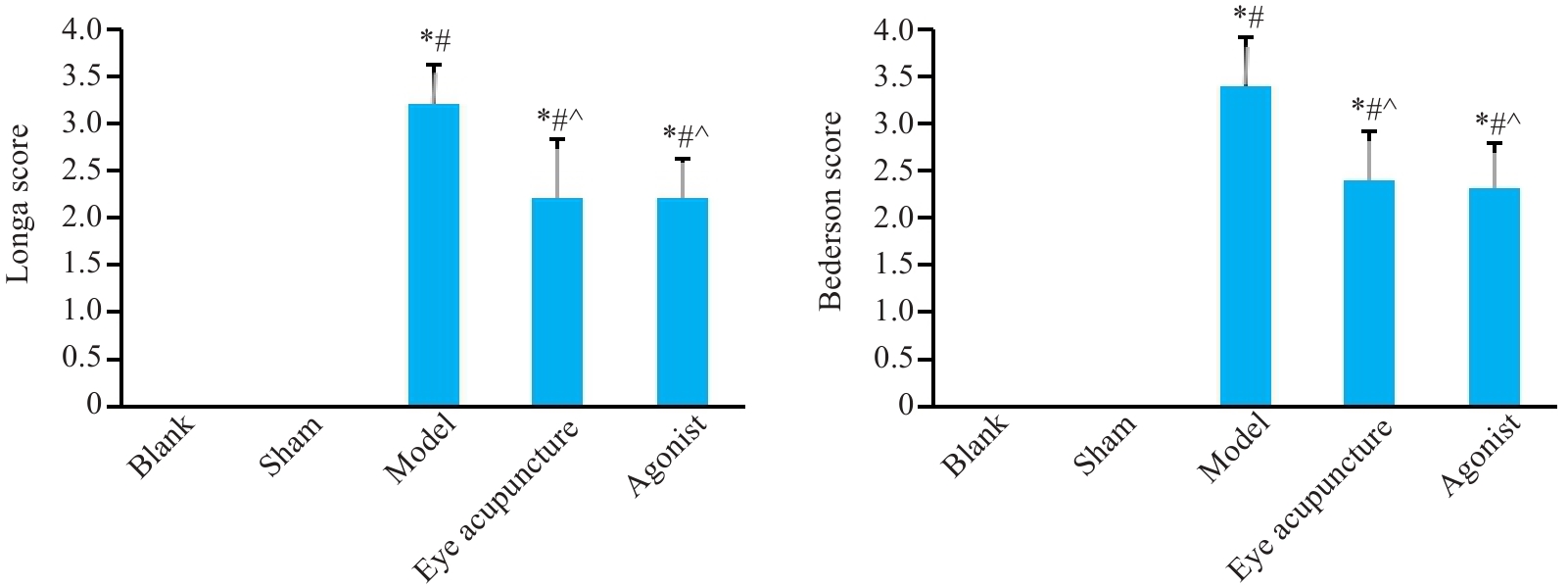
图1 神经功能评分
Fig.1 Neurological function scores of the rats in different groups (Mean±SD, n=10). *P<0.05 vs Blank group; #P<0.05 vs Sham group; ^P<0.05 vs model group.
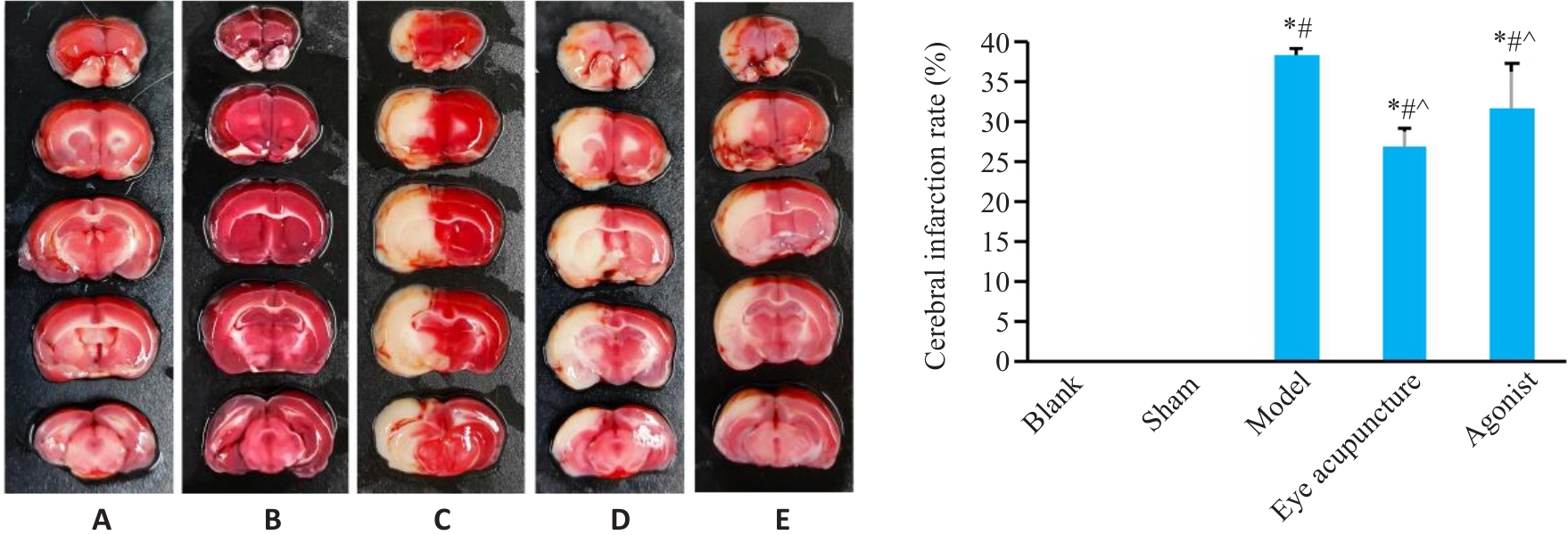
图2 脑TTC染色
Fig.2 Brain TTC staining showing cerebral infarct sizes in different groups (Mean±SD, n=3). A: Blank group; B: Sham operation group; C: Model group; D: Eye acupunctrue group; E: HIF-1α agonist group. *P<0.05 vs Blank group; #P<0.05 vs Sham group; ^P<0.05 vs model group.
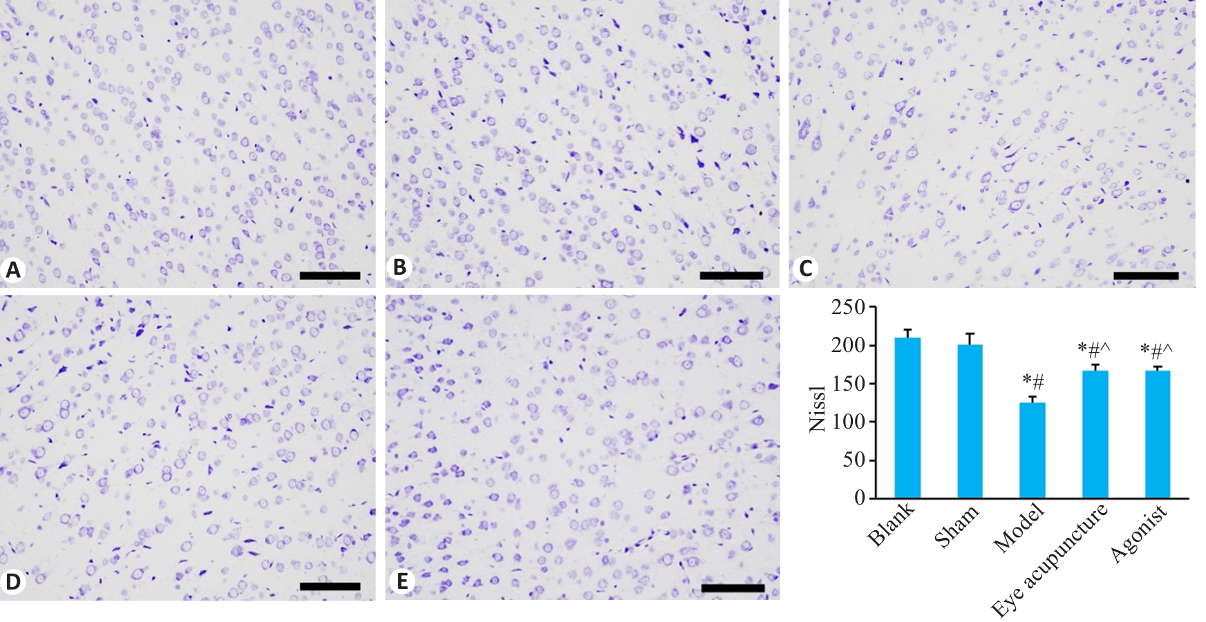
图3 大鼠脑皮层组织尼氏体染色
Fig.3 Nissl staining of the cortical tissue in different groups (Scale bar=100 μm; Mean±SD, n=3). A: Blank group; B: Sham operation group; C: Model group; D: Eye acupunctrue group; E: HIF-1α agonist group. *P<0.05 vs Blank group; #P<0.05 vs Sham group; ^P<0.05 vs model group.
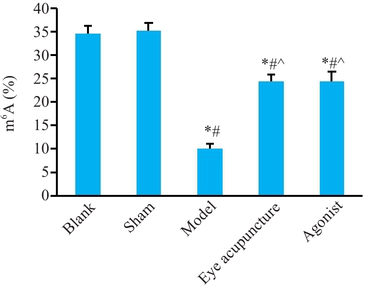
图5 大鼠脑皮层组织m6A甲基化修饰
Fig.5 m6A methylation level in rat cerebral cortex (Mean±SD, n=3). *P<0.05 vs Blank group; #P<0.05 vs Sham group; ^P<0.05 vs model group.
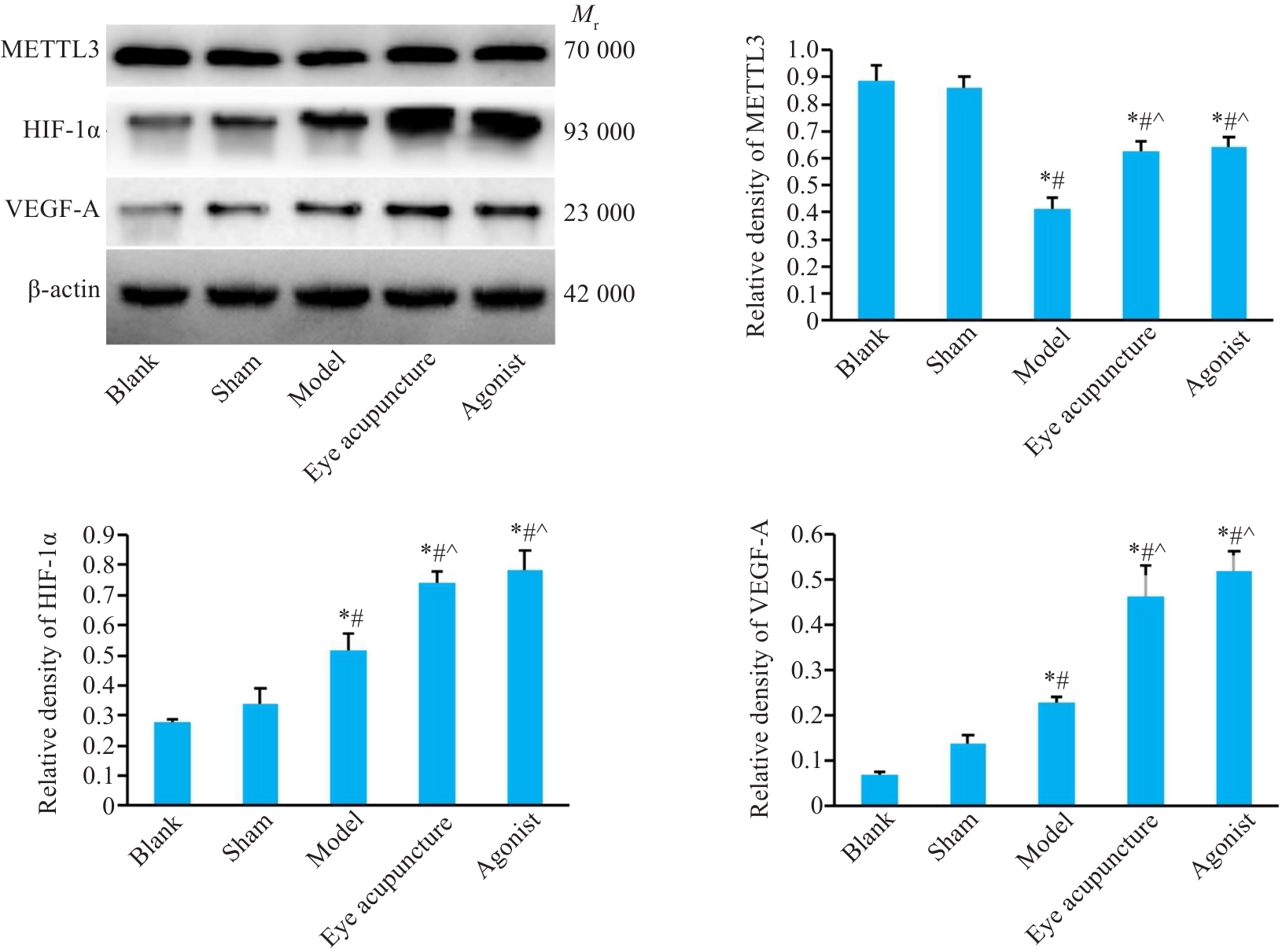
图6 大鼠脑皮层组织METTL3、HIF-1α/VEGF-A的蛋白表达
Fig.6 Expression of METTL3 and HIF-1α/VEGF-A in rat cerebral cortex detected by Western blotting (Mean±SD, n=3). *P<0.05 vs Blank group; #P<0.05 vs Sham group; ^P<0.05 vs model group.

图7 大鼠脑皮层组织METTL3、HIF-1α/VEGF-A的mRNA表达
Fig.7 mRNA expression of METTL3 and HIF-1α/VEGF-A in rat cerebral cortex detected by RT-qPCR (Mean±SD, n=3). *P<0.05 vs Blank group; #P<0.05 vs Sham group; ^P<0.05 vs model group.
| 1 | Collaborators G 2 S. Global, regional, and national burden of stroke and its risk factors, 1990-2019: a systematic analysis for the Global Burden of Disease Study 2019[J]. Lancet Neurol, 2021, 20(10): 795-820. |
| 2 | 浦延鹏, 王鹏琴, 程景艳, 等. 基于数据挖掘探析眼针的优势病种及治疗方法[J]. 辽宁中医杂志, 2023, 50(4): 11-6. DOI: 10.13192/j.issn.1000-1719.2023.04.003 |
| 3 | 楚 宝, 张继杰, 董立朋, 等. 脑血管侧支循环评价的研究进展[J]. 中国全科医学, 2020, 23(5): 516-24. DOI: 10.12114/j.issn.1007-9572.2019.00.305 |
| 4 | 武家竹, 陈林玲, 邸嘉玮, 等. 针灸改善缺血性脑卒中侧支循环的血管作用机制[J]. 针灸临床杂志, 2023, 39(6): 105-9. |
| 5 | 付彩红, 吴 爽, 张 虎, 等. 针刺促进缺血性脑卒中血管新生研究进展[J]. 辽宁中医药大学学报, 2021, 23(12): 87-93. DOI: 10.13194/j.issn.1673-842x.2021.12.018 |
| 6 | Vatte S, Ugale R. HIF-1, an important regulator in potential new therapeutic approaches to ischemic stroke[J]. Neurochem Int, 2023, 170: 105605. |
| 7 | 谭佳佳, 唐丽亚, 孙晓莹, 等. 电针预处理对脑缺血再灌注损伤大鼠神经功能及缺血半暗带区VEGF、CD31表达的影响[J]. 湖南中医药大学学报, 2023, 43(9): 1679-84. |
| 8 | Xu SY, Ni SM, Zeng CL, et al. Electro-acupuncture promotes angiogenesis via exosomal miR-210 in the hypoxia-induced HUVECs mediated HIF-1α/VEGF/Notch 1 signal pathway[J]. Curr Neurovasc Res, 2022, 19(4): 406-17. |
| 9 | Zhang HS, Zhou JW, Li JX, et al. N6-methyladenosine promotes translation of VEGFA to accelerate angiogenesis in lung cancer[J]. Cancer Res, 2023, 83(13): 2208-25. |
| 10 | Yao MD, Jiang Q, Ma Y, et al. Role of METTL3-dependent N6-methyladenosine mRNA modification in the promotion of angiogenesis[J]. Mol Ther, 2020, 28(10): 2191-202. |
| 11 | Chen K, Li WD, Li XQ. The role of m6A in angiogenesis and vascular diseases[J]. iScience, 2024, 27(7): 110082. |
| 12 | 马贤德, 孙宏伟, 柴纪严, 等. 线栓法制备大鼠脑缺血再灌注模型的方法研究[J]. 中华中医药学刊, 2009, 27(6): 1200-1. |
| 13 | Longa EZ, Weinstein PR, Carlson S, et al. Reversible middle cerebral artery occlusion without craniectomy in rats[J]. Stroke, 1989, 20(1): 84-91. |
| 14 | 彭静山原著,彭筱山, 王鹏琴整理. 眼针疗法[M]. 2版. 沈阳: 辽宁科学技术出版社, 2018. |
| 15 | Bederson JB, Pitts LH, Tsuji M, et al. Rat middle cerebral artery occlusion: evaluation of the model and development of a neurologic examination[J]. Stroke, 1986, 17(3): 472-6. |
| 16 | Zhang W, Han L, Wen Y, et al. Electroacupuncture reverses endothelial cell death and promotes angiogenesis through the VEGF/Notch signaling pathway after focal cerebral ischemia-reperfusion injury[J]. Brain Behav, 2023, 13(3): e2912. |
| 17 | Wang L, Sheng G, Cui J, et al. Electroacupuncture attenuates ischemic injury after stroke and promotes angiogenesis via activation of EPO mediated Src and VEGF signaling pathways[J]. PLoS One, 2022, 17(9): e0274620. |
| 18 | Geiseler SJ, Morland C. The Janus face of VEGF in stroke[J]. Int J Mol Sci, 2018, 19(5): 1362. |
| 19 | Yang L, Yang BC, Zhang CX, et al. Protective effects of acupuncture and LGNHFD on expressions of vascular endothelial growth factor, basic fibroblast growth factor, and cluster of differentiation 34 in rats with cerebral ischemia-reperfusion injury[J]. J Tradit Chin Med, 2021, 41(3): 463-70. |
| 20 | 邵 妍, 王鹏琴, 王树东, 等. 眼针运动疗法对脑缺血再灌注大鼠脑缺血半暗带区VEGF蛋白及VEGFmRNA表达的影响[J]. 中国中医基础医学杂志, 2015, 21(4): 445-8. |
| 21 | Chen HX, Ma D, Yue FX, et al. The potential role of hypoxia-inducible factor-1 in the progression and therapy of central nervous system diseases[J]. Curr Neuropharmacol, 2022, 20(9): 1651-66. |
| 22 | Hu QM, Liu L, Zhou L, et al. Effect of fluoxetine on HIF-1α- Netrin/VEGF cascade, angiogenesis and neuroprotection in a rat model of transient middle cerebral artery occlusion[J]. Exp Neurol, 2020, 329: 113312. |
| 23 | Wang L, Li JC, Wang Y, et al. Dan-Deng-Tong-Nao softgel capsule promotes angiogenesis of cerebral microvasculature to protect cerebral ischemia reperfusion injury via activating HIF-1α-VEGFA-Notch1 signaling pathway[J]. Phytomedicine, 2023, 118: 154966. |
| 24 | Ni HZ, Li JX, Zheng JY, et al. Cardamonin attenuates cerebral ischemia/reperfusion injury by activating the HIF-1α/VEGFA pathway[J]. Phytother Res, 2022, 36(4): 1736-47. |
| 25 | Han F. N6 -methyladenosine modification in ischemic stroke: Functions, regulation, and therapeutic potential[J]. Heliyon, 2024, 10(3): e25192. |
| 26 | Wang LJ, Xue YM, Huo R, et al. N6-methyladenosine methyltransferase METTL3 affects the phenotype of cerebral arteriovenous malformation via modulating Notch signaling pathway[J]. J Biomed Sci, 2020, 27(1): 62. |
| 27 | Huang HL, Weng HY, Chen JJ. m6A modification in coding and non-coding RNAs: roles and therapeutic implications in cancer[J]. Cancer Cell, 2020, 37(3): 270-88. |
| 28 | Tian S, Li YL, Wang J, et al. Chinese Ecliptae herba (Eclipta prostrata (L.) L.) extract and its component wedelolactone enhances osteoblastogenesis of bone marrow mesenchymal stem cells via targeting METTL3-mediated m6A RNA methylation[J]. J Ethnopharmacol, 2023, 312: 116433. |
| 29 | 庞立健, 王鹏琴, 吕晓东, 等. 彭氏眼针疗法理论阐释及应用[J]. 中华中医药杂志, 2023, 38(1): 90-5. |
| 30 | 浦延鹏. 基于Wnt3a/β-catenin/LEF1信号通路探讨眼针促MCAO/R大鼠血管新生的作用机制[D]. 沈阳: 辽宁中医药大学, 2021. |
| 31 | Sen T, Sen N. Treatment with an activator of hypoxia-inducible factor 1, DMOG provides neuroprotection after traumatic brain injury[J]. Neuropharmacology, 2016, 107: 79-88. |
| 32 | 牛雨晴, 秦合伟, 李彦杰, 等. 眼针干预脑缺血模型大鼠调节血管新生因子表达促进血管新生[J]. 中国组织工程研究, 2023, 27(23): 3674-81. |
| [1] | 张安邦, 孙秀颀, 庞博, 吴远华, 时靖宇, 张宁, 叶涛. 电针预处理通过调节肠道-大脑轴及Nrf2/HO-1信号通路抑制铁死亡减轻大鼠脑缺血再灌注损伤[J]. 南方医科大学学报, 2025, 45(5): 911-920. |
| [2] | 陈洁, 刘晨旭, 王春, 李丽, 陶伟婷, 徐婧茹, 唐红辉, 黄丽. 外源性瘦素通过上调星形胶质细胞GLT-1和GLAST的表达减轻小鼠脑缺血再灌注引起的谷氨酸兴奋毒性损伤[J]. 南方医科大学学报, 2024, 44(6): 1079-1087. |
| [3] | 刘昊铭, 林子诗, 叶 靖. CaMKIIγ和CaMKIIδ通过 PI3K/Akt/Erk 信号通路减轻小鼠神经元缺血再灌注损伤[J]. 南方医科大学学报, 2024, 44(3): 563-570. |
| [4] | 孙 伟, 陈 平, 唐小杭, 谷颖敏, 田雪松. 改良的四血管间断阻塞法制作大鼠全脑缺血再灌注损伤模型[J]. 南方医科大学学报, 2023, 43(7): 1194-1203. |
| [5] | 李佩佩, 胡音琦, 刘 佳, 王丽娜, 吴元洁, 胡建鹏. 脑络欣通促进氧糖剥夺/再灌注损伤大鼠的脑微血管内皮细胞血管新生:基于激活caspase-1/Gasdermin D通路[J]. 南方医科大学学报, 2023, 43(7): 1093-1101. |
| [6] | 曹天然, 刘青芳, 潘美民, 张雪红. LncRNA SNHG8通过抑制miR-494-3p表达减轻脑缺血再灌注损伤[J]. 南方医科大学学报, 2023, 43(12): 2015-2022. |
| [7] | 王远迎, 戴兴月, 陈春彦, 程培钧, 陈松江, 黎欣悦, 毕 炫, 程 瑶, 吴昌学, 吴 宁. 苗药验方四大血可减少关节炎大鼠滑膜组织的坏死性凋亡和血管新生:基于抑制RIPK1/RIPK3/MLKL通路[J]. 南方医科大学学报, 2022, 42(12): 1774-1782. |
| [8] | 林 媛, 刘志敏, 董梦琦, 周伟杰. 肝脏血管认知更新与研究新思路[J]. 南方医科大学学报, 2022, 42(12): 1907-1910. |
| [9] | 彭 羽, 林 赟, 侯晓林, 廖晓灵, 刘进平. 亚低温治疗对脑出血模型大鼠动物行为学的影响及其机制[J]. 南方医科大学学报, 2020, 40(09): 1359-1364. |
| [10] | 刘 刚,门运政,童旭辉,王雪如,胡 淼,姜牧君,孙志鹏,董淑英. 线粒体融合与裂变在右美托咪定减轻小鼠脑缺血再灌注损伤中的作用及其机制[J]. 南方医科大学学报, 2020, 40(04): 463-468. |
| [11] | 王颖,韦颖梅,程秀,孙小锦,马琳艳,宋宜宁,周静,魏芳,刘浩. 2-脱氧葡萄糖通过激活AMPK通路抑制类风湿关节炎血管新生[J]. 南方医科大学学报, 2018, 38(08): 962-. |
| [12] | 陈雷,卢小圣,李雅兰,毛周飞,肖銮娟,禹艳红. 球状脂联素对卵巢微血管内皮细胞增殖、迁移及管状结构形成的影响[J]. 南方医科大学学报, 2017, 37(05): 614-. |
| [13] | 郑德仲,周滔,查道刚. 聚氧化乙烯改善大鼠慢性缺血后肢的血液灌注[J]. 南方医科大学学报, 2017, 37(01): 113-. |
| [14] | 樊宗兵,童旭辉,李言,于丽,陈银玲,刘浩昂,董淑英. 丙泊酚保护脑缺血再灌注损伤可能与缝隙连接功能的抑制相关[J]. 南方医科大学学报, 2015, 35(12): 1678-. |
| [15] | 侯德仁,沙地克·沙吾提,邓锦凤,刘建锋,胡中扬,周军,周丽,刘远新. 意向运动疗法对大鼠脑缺血再灌注损伤后NT-3 及GAP-43 表达的影响[J]. 南方医科大学学报, 2011, 31(08): 1401-. |
| 阅读次数 | ||||||
|
全文 |
|
|||||
|
摘要 |
|
|||||