南方医科大学学报 ›› 2025, Vol. 45 ›› Issue (2): 422-436.doi: 10.12122/j.issn.1673-4254.2025.02.23
曾智雄1( ), 王永波2, 林宗悦2, 边兆英1,3, 马建华1()
收稿日期:2024-10-15
出版日期:2025-02-20
发布日期:2025-03-03
通讯作者:
马建华
E-mail:zxzeng@smu.edu.cn;jhma@smu.edu.cn
作者简介:曾智雄,在读硕士研究生,E-mail: zxzeng@smu.edu.cn
基金资助:
Zhixiong ZENG1(), Yongbo WANG2, Zongyue LIN2, Zhaoying BIAN1,3, Jianhua MA1()
Received:2024-10-15
Online:2025-02-20
Published:2025-03-03
Contact:
Jianhua MA
E-mail:zxzeng@smu.edu.cn;jhma@smu.edu.cn
Supported by:摘要:
目的 为了去除患者在牙科锥形束计算机断层扫描(CBCT)扫描过程中发生躯体运动导致的伪影,提升重建图像质量,提出一种基于分段反投影张量退化特征编码的运动伪影校正模型(SBP-MAC)。 方法 该模型由一个生成器和一个退化编码器构成。将分段有限角度重建的子图像堆叠成张量并作为模型输入;用退化编码器提取张量中空间变化的运动信息,自适应调制生成器的各级跳跃连接特征,从而指导模型校正不同运动波形导致的伪影;最后设计伪影一致性损失来简化生成器的学习任务。 结果 该模型能有效地去除运动伪影,提升重建图像质量。在仿真数据上的峰值信噪比提升了8.28%,结构相似度提升了2.29%,均方根误差降低了23.84%;在真实数据上的专家评分最高4.4221(5分制),与所有对比方法之间的评分差异具有统计学意义(P<0.05)。 结论 本文提出的SBP-MAC模型能够有效提取张量中空间变化的运动信息,实现从张量域到图像域的自适应伪影校正,提升牙科CBCT图像质量。
曾智雄, 王永波, 林宗悦, 边兆英, 马建华. 基于分段反投影张量退化特征编码的牙科锥形束计算机断层扫描运动伪影校正[J]. 南方医科大学学报, 2025, 45(2): 422-436.
Zhixiong ZENG, Yongbo WANG, Zongyue LIN, Zhaoying BIAN, Jianhua MA. A segmented backprojection tensor degradation feature encoding model for motion artifacts correction in dental cone beam computed tomography[J]. Journal of Southern Medical University, 2025, 45(2): 422-436.
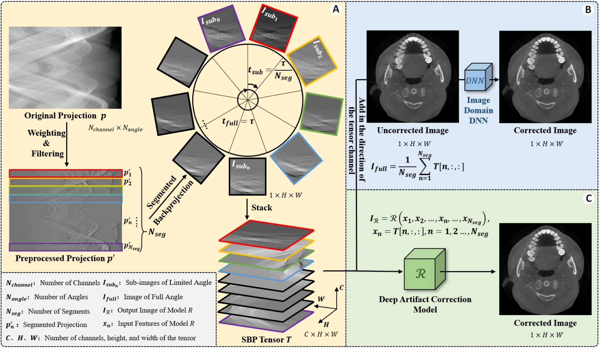
图1 分段反投影的示意图
Fig.1 Schematic diagram of the segmented backprojection tensor. A: Origin of the segmented backprojection tensor. B: Training idea of DNN in the traditional image domain. C: Training idea of the proposed model.
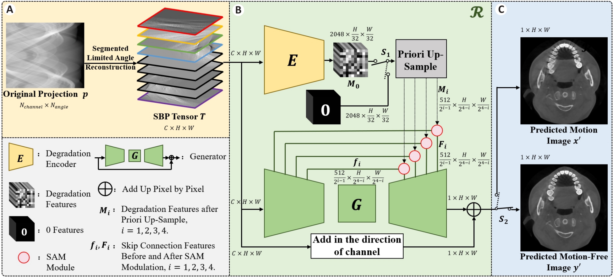
图2 SBP-MAC模型的流程框图
Fig.2 Flow chart of the SBP-MAC model. A: Preparation of the input data of the model. B: Composition of the proposed model. C: Output results of the model.
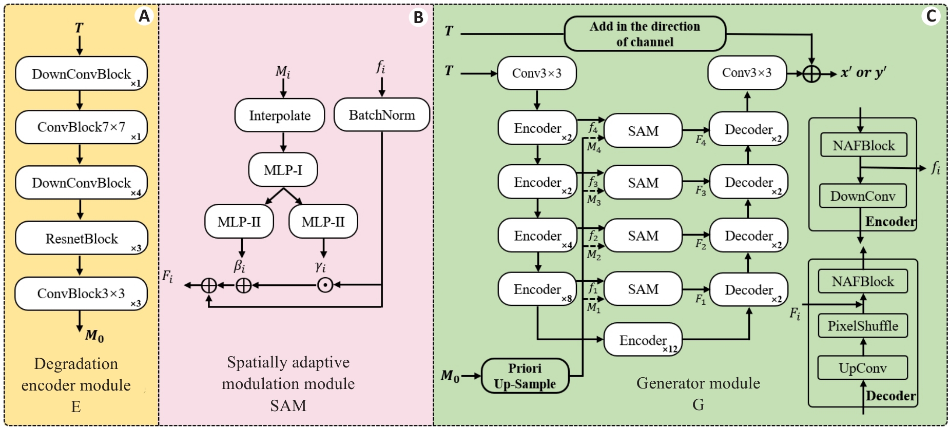
图3 关键模块细节说明
Fig.3 Detailed archetecture of the key modules. A: Degradation Encoder Module (E). B: Spatial Adaptive Modulation Module (SAM). C: Generator Module (G). The number of stacks for each smaller module is indicated at the bottom right corner.
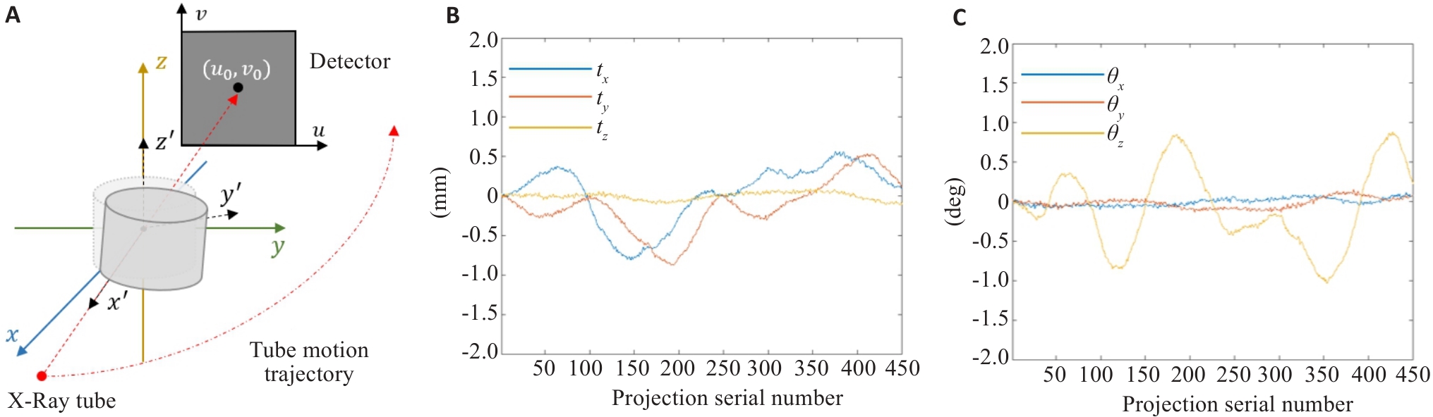
图4 CBCT扫描及运动波形示意图
Fig.4 Schematic diagram of CBCT scanning and motion waveforms. A: Schematic diagram of CBCT scanning. B: Translational motion waveform used in the simulation. C: Rotational motion waveform used in the simulation.
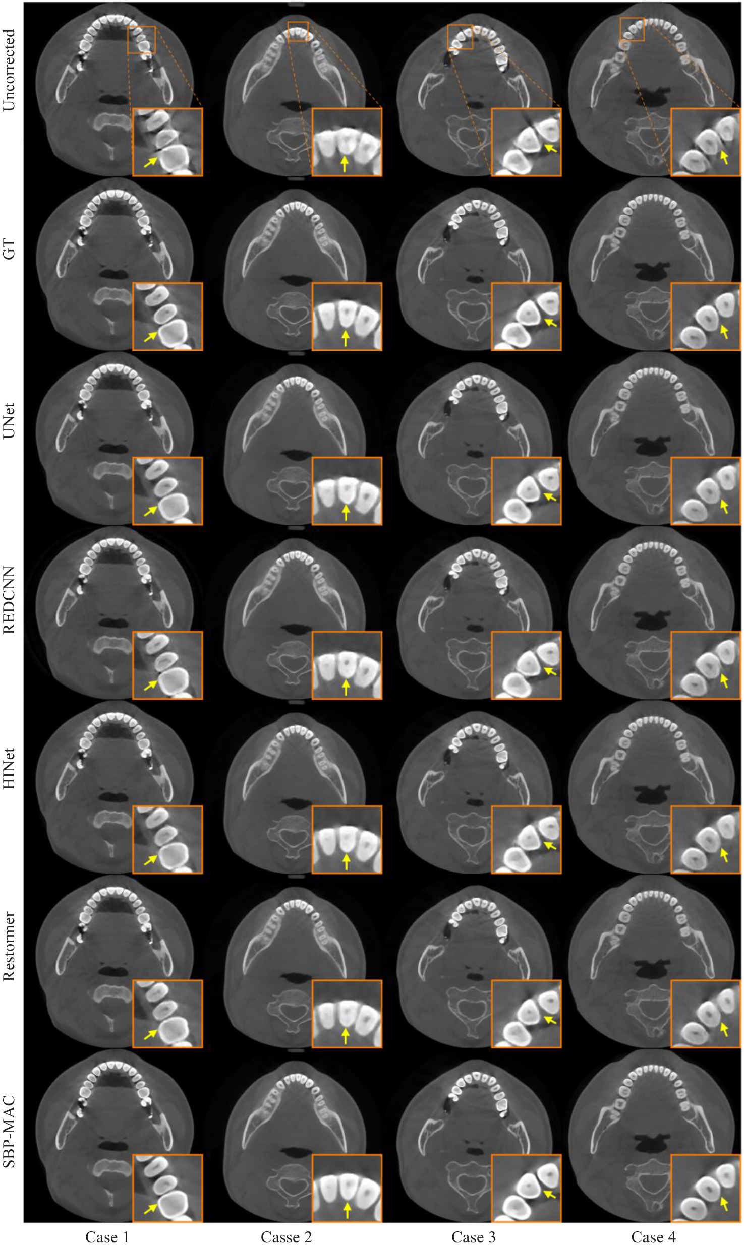
图5 不同伪影校正方法在仿真数据上的横断面结果
Fig.5 Transverse-section results of different motion artifact correction methods on simulation data.
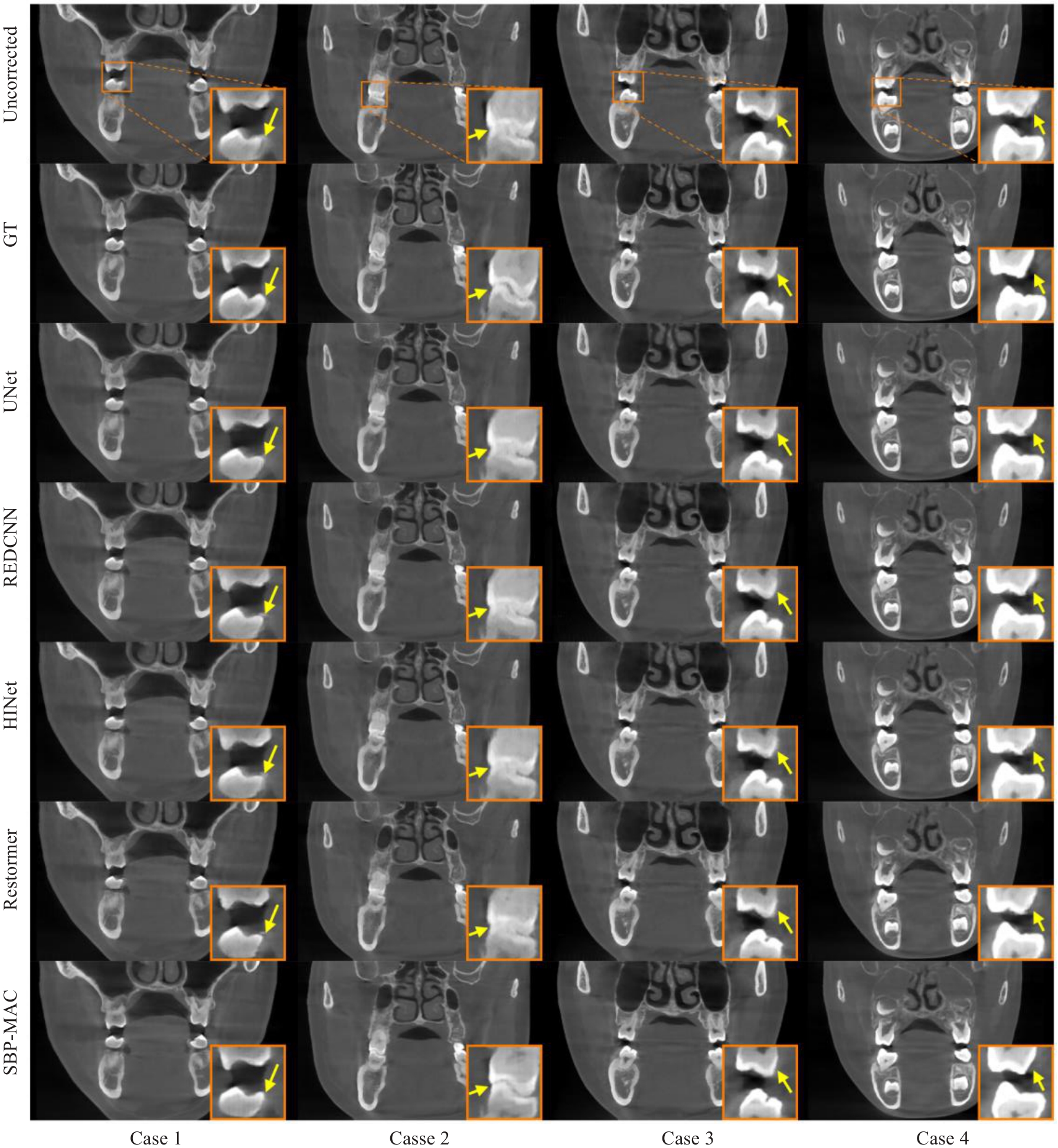
图6 不同伪影校正方法在仿真数据上的冠状面结果
Fig.6 Coronal-section results of different motion artifact correction methods on simulation data.
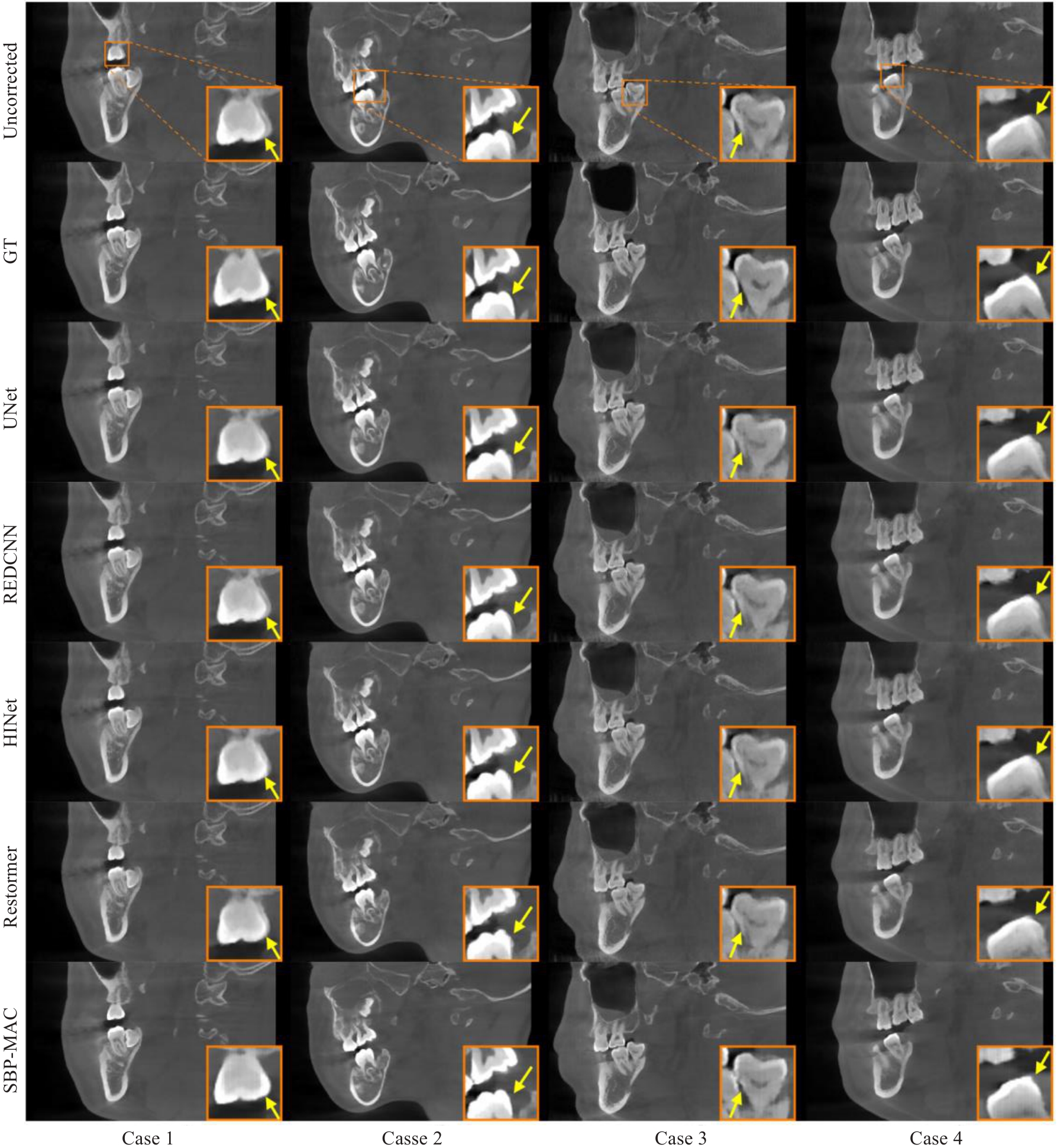
图7 不同伪影校正方法在仿真数据上的矢状面结果
Fig.7 Sagittal-section results of different motion artifact correction methods on simulation data.
| Methods | PSNR | RMSE | SSIM |
|---|---|---|---|
| Uncorrected | 29.5479±1.5036 | 8.6211±1.4713 | 0.9276±0.0191 |
| UNet | 30.2005±1.5400 | 8.0025±1.3922 | 0.9339±0.0191 |
| REDCNN | 30.0276±1.5185 | 8.1599±1.3991 | 0.9318±0.0185 |
| HINet | 30.2865±1.5343 | 7.9228±1.3729 | 0.9346±0.0183 |
| Restormer | 30.3057±1.5646 | 7.9111±1.4128 | 0.9342±0.0191 |
| SBP-MAC | 31.9936±1.9277 | 6.5657±1.4119 | 0.9547±0.0144 |
表1 不同运动伪影校正算法在仿真数据上的量化指标
Tab.1 Quantitative indicators of different motion artifact correction algorithms on simulation data (Mean±SD)
| Methods | PSNR | RMSE | SSIM |
|---|---|---|---|
| Uncorrected | 29.5479±1.5036 | 8.6211±1.4713 | 0.9276±0.0191 |
| UNet | 30.2005±1.5400 | 8.0025±1.3922 | 0.9339±0.0191 |
| REDCNN | 30.0276±1.5185 | 8.1599±1.3991 | 0.9318±0.0185 |
| HINet | 30.2865±1.5343 | 7.9228±1.3729 | 0.9346±0.0183 |
| Restormer | 30.3057±1.5646 | 7.9111±1.4128 | 0.9342±0.0191 |
| SBP-MAC | 31.9936±1.9277 | 6.5657±1.4119 | 0.9547±0.0144 |

图9 不同伪影校正方法在真实数据上的冠状面结果
Fig.10 Coronal-section results of different motion artifact correction methods on real data.
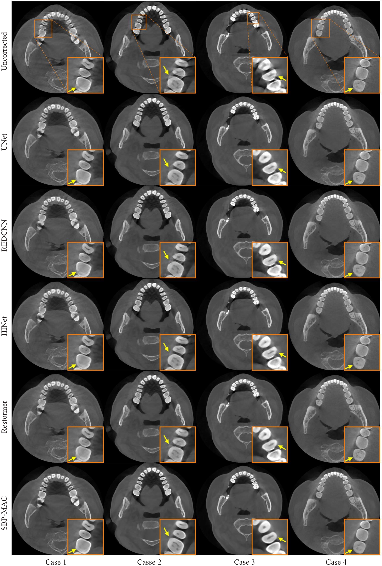
图8 不同伪影校正方法在真实数据上的横断面结果
Fig.8 Transverse-section results of different motion artifact correction methods on real data.
| Methods | Scores (Mean±SD) | P |
|---|---|---|
| UNet | 3.7263±0.5915 | 0.0018 |
| REDCNN | 3.8000±0.5416 | 0.0042 |
| HINet | 3.7895±0.4026 | 0.0012 |
| Restormer | 3.9211±0.3084 | 0.0078 |
| SBP-MAC | 4.2421±0.3548 | - |
表2 真实临床数据上的图像质量专家评分统计
Tab.2 Image quality expert score statistics on real clinical data
| Methods | Scores (Mean±SD) | P |
|---|---|---|
| UNet | 3.7263±0.5915 | 0.0018 |
| REDCNN | 3.8000±0.5416 | 0.0042 |
| HINet | 3.7895±0.4026 | 0.0012 |
| Restormer | 3.9211±0.3084 | 0.0078 |
| SBP-MAC | 4.2421±0.3548 | - |
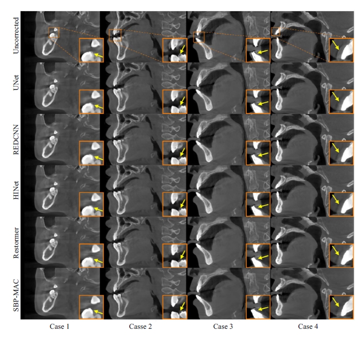
图10 不同伪影校正方法在真实数据上的矢状面结果
Fig.10 Sagittal-section results of different motion artifact correction methods on real data.
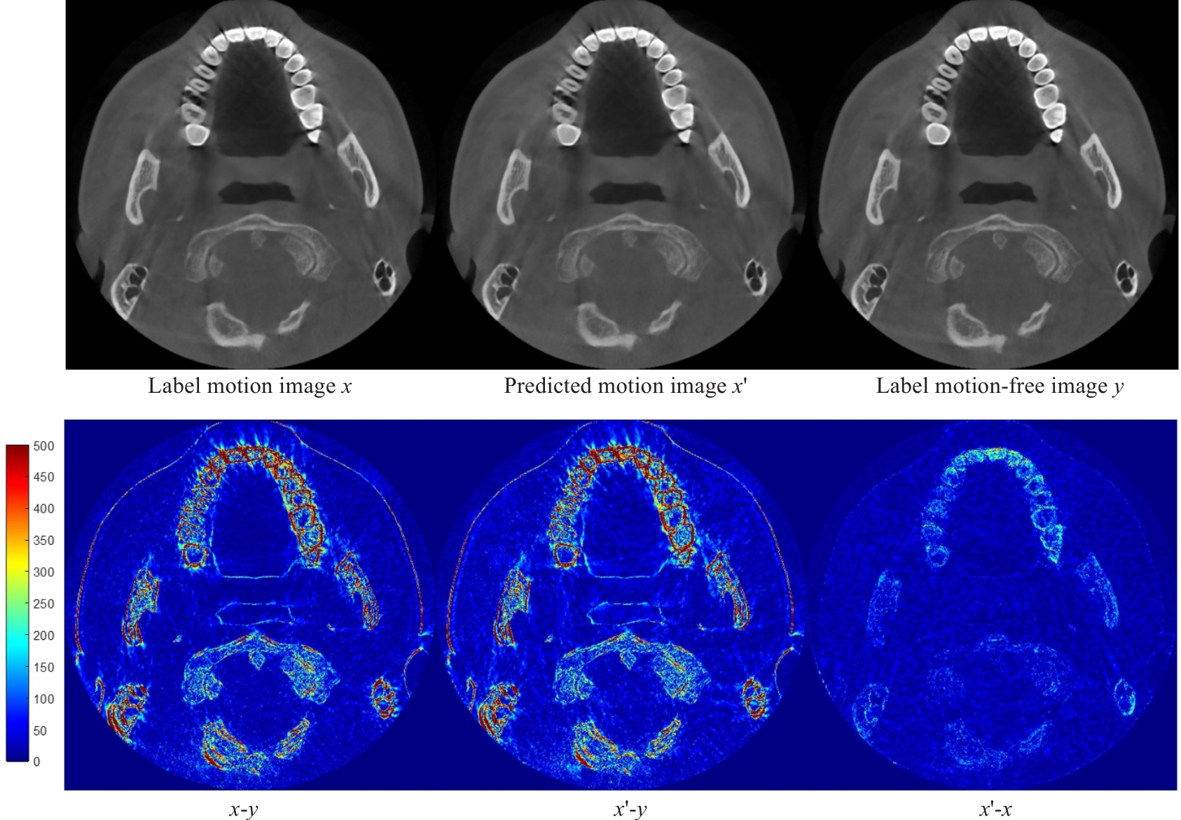
图11 有运动伪影图像生成结果展示
Fig.11 Displacement of motion artifact image generation results.
| PSNR | RMSE | SSIM | |
|---|---|---|---|
| 29.4475±1.5051 | 8.7216±1.4902 | 0.9272±0.0190 | |
| 30.9235±2.0620 | 7.4532±1.7300 | 0.9410±0.0210 | |
| 31.6159±1.7890 | 6.8346±1.3598 | 0.9497±0.0156 | |
| 31.6953±1.8010 | 6.7756±1.3774 | 0.9519±0.0150 | |
| 31.7653±1.9522 | 6.7456±1.4842 | 0.9513±0.0169 | |
| 31.9936±1.9277 | 6.5657±1.4119 | 0.9547±0.0144 | |
| 31.5523±2.0146 | 6.9245±1.5739 | 0.9504±0.0182 | |
| Uncorrected | 29.5479±1.5036 | 8.6211±1.4713 | 0.9276±0.0191 |
表3 损失函数不同超参数λ在仿真数据上的消融实验
Tab.3 Ablation experiments of different hyperparameters λ of the loss function on simulation data (Mean±SD)
| PSNR | RMSE | SSIM | |
|---|---|---|---|
| 29.4475±1.5051 | 8.7216±1.4902 | 0.9272±0.0190 | |
| 30.9235±2.0620 | 7.4532±1.7300 | 0.9410±0.0210 | |
| 31.6159±1.7890 | 6.8346±1.3598 | 0.9497±0.0156 | |
| 31.6953±1.8010 | 6.7756±1.3774 | 0.9519±0.0150 | |
| 31.7653±1.9522 | 6.7456±1.4842 | 0.9513±0.0169 | |
| 31.9936±1.9277 | 6.5657±1.4119 | 0.9547±0.0144 | |
| 31.5523±2.0146 | 6.9245±1.5739 | 0.9504±0.0182 | |
| Uncorrected | 29.5479±1.5036 | 8.6211±1.4713 | 0.9276±0.0191 |
Tensor as input | Degradation encoder guidance | Artifact consistency loss | PSNR | RMSE | SSIM |
|---|---|---|---|---|---|
| -- | -- | -- | 30.3743±1.6067 | 7.8556±1.4400 | 0.9368±0.0180 |
| √ | -- | -- | 30.7192±1.8001 | 7.5822±1.5518 | 0.9414±0.0181 |
| √ | √ | -- | 31.5523±2.0146 | 6.9245±1.5739 | 0.9504±0.0182 |
| √ | √ | √ | 31.9936±1.9277 | 6.5657±1.4119 | 0.9547±0.0144 |
| Uncorrected | 29.5479±1.5036 | 8.6211±1.4713 | 0.9276±0.0191 | ||
表4 不同关键因素在仿真数据上的消融实验
Tab.4 Ablation experiments of different key factors on simulation data (Mean±SD)
Tensor as input | Degradation encoder guidance | Artifact consistency loss | PSNR | RMSE | SSIM |
|---|---|---|---|---|---|
| -- | -- | -- | 30.3743±1.6067 | 7.8556±1.4400 | 0.9368±0.0180 |
| √ | -- | -- | 30.7192±1.8001 | 7.5822±1.5518 | 0.9414±0.0181 |
| √ | √ | -- | 31.5523±2.0146 | 6.9245±1.5739 | 0.9504±0.0182 |
| √ | √ | √ | 31.9936±1.9277 | 6.5657±1.4119 | 0.9547±0.0144 |
| Uncorrected | 29.5479±1.5036 | 8.6211±1.4713 | 0.9276±0.0191 | ||
| Fixed hyperparameter | Variable hyperparameter | PSNR | RMSE | SSIM |
|---|---|---|---|---|
| 30.5183±1.9243 | 7.7814±1.6805 | 0.9386±0.0212 | ||
| 30.6998±1.9764 | 7.6334±1.7315 | 0.9400±0.0219 | ||
| 30.7374±2.0913 | 7.6229±1.8165 | 0.9409±0.0226 | ||
| 31.9936±1.9277 | 6.5657±1.4119 | 0.9547±0.0144 | ||
| 31.2309±1.7166 | 7.1332±1.3707 | 0.9445±0.0169 | ||
| 30.9549±1.9805 | 7.4097±1.6358 | 0.9389±0.0219 | ||
| 31.8414±1.5808 | 6.6301±1.1826 | 0.9508±0.0122 | ||
| 31.9936±1.9277 | 6.5657±1.4119 | 0.9547±0.0144 | ||
| 30.4684±1.6176 | 7.7740±1.4537 | 0.9349±0.0190 | ||
| 29.5610±1.4616 | 8.6027±1.4620 | 0.9293±0.0210 | ||
| Uncorrected | 29.5479±1.5036 | 8.6211±1.4713 | 0.9276±0.0191 | |
表5 权重系数α1, α2对模型性能影响的探究性实验
Tab.5 Exploratory experiment on the impact of α1 and α2 on model performance (Mean±SD)
| Fixed hyperparameter | Variable hyperparameter | PSNR | RMSE | SSIM |
|---|---|---|---|---|
| 30.5183±1.9243 | 7.7814±1.6805 | 0.9386±0.0212 | ||
| 30.6998±1.9764 | 7.6334±1.7315 | 0.9400±0.0219 | ||
| 30.7374±2.0913 | 7.6229±1.8165 | 0.9409±0.0226 | ||
| 31.9936±1.9277 | 6.5657±1.4119 | 0.9547±0.0144 | ||
| 31.2309±1.7166 | 7.1332±1.3707 | 0.9445±0.0169 | ||
| 30.9549±1.9805 | 7.4097±1.6358 | 0.9389±0.0219 | ||
| 31.8414±1.5808 | 6.6301±1.1826 | 0.9508±0.0122 | ||
| 31.9936±1.9277 | 6.5657±1.4119 | 0.9547±0.0144 | ||
| 30.4684±1.6176 | 7.7740±1.4537 | 0.9349±0.0190 | ||
| 29.5610±1.4616 | 8.6027±1.4620 | 0.9293±0.0210 | ||
| Uncorrected | 29.5479±1.5036 | 8.6211±1.4713 | 0.9276±0.0191 | |
| PSNR | RMSE | SSIM | |
|---|---|---|---|
| 1 | 30.2443±1.8714 | 8.0235±1.7334 | 0.9318±0.0192 |
| 3 | 30.9100±2.1304 | 7.4803±1.8122 | 0.9425±0.0217 |
| 6 | 31.8551±1.7499 | 6.6448±1.3210 | 0.9536±0.0130 |
| 9 | 31.9936±1.9277 | 6.5657±1.4119 | 0.9547±0.0144 |
| 15 | 31.8019±1.8482 | 6.7010±1.4063 | 0.9516±0.0148 |
| Uncorrected | 29.5479±1.5036 | 8.6211±1.4713 | 0.9276±0.0191 |
表6 投影分段个数Nseg对模型性能影响的探究性实验
Tab.6 Exploratory experiment on the impact of Nseg on model performance (Mean±SD)
| PSNR | RMSE | SSIM | |
|---|---|---|---|
| 1 | 30.2443±1.8714 | 8.0235±1.7334 | 0.9318±0.0192 |
| 3 | 30.9100±2.1304 | 7.4803±1.8122 | 0.9425±0.0217 |
| 6 | 31.8551±1.7499 | 6.6448±1.3210 | 0.9536±0.0130 |
| 9 | 31.9936±1.9277 | 6.5657±1.4119 | 0.9547±0.0144 |
| 15 | 31.8019±1.8482 | 6.7010±1.4063 | 0.9516±0.0148 |
| Uncorrected | 29.5479±1.5036 | 8.6211±1.4713 | 0.9276±0.0191 |
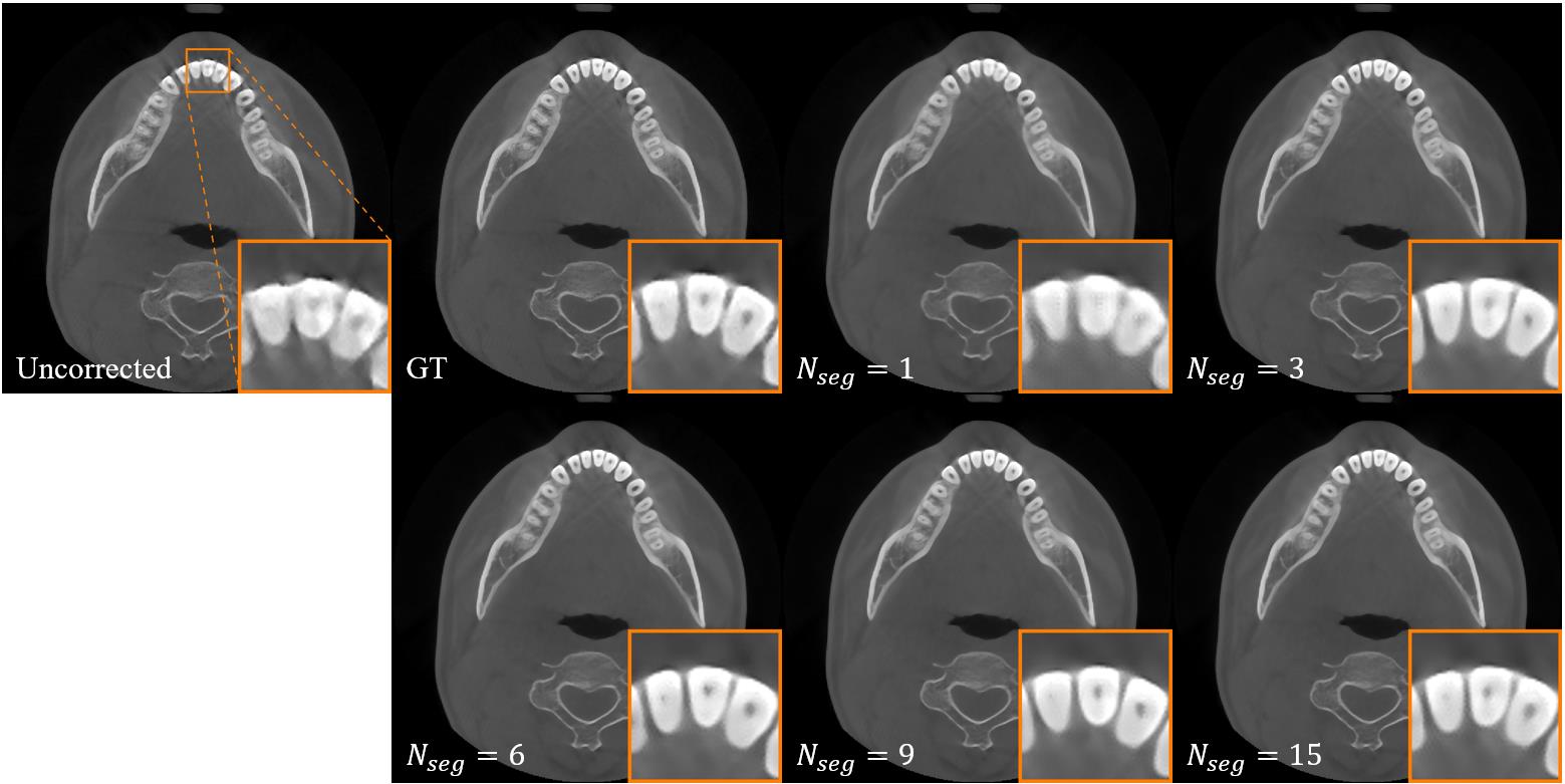
图12 不同投影分段个数Nseg取值下的伪影校正结果
Fig.12 Artifact correction results under different values of the Nseg .
| 1 | Ng SY. Comparison between conventional dental radiography and CBCT[M]// Cone Beam CT in Dentistry. Cham: Springer, 2023: 271-330. |
| 2 | Schulze RKW, Drage NA. Cone-beam computed tomography and its applications in dental and maxillofacial radiology[J]. Clin Radiol, 2020, 75(9): 647-57. |
| 3 | Nemtoi A, Czink C, Haba D, et al. Cone beam CT: a current overview of devices[J]. Dentomaxillofac Radiol, 2013, 42(8): 20120443. |
| 4 | Spin-Neto R, Wenzel A. Patient movement and motion artefacts in cone beam computed tomography of the dentomaxillofacial region: a systematic literature review[J]. Oral Surg Oral Med Oral Pathol Oral Radiol, 2016, 121(4): 425-33. |
| 5 | Keriş EY. Effect of patient anxiety on image motion artefacts in CBCT[J]. BMC Oral Health, 2017, 17(1): 73. |
| 6 | Donaldson K, O'Connor S, Heath N. Dental cone beam CT image quality possibly reduced by patient movement[J]. Dentomaxillofac Radiol, 2013, 42(2): 91866873. |
| 7 | Spin-Neto R, Hauge Matzen L, Hermann L, et al. Head motion and perception of discomfort by young children during simulated CBCT examinations[J]. Dentomaxillofac Radiol, 2021, 50(3): 20200445. |
| 8 | Spin-Neto R, Costa C, Salgado DM, et al. Patient movement characteristics and the impact on CBCT image quality and interpretability[J]. Dentomaxillofac Radiol, 2018, 47(1): 20170216. |
| 9 | Spin-Neto R, Matzen L H, Schropp L, et al. Factors affecting patient movement and re-exposure in cone beam computed tomography examination [J]. Oral Surg Oral Med Oral Pathol Oral Radiol, 2015, 119(5): 572-8. |
| 10 | Nardi C, Taliani GG, Castellani A, et al. Repetition of examination due to motion artifacts in horizontal cone beam CT: comparison among three different kinds of head support[J]. J Int Soc Prev Community Dent, 2017, 7(4): 208-13. |
| 11 | Yildizer Keris E, Demirel O, Ozdede M. Evaluation of motion artifacts in cone-beam computed tomography with three different patient positioning[J]. Oral Radiol, 2021, 37(2): 276-81. |
| 12 | Jacobson MW, Stayman JW. Compensating for head motion in slowly-rotating cone beam CT systems with optimization transfer based motion estimation[C]//2008 IEEE Nuclear Science Symposium Conference Record. Dresden, Germany. IEEE, 2008: 5240-5. |
| 13 | Kyme AZ, Fulton RR. Motion estimation and correction in SPECT, PET and CT[J]. Phys Med Biol, 2021, 66(18): 18TR02. |
| 14 | Ouadah S, Jacobson M, Stayman JW, et al. Correction of patient motion in cone-beam CT using 3D-2D registration[J]. Phys Med Biol, 2017, 62(23): 8813-31. |
| 15 | Sun T, Jacobs R, Pauwels R, et al. A motion correction approach for oral and maxillofacial cone-beam CT imaging[J]. Phys Med Biol, 2021, 66(12): 125008. |
| 16 | Berger M, Müller K, Aichert A, et al. Marker-free motion correction in weight-bearing cone-beam CT of the knee joint[J]. Med Phys, 2016, 43(3): 1235-48. |
| 17 | Niebler S, Schömer E, Tjaden H, et al. Projection-based improvement of 3D reconstructions from motion-impaired dental cone beam CT data[J]. Med Phys, 2019, 46(10): 4470-80. |
| 18 | Hernandez D, Eldib ME, Hegazy MAA, et al. A head motion estimation algorithm for motion artifact correction in dental CT imaging[J]. Phys Med Biol, 2018, 63(6): 065014. |
| 19 | Berger M, Xia Y, Aichinger W, et al. Motion compensation for cone-beam CT using Fourier consistency conditions[J]. Phys Med Biol, 2017, 62(17): 7181-215. |
| 20 | Aichert A, Berger M, Wang J, et al. Epipolar consistency in transmission imaging[J]. IEEE Trans Med Imaging, 2015, 34(11): 2205-19. |
| 21 | A'lvarez-Borrego J. Fast autofocus algorithm for automated microscopes[J]. Opt Eng, 2005, 44(6): 063601. |
| 22 | Wicklein J, Kyriakou Y, Kalender WA, et al. An online motion- and misalignment-correction method for medical flat-detector CT[C]//SPIE Proceedings", "Medical Imaging 2013: Physics of Medical Imaging. Lake Buena Vista (Orlando Area), Florida, USA. SPIE, 2013: 466-72. |
| 23 | Sisniega A, Stayman JW, Yorkston J, et al. Motion compensation in extremity cone-beam CT using a penalized image sharpness criterion[J]. Phys Med Biol, 2017, 62(9): 3712-34. |
| 24 | Zhang Y, Zhang LY. A rigid motion artifact reduction method for CT based on blind deconvolution[J]. Algorithms, 2019, 12(8): 155. |
| 25 | Gao C, Feng AQ, Liu XT, et al. A fully differentiable framework for 2D/3D registration and the projective spatial transformers[J]. IEEE Trans Med Imag, 2024, 43(1): 275-85. |
| 26 | Ali ASRA, Landi C, Sarti C, et al. Non-iterative compensation for patient motion in dental CBCT imaging[C]//2023 IEEE Nuclear Science Symposium, Medical Imaging Conference and International Symposium on Room-Temperature Semiconductor Detectors (NSS MIC RTSD). Vancouver, BC, Canada. IEEE, 2023: 1. |
| 27 | Preuhs A, Manhart M, Roser P, et al. Appearance learning for image-based motion estimation in tomography[J]. IEEE Trans Med Imaging, 2020, 39(11): 3667-78. |
| 28 | Thies M, Wagner F, Maul N, et al. A gradient-based approach to fast and accurate head motion compensation in cone-beam CT[J]. IEEE Trans Med Imaging, 2024, doi: 10.1109/TMI.2024.3474250 |
| 29 | Amirian M, Montoya-Zegarra JA, Herzig I, et al. Mitigation of motion-induced artifacts in cone beam computed tomography using deep convolutional neural networks[J]. Med Phys, 2023, 50(10): 6228-42. |
| 30 | Ko Y, Moon S, Baek J, et al. Rigid and non-rigid motion artifact reduction in X-ray CT using attention module[J]. Med Image Anal, 2021, 67: 101883. |
| 31 | Li DS, Zhang Y, Cheung KC, et al. Learning degradation representations for Image deblurring[M]//Lecture Notes in Computer Science. Cham: Springer Nature Switzerland, 2022: 736-53. |
| 32 | Chen LY, Chu XJ, Zhang XY, et al. Simple baselines for Image restoration[M]//Lecture Notes in Computer Science. Cham: Springer Nature Switzerland, 2022: 17-33. |
| 33 | Park T, Liu MY, Wang TC, et al. Semantic image synthesis with spatially-adaptive normalization[C]//2019 IEEE/CVF Conference on Computer Vision and Pattern Recognition (CVPR). Long Beach, CA, USA. IEEE, 2019: 2332-41. |
| 34 | He KM, Zhang XY, Ren SQ, et al. Deep residual learning for image recognition[C]//2016 IEEE Conference on Computer Vision and Pattern Recognition (CVPR). Las Vegas, NV, USA. IEEE, 2016: 770-8. |
| 35 | Mechrez R, Talmi I, Shama F, et al. Maintaining natural image statistics with the contextual loss[C]//Asian Conference on Computer Vision. Cham: Springer, 2019: 427-443. |
| 36 | Simonyan K, Zisserman A. Very deep convolutional networks for large-scale image recognition[EB/OL]. 2014: arXiv: 1409.1556. |
| 37 | Ronneberger O, Fischer P, Brox T. U-net: Convolutional networks for biomedical image segmentation [C]//2015 Medical Image Computing and Computer Assisted Intervention (MICCAI). Munich, Germany. Springer International Publishing, 2015: 234-41. |
| 38 | Chen H, Zhang Y, Kalra MK, et al. Low-dose CT with a residual encoder-decoder convolutional neural network[J]. IEEE Trans Med Imaging, 2017, 36(12): 2524-35. |
| 39 | Chen LY, Lu X, Zhang J, et al. HINet: half instance normalization network for image restoration[C]//2021 IEEE/CVF Conference on Computer Vision and Pattern Recognition Workshops (CVPRW). Nashville, TN, USA. IEEE, 2021: 182-92. |
| 40 | Zamir SW, Arora A, Khan S, et al. Restormer: efficient transformer for high-resolution image restoration[C]//2022 IEEE/CVF Conference on Computer Vision and Pattern Recognition (CVPR). New Orleans, LA, USA. IEEE, 2022: 5718-29. |
| [1] | 林宗悦, 王永波, 边兆英, 马建华. 基于深度模糊学习的牙科CBCT运动伪影校正算法[J]. 南方医科大学学报, 2024, 44(6): 1198-1208. |
| 阅读次数 | ||||||
|
全文 |
|
|||||
|
摘要 |
|
|||||