
南方医科大学学报 ›› 2026, Vol. 46 ›› Issue (9): 1993-2005.doi: 10.12122/j.issn.1673-4254.2026.09.01
• •
袁权( ), 张东东, 王景涛, 宋汉君()
收稿日期:2026-04-16
出版日期:2026-09-20
发布日期:2026-09-30
通讯作者:
宋汉君
E-mail:18245506700@163.com;songhanjun1973@163.com
作者简介:袁 权,在读硕士研究生,E-mail: 18245506700@163.com
基金资助:
Quan YUAN(), Dongdong ZHANG, Jingtao WANG, Hanjun SONG()
Received:2026-04-16
Online:2026-09-20
Published:2026-09-30
Contact:
Hanjun SONG
E-mail:18245506700@163.com;songhanjun1973@163.com
Supported by:摘要:
目的 采用社交隔离(SI)模型,探讨SI会诱导小鼠出现哪些异常行为和SI对小鼠内侧前额叶皮层(mPFC)神经元活性的影响。 方法 选用雄性C57BL/6J小鼠,随机分为8组(8只/组),对照组(Ctrl)小鼠正常饲养,SI组小鼠单笼饲养2周来构建SI小鼠模型,Ctrl-mCherry组小鼠注射对照病毒,Ctrl-hM3Dq组小鼠注射化学遗传激活病毒,Ctrl-hChR2组小鼠注射光遗传激活病毒,SI-mCherry组小鼠注射对照病毒,SI-hM4Di组小鼠注射化学遗传抑制病毒,SI-NpHR组小鼠注射光遗传抑制病毒。造模结束后,采用相关行为学检测评估小鼠的行为;使用c-Fos免疫荧光染色对Ctrl小鼠和SI小鼠mPFC脑区进行染色,后用c-Fos与代表兴奋性神经元的特异性抗体CaMKIIα以及代表抑制性神经元的特异性抗体GABA的共染色,确定神经元的类型,结合在体光纤记录实时监测两组小鼠在行为过程中神经元的变化;采用化学遗传和光遗传学调控Ctrl组小鼠mPFC Glu能神经元活性,行为学检测明确对两组小鼠社交行为的影响,以探究激活mPFC Glu能神经元是否可以诱导Ctrl组小鼠出现社交回避以及抑制mPFC Glu能神经元是否可以挽救SI小鼠的社交回避。 结果 行为学测试显示,2周的社交隔离不会导致小鼠出现焦虑抑郁样行为,没有损害小鼠的物体识别记忆和工作记忆,不会影响小鼠的社交能力和社交记忆,但会导致小鼠出现社交回避行为(P<0.001);免疫荧光结果显示,SI组小鼠mPFC脑区c-Fos阳性神经元增加(P<0.01),且增加的大部分是兴奋性谷氨酸能神经元(P<0.05);在体光纤记录结果显示,在社交互动测试(SIT)第二阶段中,SI小鼠mPFC Glu能神经元的钙活性显著增加(P<0.001);化学遗传和光遗传学激活Ctrl小鼠mPFC Glu能神经元活性诱导小鼠出现社交回避(P<0.01),抑制可缓解SI小鼠的社交回避(P<0.05)。 结论 2周的社交隔离导致小鼠出现社交回避行为,使mPFC Glu能神经元活性增加。
袁权, 张东东, 王景涛, 宋汉君. 社交隔离诱导小鼠社交回避:mPFC谷氨酸能神经元的介导作用[J]. 南方医科大学学报, 2026, 46(9): 1993-2005.
Quan YUAN, Dongdong ZHANG, Jingtao WANG, Hanjun SONG. Social isolation induces social avoidance in mice: the mediating role of the medial prefrontal cortex glutamatergic neurons[J]. Journal of Southern Medical University, 2026, 46(9): 1993-2005.
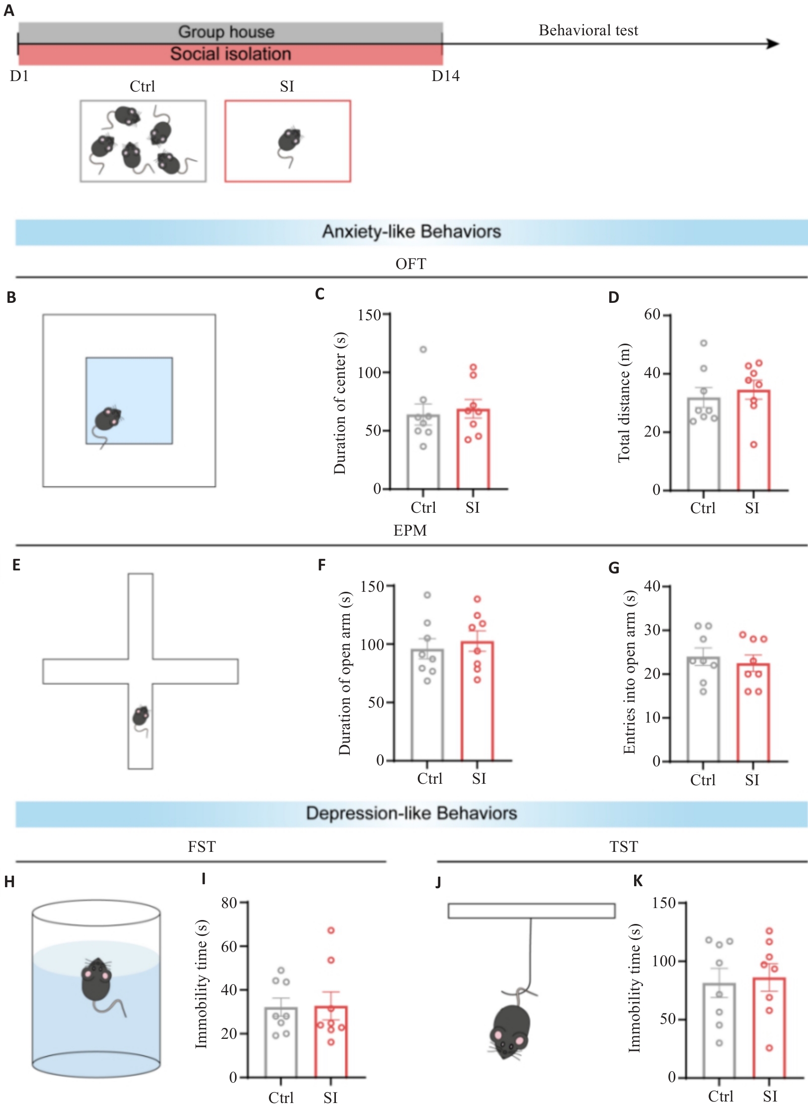
图1 社交隔离未导致焦虑抑郁样行为
Fig.1 Social isolation (SI) does not cause anxiety- or depressive-like behaviors in mice. A: Schematic diagram of SI modeling in mice (n=8). B: Diagram of open field test (OFT) of the mice. C: Statistical plot of exploration time in the central areas of the mice during OFT. D: Statistical plot of total distance traveled of the mice in OFT. E: Diagram of elevated plus-maze (EPM) test of the mice. F: Statistical plot of exploration time in the open arms of the mice in EPM test. G: Statistical plot of open arm entry frequency of the mice in EPM test. H: Diagram of forced swimming test (FST) of the mice. I: Statistical plot of inactivity duration of the mice during the final 4 min in FST. J: Behavioral patterns of the mice in tail suspension test (TST). K: Statistical plot of inactivity duration of the mice during the final 4 min in TST.
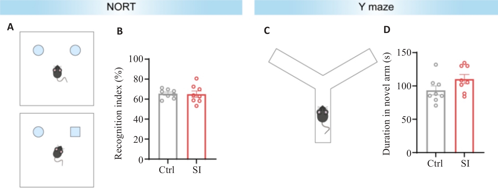
图2 社交隔离未影响物体识别和工作记忆
Fig.2 SI does not affect object recognition and working memory of mice. A: Diagram of novel object recognition test (NORT) of the mice. B: Statistical chart of cognitive indices in the two groups of mice during the second phase of NORT. C: Diagram of Y-maze test of the mice. D: Statistical chart of exploration time of the mice in the novel arm in Y-maze test (n=8).
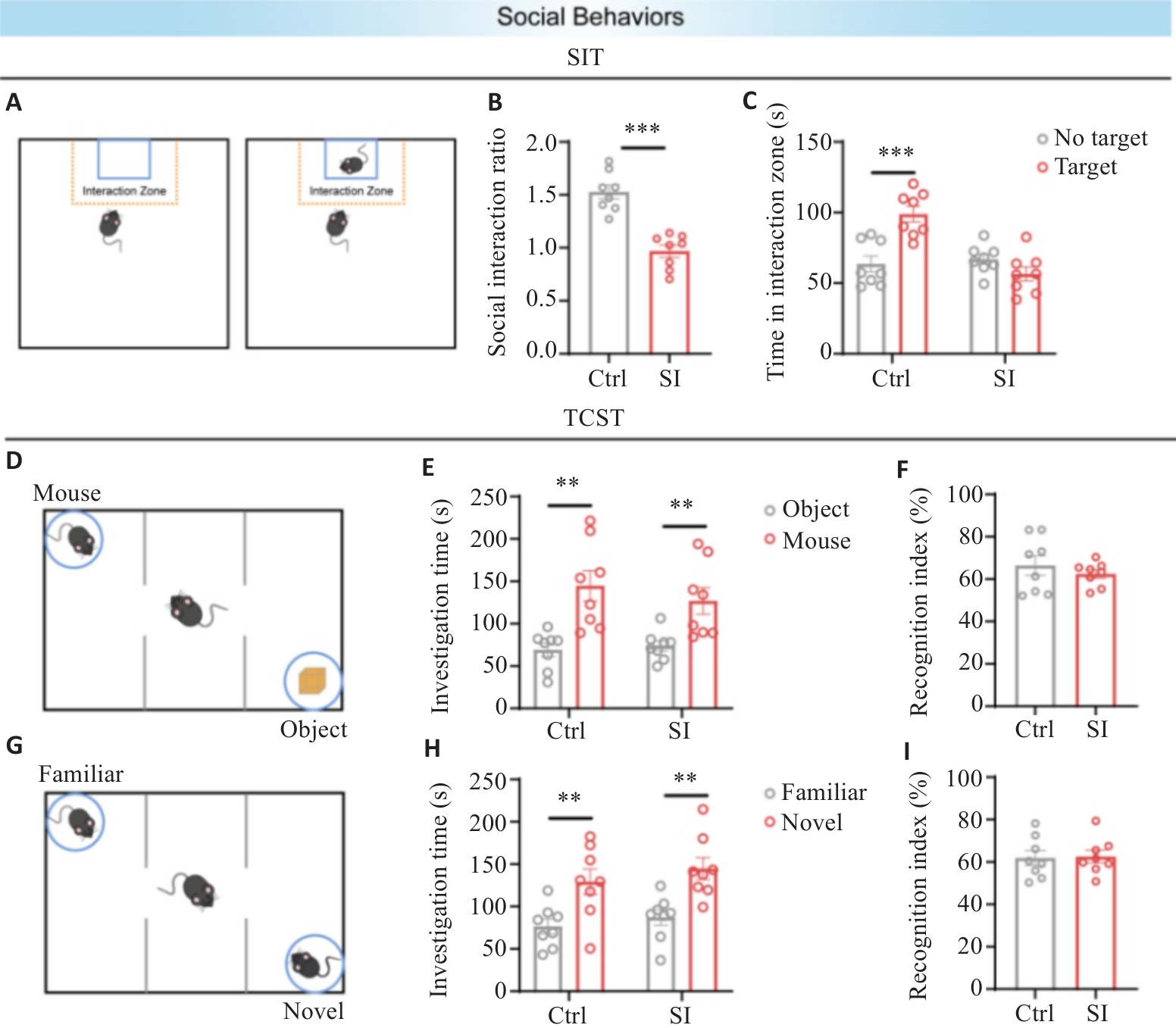
图3 社交隔离不影响社交能力和社交记忆, 但会出现社交回避
Fig.3 SI does not affect social competence or social memory, but may cause social avoidance in mice. A: Diagram of social interaction test (SIT) of the mice. B: Statistical chart of social interaction ratios of the mice in SIT. C: Statistical chart of time spent by target mice in the social interaction zone in the two groups. D: Diagram of TCST social competence stage. E: Statistical chart of exploration time of the mice toward the social mice and objects. F: Statistical chart of social preference index of the social mice in the two groups. G: Diagram of TCST social memory stage. H: Statistical chart of exploration time toward familiar mice and new mice in the two groups. I: Statistical chart of social preference index of the new mice in the two groups. **P<0.01, ***P<0.001 (n=8).
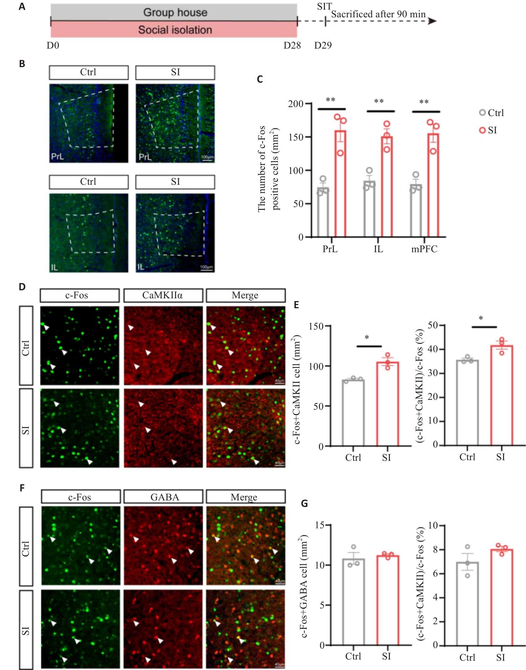
图4 社交隔离导致mPFC Glu能神经元c-Fos表达增加
Fig.4 SI leads to increased c-Fos expression in the glutamatergic neurons in the medial prefrontal cortex (mPFC) of mice. A: Flowchart of c-Fos staining experiments in mice. B: Representative immunofluorescence images of c-Fos staining in PrL and IL subregions of the mPFC (Scale bar=100 μm). C: Statistical plot of average density of c-Fos-positive neurons in the PrL, IL subregions and mPFC brain region of the mice. D: Representative immunofluorescence images of co-staining for c-Fos and CaMKIIα in the mPFC brain region of the mice (Scale bar=40 μm). White arrows indicate co-localized cells expressing c-Fos and CaMKIIα. E: Statistical plot of co-localization of c-Fos- and CaMKIIα-positive neurons in the two groups. F: Representative immunofluorescence images of co-staining for c-Fos and GABA in the mPFC brain region of the mice (Scale bar=40 μm). White arrows indicate co-localized cells expressing c-Fos and GABA. G: Statistical plot of co-localization of c-Fos- and GABA-positive neurons in the two groups. *P<0.05, **P<0.01 (n=3).
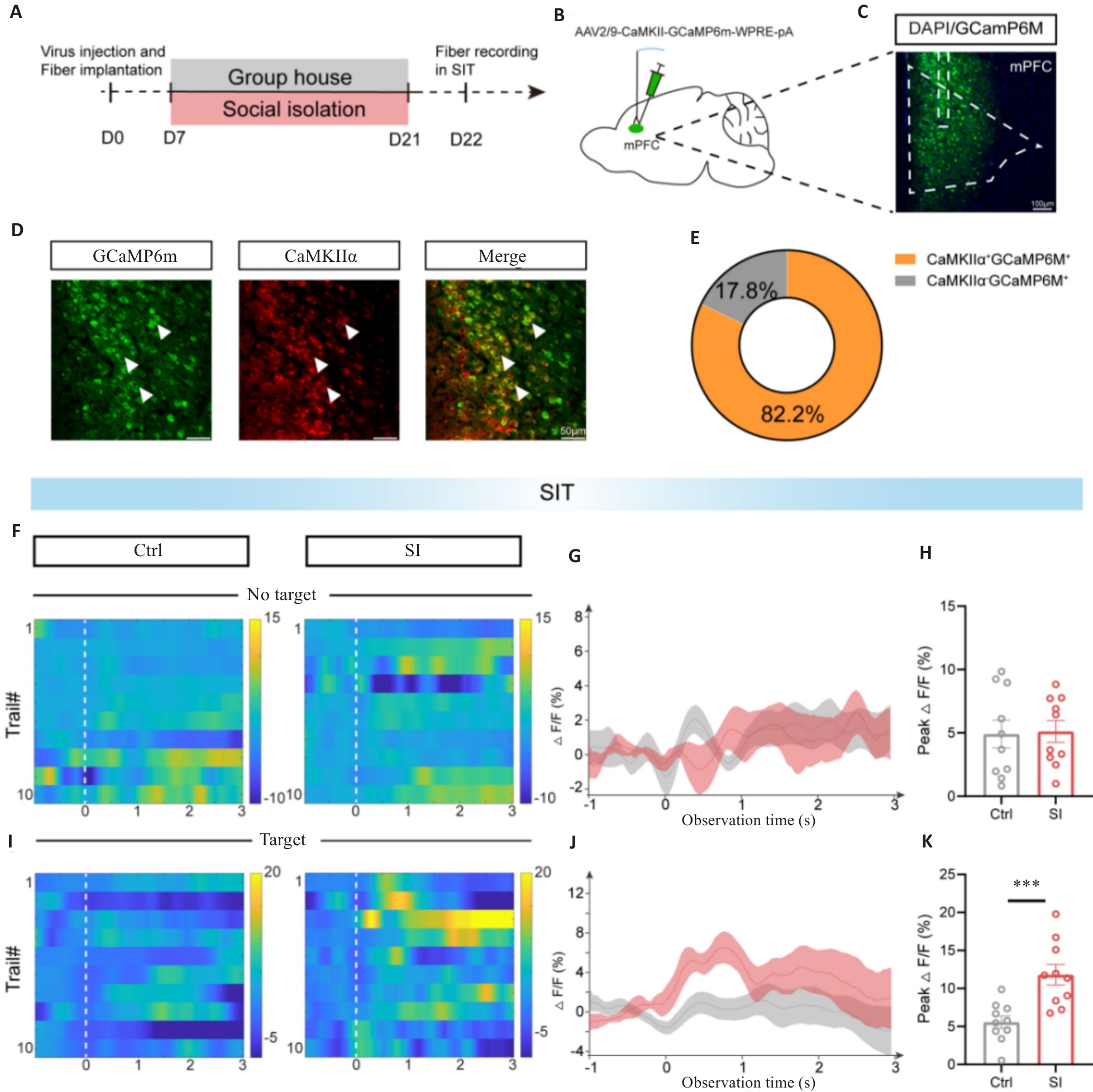
图5 SI小鼠在SIT中mPFC Glu能神经元钙活性增加
Fig.5 Increased calcium activity in mPFC glutamatergic neurons in SI mice during SIT. A: Flowchart of optical fiber recording in the two groups of mice. B: Schematic diagram of calcium indicator GCaMP6m injection and fiber embedding in mPFC neurons expressing CaMKIIα. C: Schematic diagram of viral injection sites (Scale bar=100 μm). D: Co-staining of GCaMP6m (green) and CaMKIIα (red). White arrows indicate co-localized cells expressing GCaMP6m and CaMKIIα (Scale bar=50 μm). E: Pie chart showing co-labeling rates of GCaMP6m and CaMKIIα. F: Heat map illustrating Ca2+ signal changes during optical fiber recording in the first stage of SIT in the two groups of mice. G: Curve plots of average Ca2+ fluorescence signals in the first stage in the two groups. H: Statistical plot of ΔF/F peak signals in the first stage in the two groups. I: Heat map illustrating Ca2+ signal changes during optical fiber recording in the second stage of SIT in the two groups. J: Curve plots of average Ca2+ fluorescence signals in the second stage in the two groups. K: Statistical plot of ΔF/F peak signals in the second stage in the two groups. ***P<0.001 (n=5).
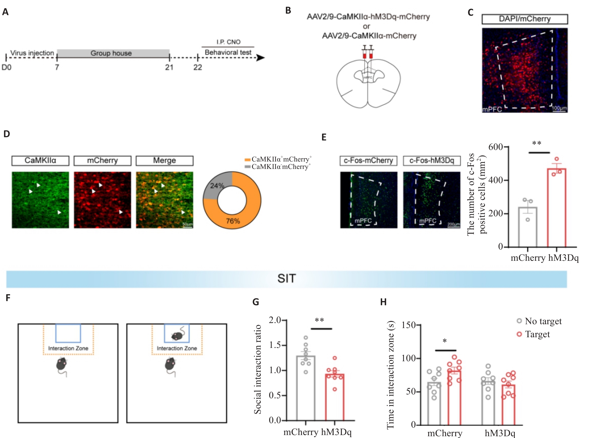
图6 化学遗传学激活mPFC Glu能神经元诱导社交回避
Fig.6 Chemical genetics activation of mPFC glutamatergic neurons induces social avoidance in mice. A: Experimental flowchart of chemogenetic activation of mPFC glutamatergic neurons. B: Schematic diagram of viral injection protocol and injection sites. C: Fluorescence image showing viral expression of hM3Dq in mPFC (Scale bar=100 μm). D: Fluorescence image of co-staining with hM3Dq-mCherry (red) and CaMKIIα (green) in mPFC sections. White arrows indicate co-localized cells, and the pie chart displays co-localization rates of hM3Dq and CaMKIIα (Scale bar= 50 μm). E: Representative immunofluorescence images of c-Fos staining in mPFC regions and statistical plot of average c-Fos density in mPFC of the mice (Scale bar=200 μm; n=3). F: SIT diagram. G: Statistical plot of social interaction rates during social interaction tests in SIT in the two groups. H: Statistical plot of time spent in social interaction areas by target mice in SIT in the two groups. *P<0.05, **P<0.01 (n=8).
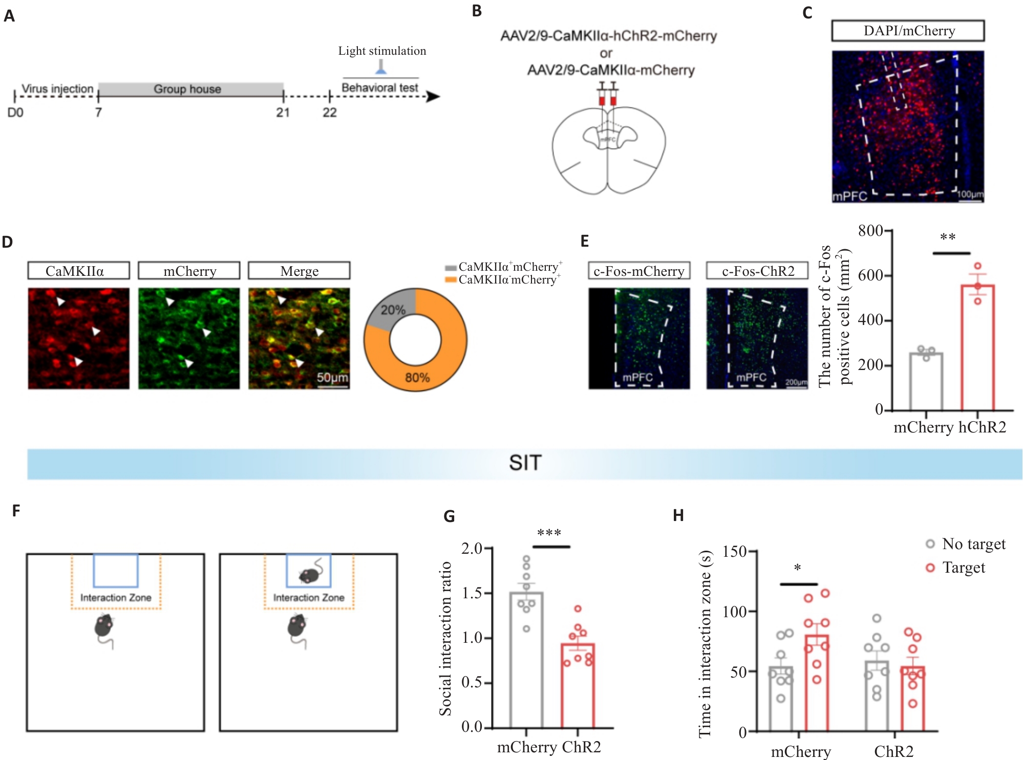
图7 光遗传学激活mPFC Glu能神经元诱导了社交回避
Fig.7 Optogenetic activation of mPFC glutamatergic neurons induces social avoidance in mice. A: Experimental flowchart for optogenetic activation of mPFC glutamatergic neurons. B: Schematic diagram of viral injection protocol and injection sites. C: Fluorescence image showing viral expression of hChR2 in mPFC (Scale bar=100 μm). D: Fluorescence image of co-staining hChR2-mCherry (red) and CaMKIIα (green) in mPFC sections (Scale bar=50 μm). White arrows indicate co-localized cells. The pie chart displays co-localization rates of hChR2 and CaMKIIα. E: Representative immunofluorescence images of c-Fos staining in mPFC regions and statistical plot of average c-Fos density in the mPFC of the mice (Scale bar=200 μm; n=3). F: SIT pattern diagram. G: Statistical plot of social interaction rates during social interaction tests in SIT in the two groups. H: Statistical plot of time spent in social interaction areas by target mice in SIT in the two groups. *P<0.05, **P<0.01, ***P<0.001 (n=8).
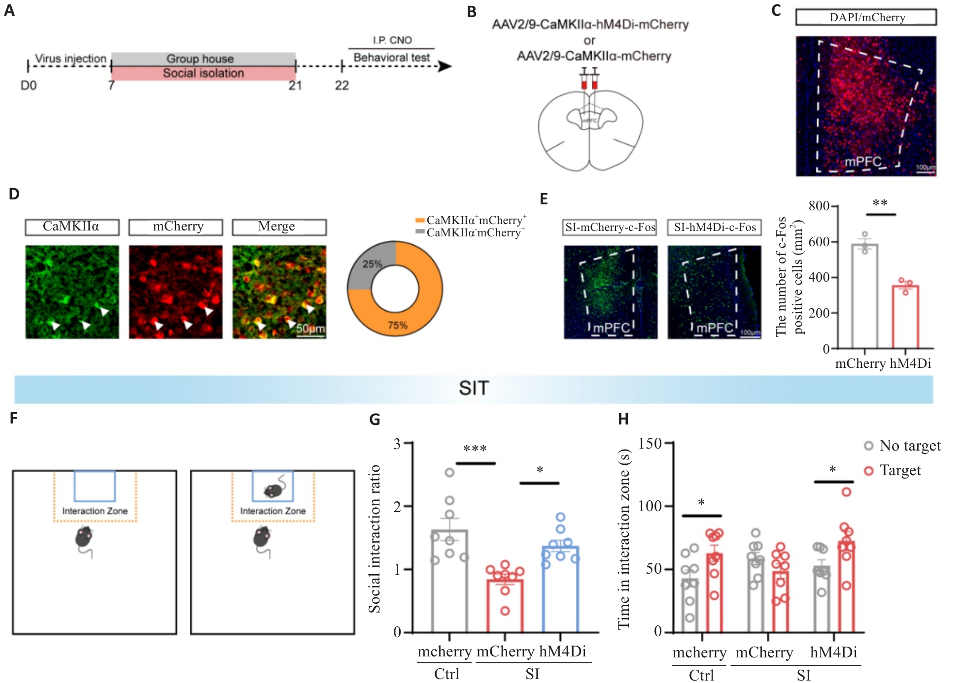
图8 化学遗传学抑制mPFC Glu能神经元缓解SI小鼠社交回避
Fig.8 Chemical genetics inhibition of mPFC glutamatergic neurons alleviates social avoidance in SI mice. A: Flowchart of the experimental protocol for chemogenetic inhibition of mPFC glutamatergic neurons. B: Schematic diagram of the viral injection protocol and injection sites. C: Fluorescence image showing the localization of hM4Di virus expression in the mPFC (Scale bar= 100 μm). D: Fluorescence image of co-staining with hM4Di-mCherry (red) and CaMKIIα (green) in mPFC sections (Scale bar= 50 μm). White arrows indicate co-localized cells. The pie chart shows the co-localization rate of hM4Di and CaMKIIα. E: Representative immunofluorescence images of c-Fos staining in the mPFC regions (Scale bar=100 μm) and statistical plot of the average c-Fos density in the mPFC in the two groups (n=3). F: SIT protocol diagram. G: Statistical plot of social interaction rates among the 3 groups during the social interaction test in SIT. H: Statistical plot of time spent by target mice in the social interaction regions in SIT. *P<0.05, **P<0.01, ***P<0.001 (n=8).
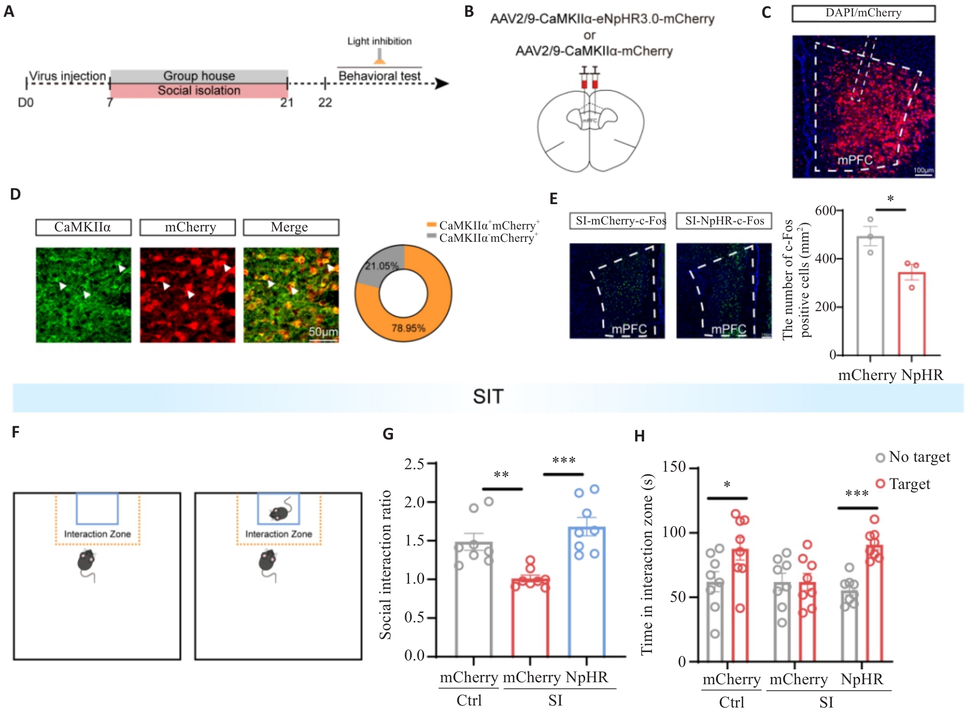
图9 光遗传学抑制mPFC Glu能神经元缓解SI小鼠社交回避
Fig.9 Optogenetic inhibition of mPFC glutamatergic neurons alleviates social avoidance in SI mice. A: Flowchart of the optogenetic inhibition of mPFC glutamatergic neurons. B: Schematic diagram of viral injection protocol and injection sites. C: Fluorescence image showing the injection site of NpHR virus in the mPFC (Scale bar=100 μm). D: Fluorescence image demonstrating co-staining of NpHR-mCherry (red) and CaMKIIα (green) in mPFC sections (Scale bar=50 μm). White arrows indicate co-localized cells. The pie chart shows the co-localization rate of NpHR and CaMKIIα. E: Representative immunofluorescence images of c-Fos staining in the mPFC regions (Scale bar=100 μm) and average c-Fos density in the mPFC in the two groups (n=3). F: SIT protocol diagram. G: Statistical plot of social interaction rates among the 3 groups during the social interaction test in the SIT. H: Statistical plot of time spent by target mice in the social interaction regions in SIT. *P<0.05, **P<0.01, ***P<0.001 (n=8).
| [1] | Albasheer O, Abdelwahab SI, Zaino MR, et al. The impact of social isolation and loneliness on cardiovascular disease risk factors: a systematic review, meta-analysis, and bibliometric investigation[J]. Sci Rep, 2024, 14: 12871. doi:10.1038/s41598-024-63528-4 |
| [2] | Holt-Lunstad J. Social connection as a critical factor for mental and physical health: evidence, trends, challenges, and future implications[J]. World Psychiatry, 2024, 23(3): 312-32. doi:10.1002/wps.21224 |
| [3] | Ran ZR, Wei JJ, Yang GJ, et al. Prevalence of social isolation in the elderly: a systematic review and meta-analysis[J]. Geriatr Nurs, 2024, 58: 87-97. doi:10.1016/j.gerinurse.2024.05.008 |
| [4] | 赵嘉盈, 纪小小, 潘玉峰, 等. 社交隔离调控本能行为的神经机制研究进展[J]. 生理学报, 2024, 76(2): 309-18. |
| [5] | Boyraz G, Legros DN, Tigershtrom A. COVID-19 and traumatic stress: The role of perceived vulnerability, COVID-19-related worries, and social isolation[J]. J Anxiety Disord, 2020, 76: 102307. doi:10.1016/j.janxdis.2020.102307 |
| [6] | Adepoju OE, Ojinnaka CO, Pieratt J, et al. Racial and ethnic differences in predictors of participation in an intergenerational social connectedness intervention for older adults[J]. BMC Geriatr, 2024, 24(1): 70. doi:10.1186/s12877-024-04679-x |
| [7] | Li WH, Wang ZK, Syed S, et al. Chronic social isolation signals starvation and reduces sleep in Drosophila [J]. Nature, 2021, 597(7875): 239-44. doi:10.1038/s41586-021-03837-0 |
| [8] | Xiong Y, Hong HL, Liu CR, et al. Social isolation and the brain: effects and mechanisms[J]. Mol Psychiatry, 2023, 28(1): 191-201. doi:10.1038/s41380-022-01835-w |
| [9] | 马 雪, 董莹莹, 刘 鹏, 等. 成年期社会隔离对雄性BALB/C小鼠神经行为学的影响[J]. 中国医药导报, 2016, 13(18): 13-6, 41. |
| [10] | Clemenza K, Weiss SH, Cheslack K, et al. Social isolation is closely linked to a marked reduction in physical activity in male mice[J]. J Neurosci Res, 2021, 99(4): 1099-107. doi:10.1002/jnr.24777 |
| [11] | Zelikowsky M, Hui M, Karigo T, et al. The neuropeptide Tac2 controls a distributed brain state induced by chronic social isolation stress[J]. Cell, 2018, 173(5): 1265-79.e19. doi:10.1016/j.cell.2018.03.037 |
| [12] | Carlén M. What constitutes the prefrontal cortex?[J]. Science, 2017, 358(6362): 478-82. doi:10.1126/science.aan8868 |
| [13] | Liu H, Jia X, Huang ZQ, et al. A top-down neural circuit for affective-motivational responses of pain relief induced by electroacupuncture[J]. Chin Med, 2026, 21(1): 73. doi:10.1186/s13020-026-01349-5 |
| [14] | Yang SS, Mack NR, Shu YS, et al. Prefrontal GABAergic interneurons gate long-range afferents to regulate prefrontal cortex-associated complex behaviors[J]. Front Neural Circuits, 2021, 15: 716408. doi:10.3389/fncir.2021.716408 |
| [15] | 克丽比努尔·艾尔肯. 生命早期社交孤立破坏社会功能及内侧前额叶皮层调控机制[J]. 社会科学前沿, 2024(4): 398-405. doi:10.12677/ass.2024.134314 |
| [16] | Fruehwirth JC, Biswas S, Perreira KM. The Covid-19 pandemic and mental health of first-year college students: Examining the effect of Covid-19 stressors using longitudinal data[J]. PLoS One, 2021, 16(3): e0247999. doi:10.1371/journal.pone.0247999 |
| [17] | Cauberghe V, Van Wesenbeeck I, De Jans S, et al. How adolescents use social media to cope with feelings of loneliness and anxiety during COVID-19 lockdown[J]. Cyberpsychol Behav Soc Netw, 2021, 24(4): 250-7. doi:10.1089/cyber.2020.0478 |
| [18] | Smith L, Jacob L, Yakkundi A, et al. Correlates of symptoms of anxiety and depression and mental wellbeing associated with COVID-19: a cross-sectional study of UK-based respondents[J]. Psychiatry Res, 2020, 291: 113138. doi:10.1016/j.psychres.2020.113138 |
| [19] | Sepúlveda-Loyola W, Rodríguez-Sánchez I, Pérez-Rodríguez P, et al. Impact of social isolation due to COVID-19 on health in older people: mental and physical effects and recommendations[J]. J Nutr Health Aging, 2020, 24(9): 938-47. doi:10.1007/s12603-020-1500-7 |
| [20] | Liu CH, Zhang E, Wong GTF, et al. Factors associated with depression, anxiety, and PTSD symptomatology during the COVID-19 pandemic: Clinical implications for U.S. young adult mental health[J]. Psychiatry Res, 2020, 290: 113172. doi:10.1016/j.psychres.2020.113172 |
| [21] | Morrissette M. School closures and social anxiety during the COVID-19 pandemic[J]. J Am Acad Child Adolesc Psychiatry, 2021, 60(1): 6-7. doi:10.1016/j.jaac.2020.08.436 |
| [22] | Wu X, Ding ZB, Fan TT, et al. Childhood social isolation causes anxiety-like behaviors via the damage of blood-brain barrier in amygdala in female mice[J]. Front Cell Dev Biol, 2022, 10: 943067. doi:10.3389/fcell.2022.943067 |
| [23] | Fosnocht AQ, Lucerne KE, Ellis AS, et al. Adolescent social isolation increases cocaine seeking in male and female mice[J]. Behav Brain Res, 2019, 359: 589-96. doi:10.1016/j.bbr.2018.10.007 |
| [24] | Lin S, Li X, Chen YH, et al. Social isolation during adolescence induces anxiety behaviors and enhances firing activity in BLA pyramidal neurons via mGluR5 upregulation[J]. Mol Neurobiol, 2018, 55(6): 5310-20. doi:10.1007/s12035-017-0766-1 |
| [25] | Yin T, Yuan JY, Liu L, et al. Confronting the anxiety of Generation Z: electroacupuncture therapy regulates oxidative stress and microglia activity in amygdala-basolateral of socially isolated mice[J]. Front Psychiatry, 2025, 15: 1496201. doi:10.3389/fpsyt.2024.1496201 |
| [26] | Locci A, Geoffroy P, Miesch M, et al. Social isolation in early versus late adolescent mice is associated with persistent behavioral deficits that can be improved by neurosteroid-based treatment[J]. Front Cell Neurosci, 2017, 11: 208. doi:10.3389/fncel.2017.00208 |
| [27] | Wang XN, Zhan Y. Regulation of social recognition memory in the hippocampal circuits[J]. Front Neural Circuits, 2022, 16: 839931. doi:10.3389/fncir.2022.839931 |
| [28] | Zhao JL, Ye LH, Liu ZY, et al. Protective effects of resveratrol on adolescent social isolation-induced anxiety-like behaviors via modulating nucleus accumbens spine plasticity and mitochondrial function in female rats[J]. Nutrients, 2022, 14(21): 4542. doi:10.3390/nu14214542 |
| [29] | Hu YY, Ding XS, Yang G, et al. Analysis of the influences of social isolation on cognition and the therapeutic potential of deep brain stimulation in a mouse model[J]. Front Psychiatry, 2023, 14: 1186073. doi:10.3389/fpsyt.2023.1186073 |
| [30] | Halldorsdottir T. Youth mental health crisis: a translational view of the COVID-19 pandemic[J]. Neurobiol Stress, 2025, 37: 100738. doi:10.1016/j.ynstr.2025.100738 |
| [31] | Khodr CE, Chen LH, Dave S, et al. Combined chronic blockade of hyper-active L-type calcium channels and NMDA receptors ameliorates HIV-1 associated hyper-excitability of mPFC pyramidal neurons[J]. Neurobiol Dis, 2016, 94: 85-94. doi:10.1016/j.nbd.2016.06.008 |
| [32] | Reeves KC, Shah N, Muñoz B, et al. Opioid receptor-mediated regulation of neurotransmission in the brain[J]. Front Mol Neurosci, 2022, 15: 919773. doi:10.3389/fnmol.2022.919773 |
| [33] | Li H, Zhao Z, Jiang SF, et al. Brain circuits that regulate social behavior[J]. Mol Psychiatry, 2025, 30(7): 3240-56. doi:10.1038/s41380-025-03037-6 |
| [34] | Shirenova SD, Khlebnikova NN, Krupina NA. Long-term social isolation reduces expression of the BDNF precursor and prolyl endopeptidase in the rat brain[J]. Biochemistry, 2021, 86(6): 704-15. doi:10.1134/s0006297921060080 |
| [35] | Yang XK, Shi YL, Li M, et al. Divergent BLA engram circuits orchestrate social preference dynamics in bystander male mice with self-experienced stress[J]. Proc Natl Acad Sci USA, 2026, 123(11): e2516904123. doi:10.1073/pnas.2516904123 |
| [36] | Park G, Ryu C, Kim S, et al. Social isolation impairs the prefrontal-nucleus accumbens circuit subserving social recognition in mice[J]. Cell Rep, 2021, 35(6): 109104. doi:10.1016/j.celrep.2021.109104 |
| [37] | Lai CH, Park G, Xu P, et al. Functional specialization of mPFC-BLA and mPFC-NAc pathways in affective state representation[J/OL]. eLife, 2026. DOI:10.7554/eLife.105528 . |
| [38] | Cui WP, Shen C, Xiong WC, et al. Prefrontal ErbB4-positive interneurons for avoidance[J]. Cell Rep, 2025, 44(5): 115628. doi:10.1016/j.celrep.2025.115628 |
| [39] | Liu L, Xu HF, Wang J, et al. Cell type-differential modulation of prefrontal cortical GABAergic interneurons on low gamma rhythm and social interaction[J]. Sci Adv, 2020, 6(30): eaay4073. doi:10.1126/sciadv.aay4073 |
| [40] | Xu HF, Liu L, Tian YY, et al. A disinhibitory microcircuit mediates conditioned social fear in the prefrontal cortex[J]. Neuron, 2019, 102(3): 668-82.e5. doi:10.1016/j.neuron.2019.02.026 |
| [1] | 张倬宁, 郝新宇, 曹福羊, 郭永馨, 郭舒婷, 蔡辰, 米卫东, 仝黎. 艾司氯胺酮通过激活小鼠mPFC区谷氨酸能神经元发挥抗抑郁样作用[J]. 南方医科大学学报, 2026, 46(3): 489-496. |
| [2] | 李言响, 郭永馨, 曹福羊, 郭舒婷, 薛丁豪, 周志康, 郝新宇, 仝 黎, 傅 强. 抑制dmPAG区谷氨酸能神经元可减轻创伤后应激障碍小鼠的过度防御反应[J]. 南方医科大学学报, 2024, 44(3): 420-427. |
| [3] | 刘冬播, 陈泽文, 王云, 李昕鹏, 赵彭玉, 郑浩贤. 慢性不可预知温和应激诱导的抑郁小鼠中央杏仁核和边缘前皮质神经网络的可塑性变化[J]. 南方医科大学学报, 2024, 44(11): 2082-2091. |
| [4] | 倪 虹,丁海虎,陶 静,王元元,陶明飞,黄 丽. 嗅觉剥夺对小鼠大脑桶状皮层谷氨酸能神经元动作电位及Ankyrin-G表达的影响[J]. 南方医科大学学报, 2020, 40(02): 262-267. |
| [5] | 范玲玲,邓博,闫君宝,胡志红,任爱红,胡咏梅,杨东伟. 5-羟色胺-7受体激动剂对帕金森病模型大鼠内侧前额叶皮层锥体神经元兴奋性的影响[J]. 南方医科大学学报, 2016, 36(06): 756-. |
| 阅读次数 | ||||||
|
全文 |
|
|||||
|
摘要 |
|
|||||
