
南方医科大学学报 ›› 2026, Vol. 46 ›› Issue (10): 2338-2356.doi: 10.12122/j.issn.1673-4254.2026.10.05
• 基础研究 • 上一篇
谭慧心1( ), 金军国2, 杨曼其3, 王京丽3, 周凤华4(), 曾平1(), 孙学刚4()
收稿日期:2026-04-11
出版日期:2026-10-20
发布日期:2026-10-26
通讯作者:
周凤华,曾平,孙学刚
E-mail:tanhuixinx@163.com;Wendyzhou515@126.com;zengping@gdph.org.cn;sun_smu@126.com
作者简介:谭慧心,在读硕士研究生,E-mail: tanhuixinx@163.com
基金资助:
Huixin TAN1(), Junguo JIN2, Manqi YANG3, Jingli WANG3, Fenghua ZHOU4(), Ping ZENG1(), Xuegang SUN4()
Received:2026-04-11
Online:2026-10-20
Published:2026-10-26
Contact:
Fenghua ZHOU, Ping ZENG, Xuegang SUN
E-mail:tanhuixinx@163.com;Wendyzhou515@126.com;zengping@gdph.org.cn;sun_smu@126.com
Supported by:摘要:
目的 探讨线粒体磷酸甘油酸变位酶5(PGAM5)缺失对肝窦内皮细胞(LSECs)内吞功能及循环脂质重塑的影响,及其与动脉粥样硬化(AS)的关联。 方法 采用尾静脉注射AAV-PCSK9联合西方饮食(WD)构建AS小鼠模型。实验共分为5组:阴性对照组(NC组,普通饲料喂养)、普通喂养组(ND组,注射AAV-PCSK9后普通饲料喂养)、WD组(注射AAV-PCSK9后WD喂养)、阿托伐他汀灌胃组(ATOR组,注射AAV-PCSK9后WD喂养并给予阿托伐他汀钙灌胃)及PGAM5 KO组(PGAM5-/-小鼠,注射AAV-PCSK9后WD喂养)。通过油红O、HE及Masson染色评估主动脉斑块形成及肝脏脂质沉积情况,检测血浆脂质水平变化。使用肝脏蛋白质组学和单细胞RNA测序来筛选通路并分析细胞亚群。通过血浆代谢组与脂质组学筛选关键差异代谢物;体外在永生化人肝窦内皮细胞(HLSECs)中敲低PGAM5,以评估在代谢物干预下的氧化低密度脂蛋白(oxLDL)摄取情况。 结果 AS状态下PGAM5在肝脏组织中整体表达水平升高(P<0.01)。与NC组相比,WD组小鼠主动脉斑块负荷显著增加(P<0.0001)、肝脏脂肪变性显著加重(P<0.0001)及LDL-C异常(P<0.0001)。与WD组相比,PGAM5 KO组小鼠主动脉脂质沉积明显减少(P<0.0001),斑块稳定性增强,血浆低密度脂蛋白胆固醇(LDL-C)水平显著降低(P<0.01),肝脏脂肪变性明显改善(P<0.0001)。肝脏蛋白质组学分析显示差异蛋白显著富集于脂肪酸代谢及内吞作用相关通路。肝脏单细胞RNA测序分析显示,PGAM5缺失后LSECs的相对比例发生改变。血浆代谢组学分析发现,PGAM5-/-小鼠血浆的棕榈酰肉碱(P<0.001)和花生四烯酸(P<0.01)等脂质代谢物水平显著降低。血浆脂质组学分析显示PGAM5缺失后血浆脂质谱发生改变。体外实验表明,敲低PGAM5可增强HLSECs对oxLDL的摄取能力,AA或L-PC处理可抑制该过程。 结论 PGAM5缺失伴随LSECs内吞增强和循环脂质重塑,可能有助于改善AS。
谭慧心, 金军国, 杨曼其, 王京丽, 周凤华, 曾平, 孙学刚. PGAM5缺失增强肝窦内皮细胞内吞功能并重塑循环脂质代谢改善脂质沉积[J]. 南方医科大学学报, 2026, 46(10): 2338-2356.
Huixin TAN, Junguo JIN, Manqi YANG, Jingli WANG, Fenghua ZHOU, Ping ZENG, Xuegang SUN. PGAM5 deficiency attenuates atherosclerosis, enhances oxidized LDL uptake by liver sinusoidal endothelial cells and reshapes circulating lipidome in mice[J]. Journal of Southern Medical University, 2026, 46(10): 2338-2356.
| Primers | Sequence | Length (bp) |
|---|---|---|
| β-actin | Upstream5'-CACCCAGCACAATGAAGATC AAGAT-3' Downstream5'CCAGTTTTTAAATCCTGAGT CAAGC-3' | 317 |
| PGAM5 | Upstream5'-CTCAATAATGGCAGCATCACCC-3' Downstream5'-ATCTTGTCGGGAGGCATGAA-3' | 98 |
表1 引物序列
Tab.1 Primer sequences
| Primers | Sequence | Length (bp) |
|---|---|---|
| β-actin | Upstream5'-CACCCAGCACAATGAAGATC AAGAT-3' Downstream5'CCAGTTTTTAAATCCTGAGT CAAGC-3' | 317 |
| PGAM5 | Upstream5'-CTCAATAATGGCAGCATCACCC-3' Downstream5'-ATCTTGTCGGGAGGCATGAA-3' | 98 |
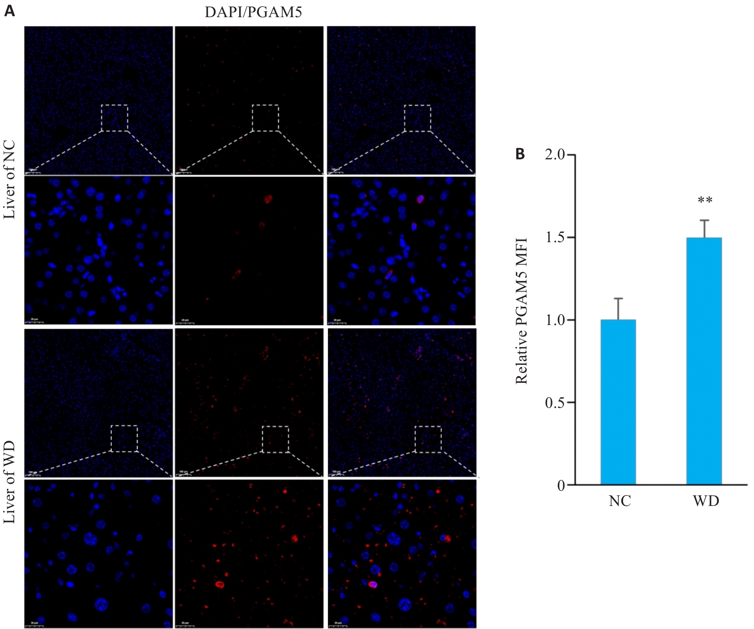
图1 动脉粥样硬化小鼠肝脏中PGAM5表达升高
Fig.1 Increased hepatic PGAM5 expression in atherosclerotic mice. A: Immunofluorescence staining with DAPI (blue) and PGAM5 (red) of mouse liver tissues (Original magnification: ×100; inset magnification: ×600). B: Quantitative analysis of relative mean fluorescence intensity (MFI) of PGAM5 in mouse liver tissues (n=4). **P<0.01 vs NC group.
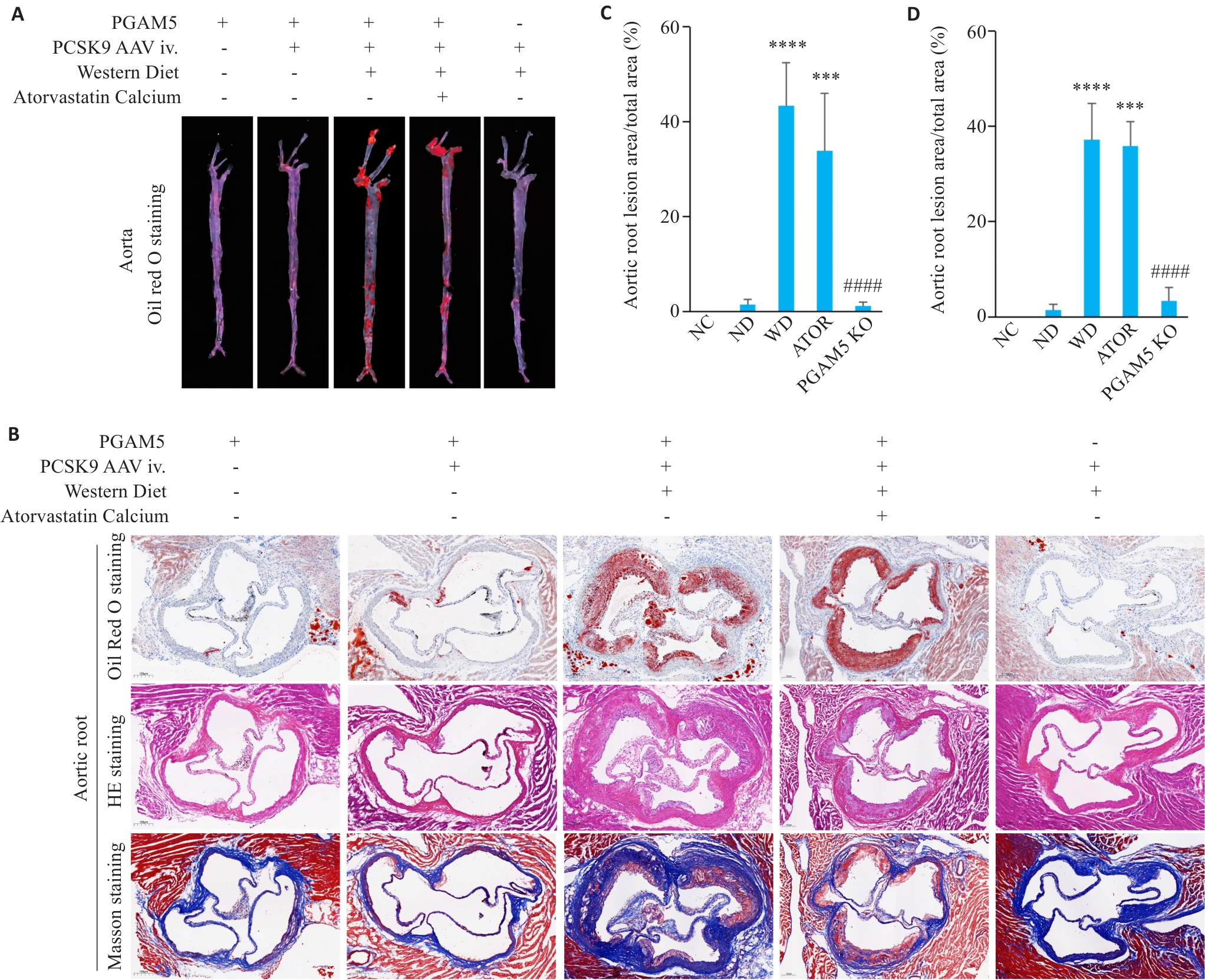
图2 PGAM5缺失改善动脉粥样硬化表型
Fig.2 PGAM5 deficiency attenuates atherosclerosis in mice. A: Oil Red O staining of mouse whole aorta. B: Oil Red O, HE, and Masson staining of mouse aortic root sections (×60). C: Quantification of aortic root lipid deposition with the lesion area presented as percentage of total vessel area (NC, n=3; ND, n=4; WD, n=6; ATOR, n=5; PGAM5 KO, n=3). D: Quantification of aortic root lesion area in HE staining (NC, n=5; ND, n=4; WD, n=6; ATOR, n=5; PGAM5 KO, n=5). ***P<0.001, ****P<0.0001 vs NC group; ####P<0.0001 vs WD group.
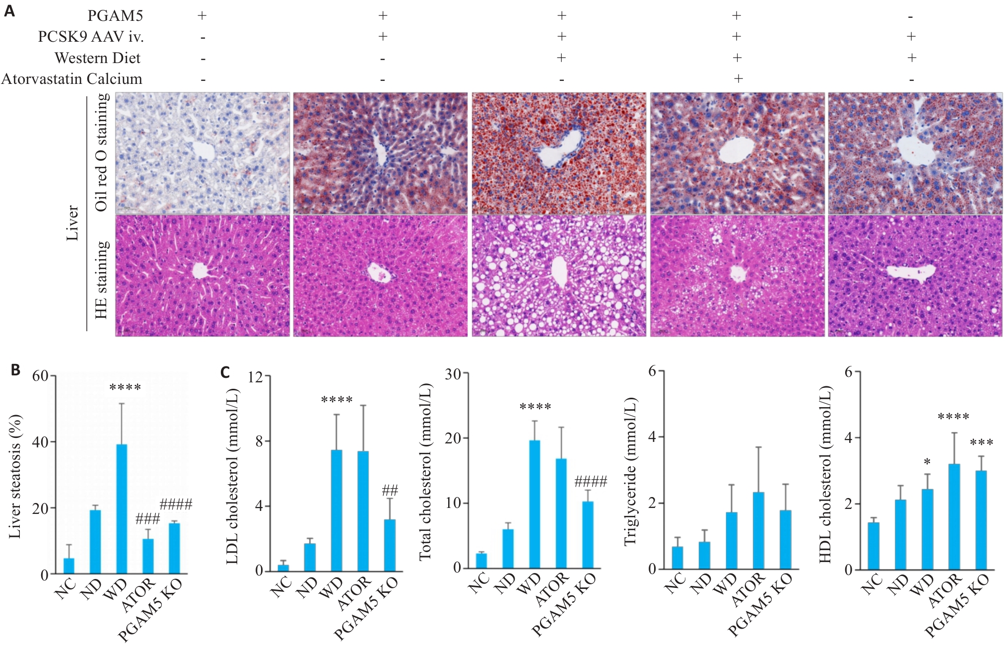
图3 PGAM5缺失改善动脉粥样硬化小鼠的肝脏脂质沉积和血浆脂质异常
Fig.3 PGAM5 deficiency alleviates hepatic lipid deposition and plasma lipid abnormalities in atherosclerotic mice. A: Oil Red O staining and HE staining of mouse liver tissues (×20). B: Quantitative analysis of hepatic lipid accumulation area (expressed as the percentage of lipid-positive area relative to total tissue area) (NC, n=7; ND, n=3; WD, n=7; ATOR, n=3; PGAM5 KO, n=5). C: Plasma levels of LDL-C (n=6), TC (n=6), TG (n=6), and HDL-C (NC, n=7; ND, n=6; WD, n=6; ATOR, n=6; PGAM5 KO, n=6) in mice of each group. *P<0.05, ***P<0.001, ****P<0.0001 vs NC group; ##P<0.01, ###P<0.001, ####P<0.0001 vs WD group.
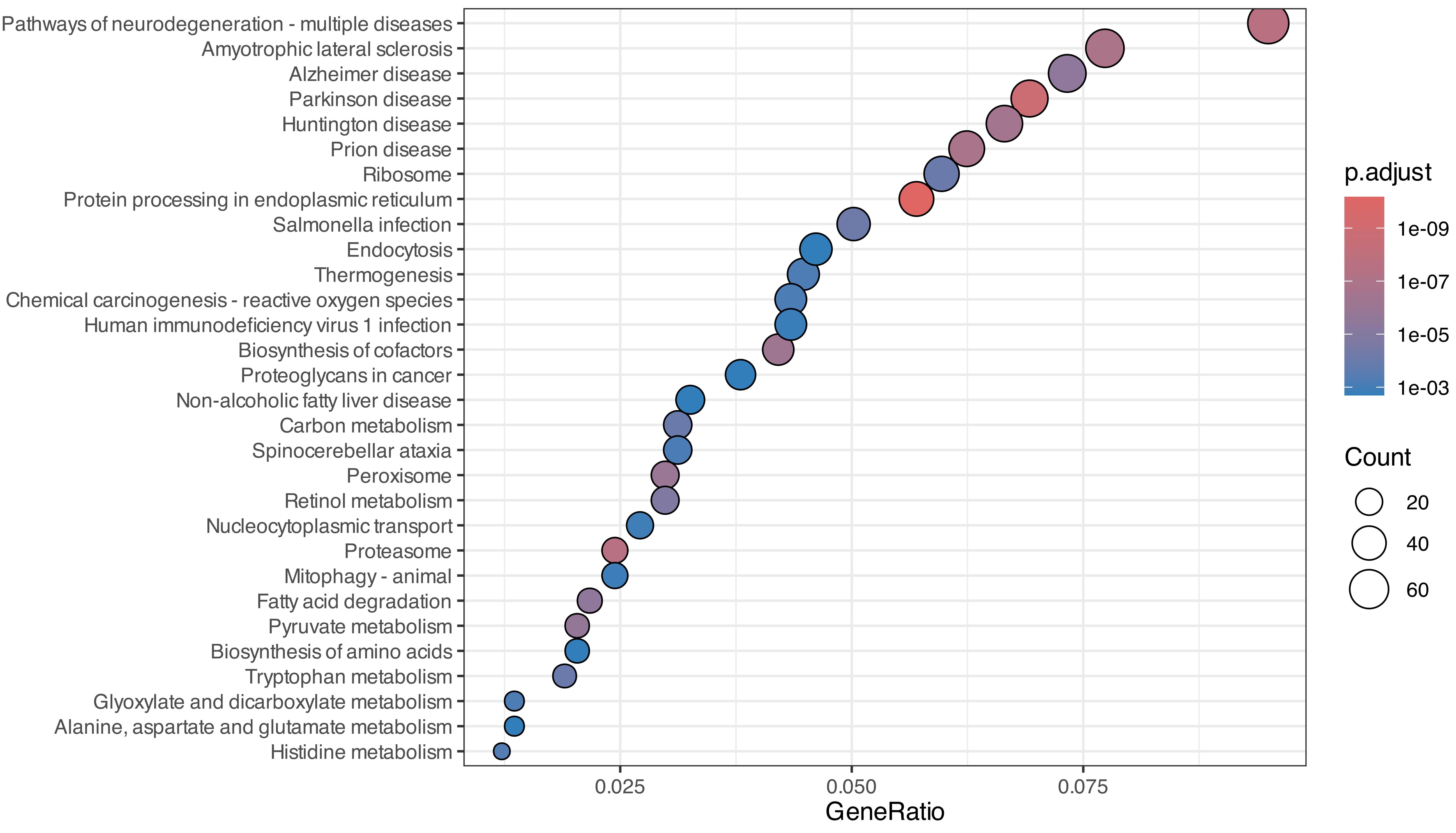
图4 两组肝脏差异蛋白KEGG通路富集分析
Fig.4 Enrichment analysis of differential liver proteins between the two groups using KEGG pathways.
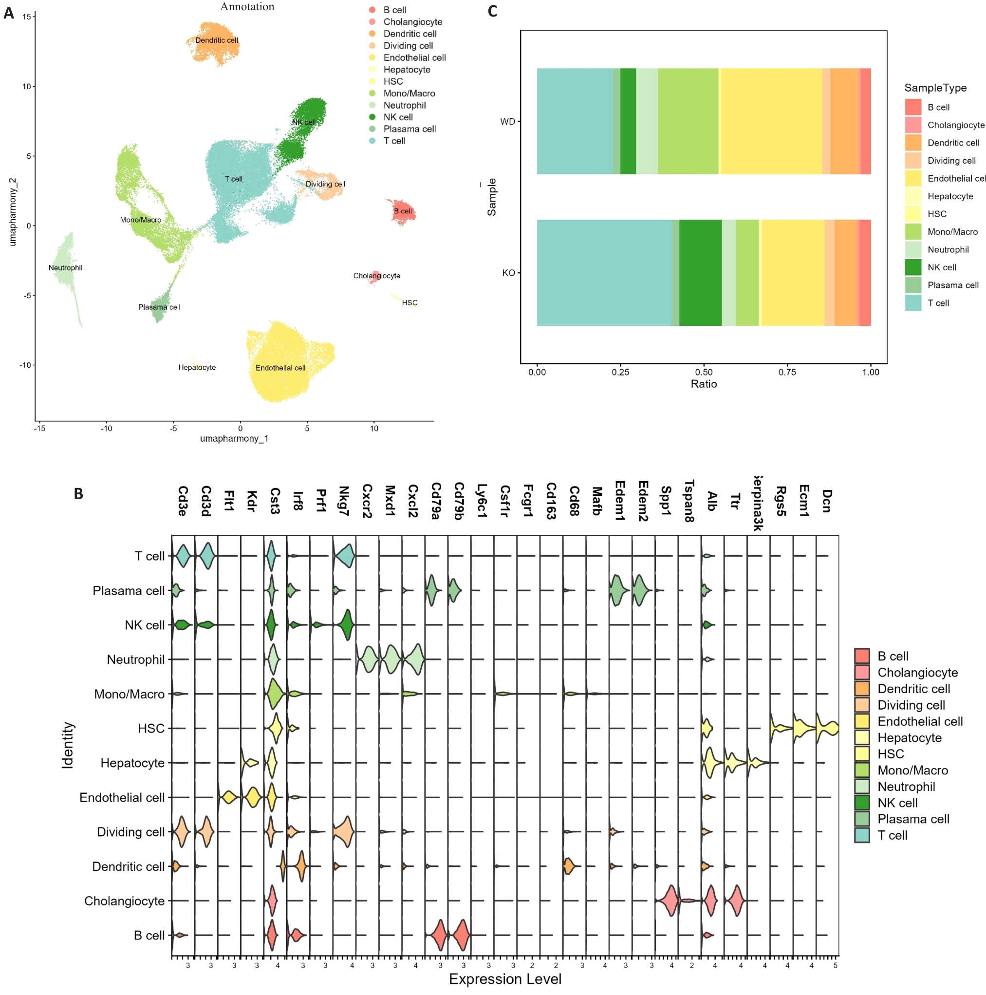
图5 小鼠肝脏单细胞RNA测序分析
Fig.5 Single-cell RNA sequencing analysis of mouse liver tissues. A: UMAP visualization of major hepatic cell clusters in WD and PGAM5 KO groups. B: Marker gene expression used for cell-type annotation. C: Proportional distribution of major cell populations between groups (n=4).
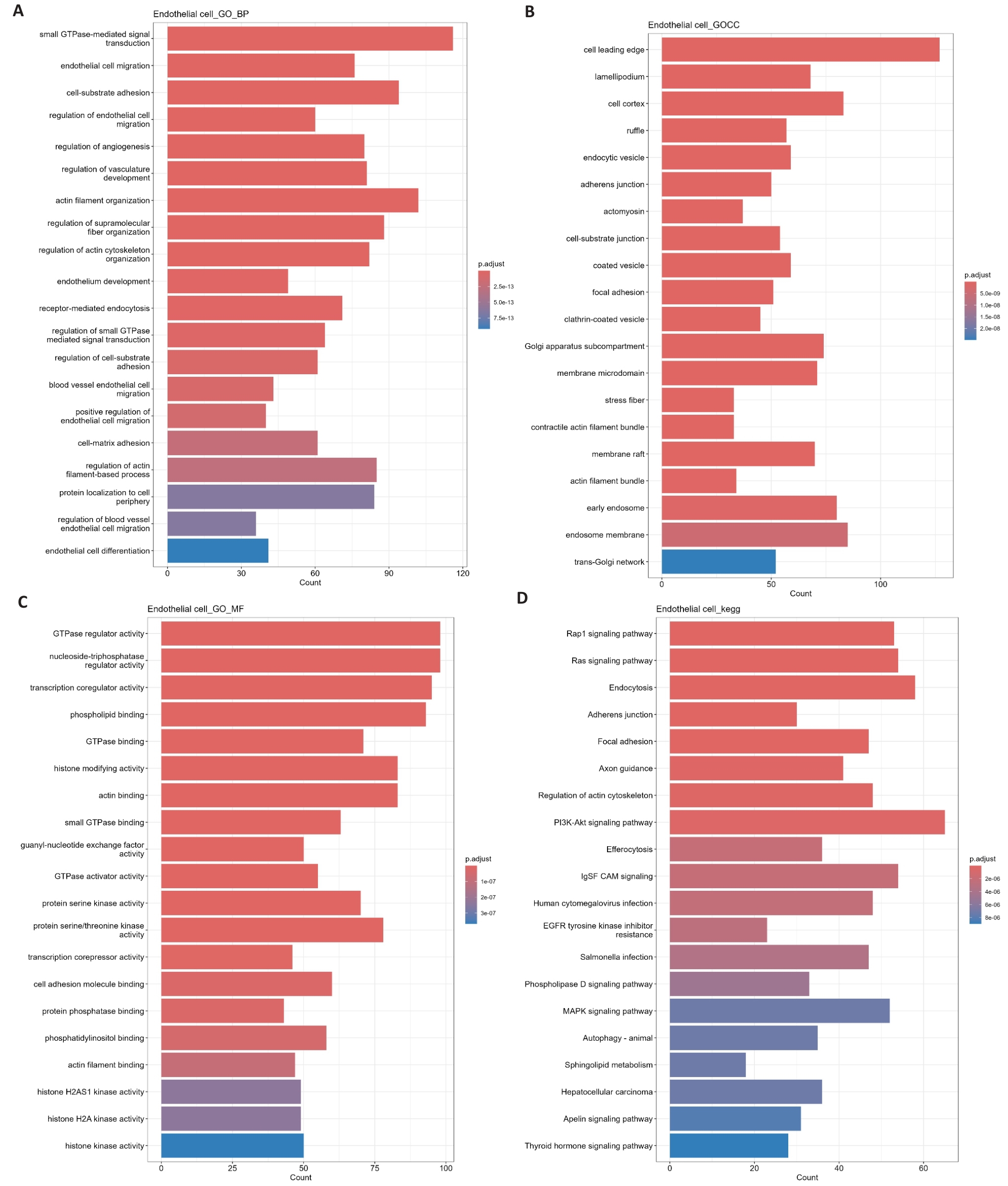
图6 内皮细胞与肝脏其他细胞差异基因的GO与KEGG富集分析
Fig.6 GO and KEGG enrichment analysis of differential genes between endothelial cells and other hepatic cell populations. A-C: Gene Ontology (GO) enrichment analysis of differentially expressed genes in ECs, including biological process (A), cellular component (B), and molecular function (C). D: Kyoto Encyclopedia of Genes and Genomes (KEGG) pathway enrichment analysis of differentially expressed genes in ECs.
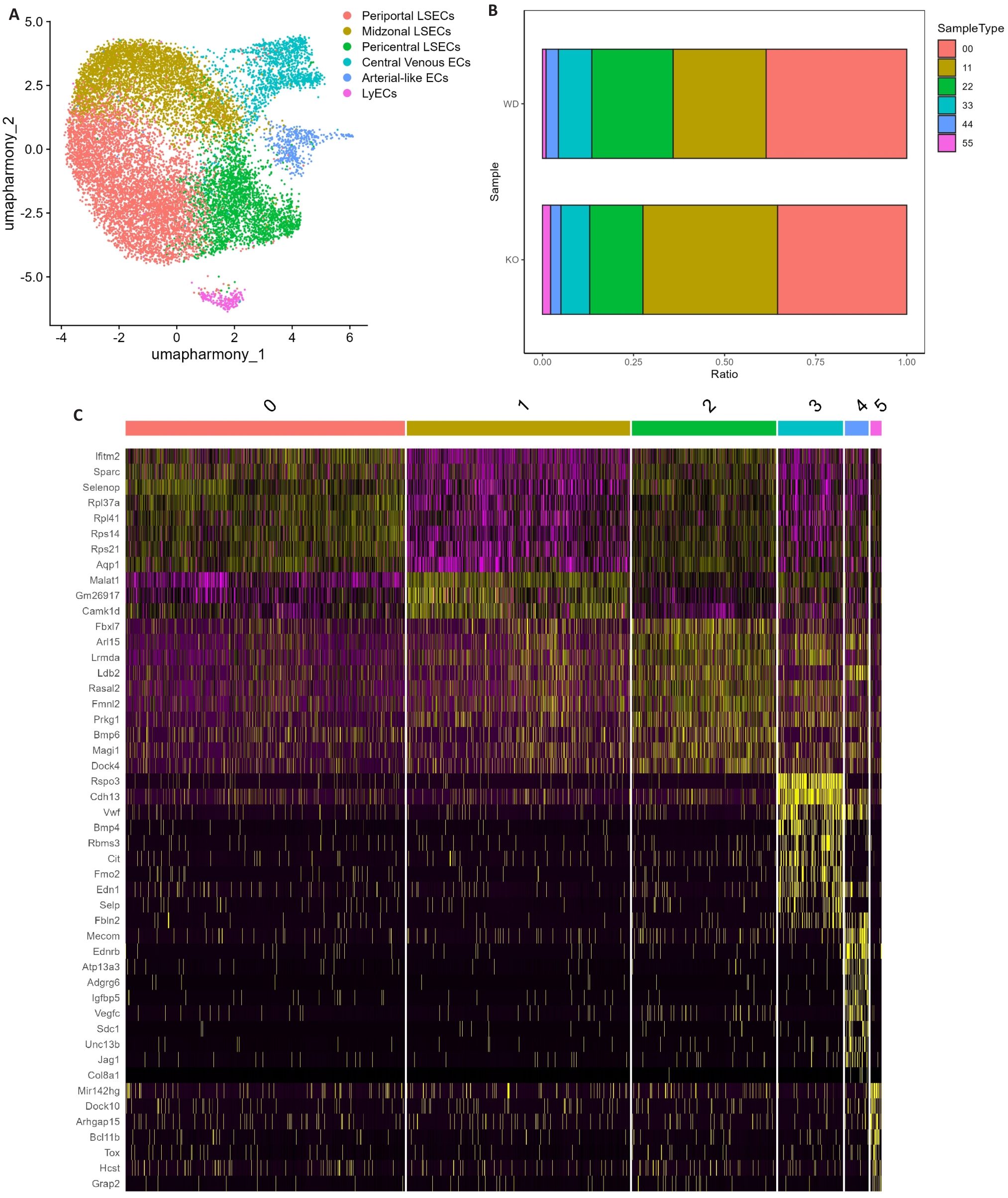
图7 肝脏内皮细胞亚群的转录特征
Fig.7 Transcriptional characteristics of hepatic endothelial subpopulations. A: UMAP dimensionality reduction plot of liver endothelial cells subpopulations. B: Proportional distribution of different endothelial cells subpopulations across samples. C: Heatmap of differentially expressed genes among endothelial cells subpopulations.
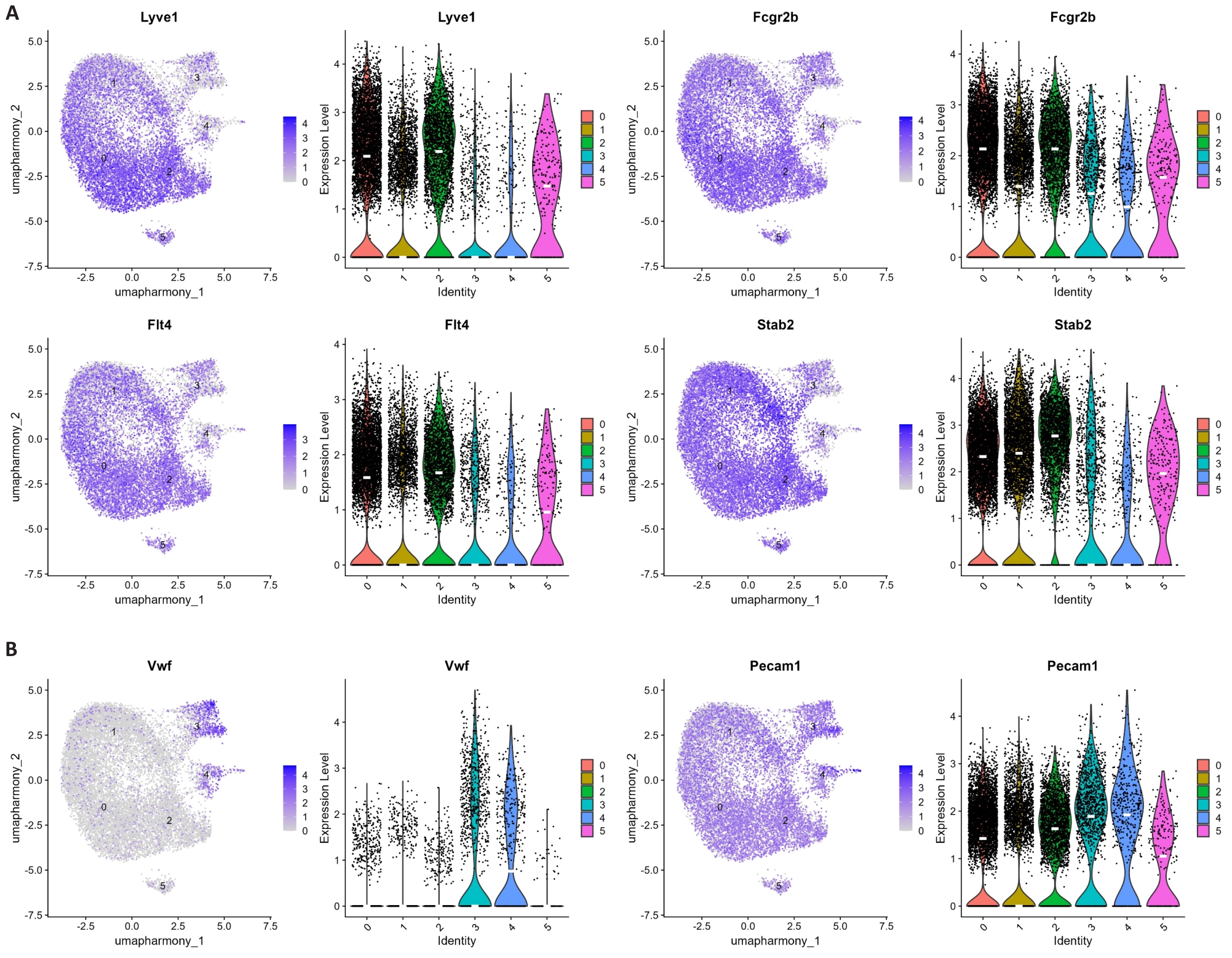
图8 肝脏内皮细胞亚群标志基因表达图谱
Fig.8 Expression of marker genes in hepatic endothelial subpopulations. A: UMAP visualization and violin plots of Lyve1, Fcgr2b, Flt4, and Stab2 expression across clusters. B: UMAP visualization and violin plots of Vwf and Pecam1 expression across clusters.
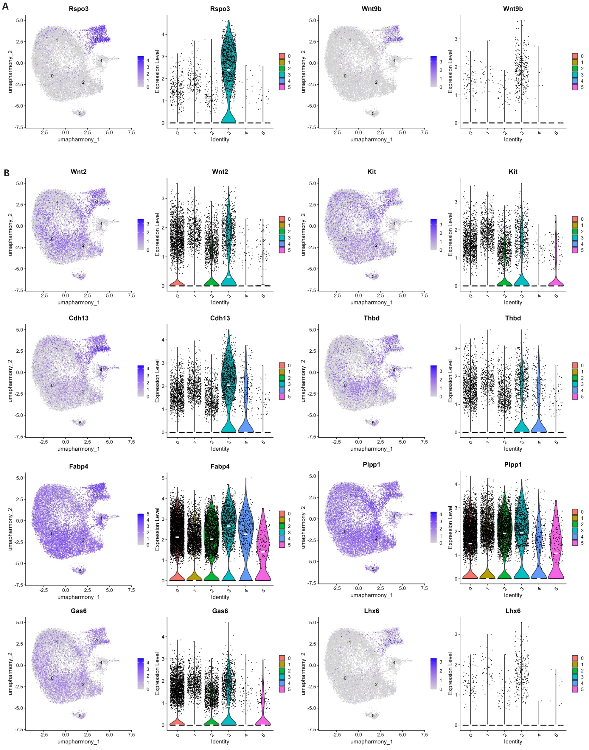
图9 中心静脉内皮细胞标志基因表达图谱
Fig.9 Expression of central vein endothelial cells marker genes. A: UMAP visualization and violin plots of Rspo3 and Wnt9b expression across clusters. B: UMAP visualization and violin plots of Wnt2, Kit, Cdh13, Thbd, Fabp4, Plpp1, Gas6, and Lhx6 expression across clusters.
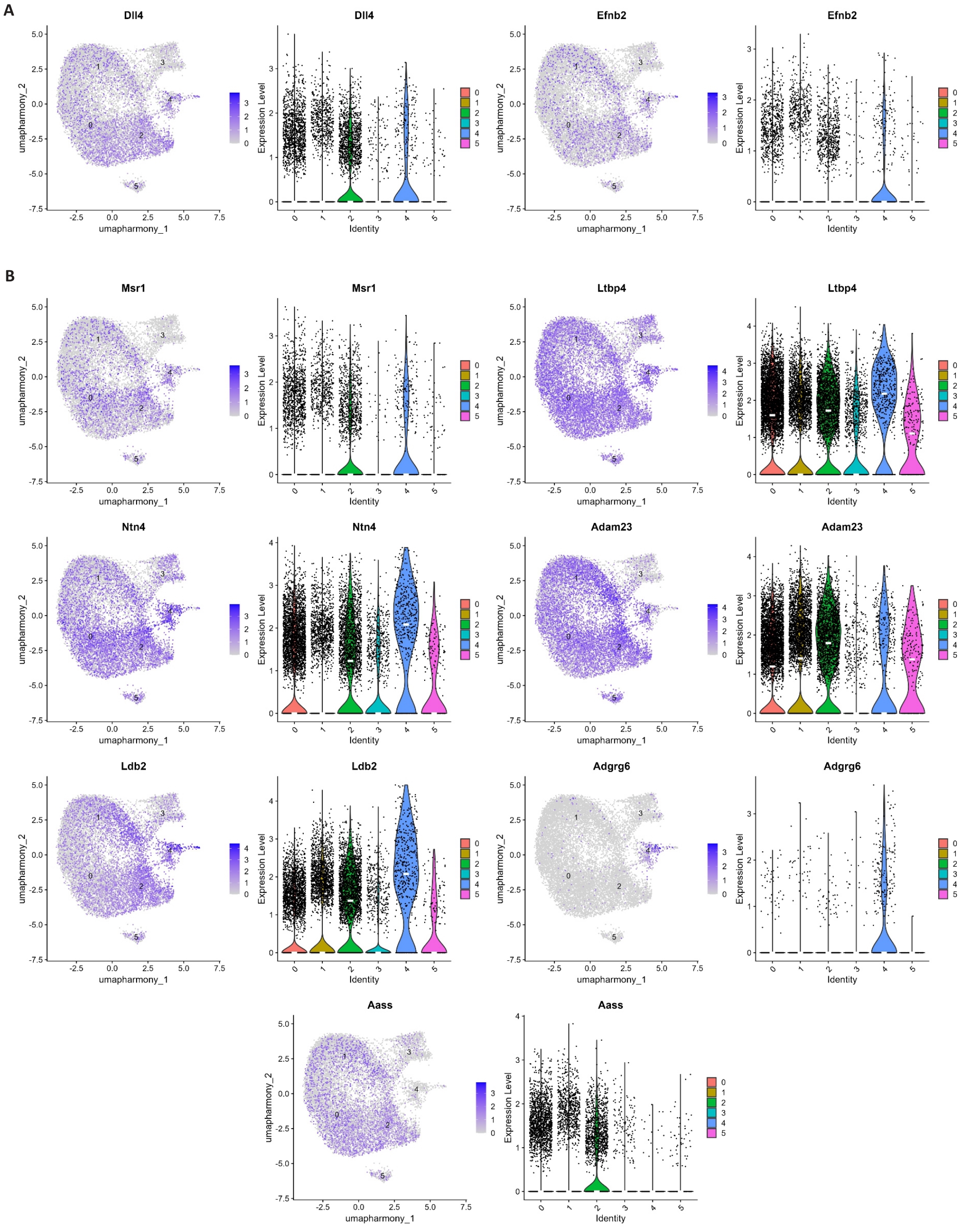
图10 动脉样内皮细胞标志基因表达图谱
Fig.10 Expression profile of marker genes in arterial-like endothelial cells. A: UMAP visualization and violin plots of Dll4 and Efnb2 expression across clusters. B: UMAP visualization and violin plots of Msr1, Ltbp4, Ntn4, Adam23, Ldb2, Adgrg6, and Aass expression across clusters.
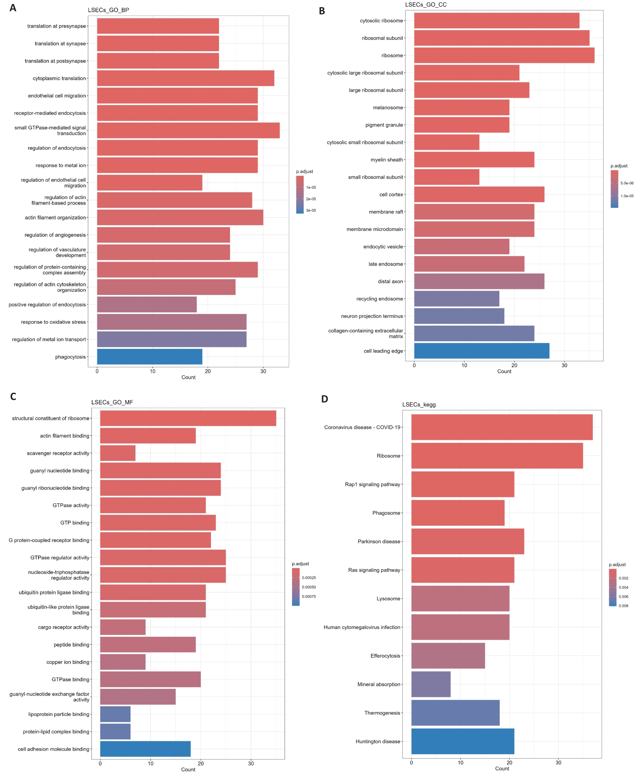
图11 肝窦内皮细胞差异基因的GO与KEGG富集分析
Fig.11 GO/KEGG enrichment analysis of differential genes in LSECs. A-C: Gene Ontology enrichment analysis of differentially expressed genes in LSECs, including biological process (A), cellular component (B), and molecular function (C). D: KEGG pathway enrichment analysis of differentially expressed genes in LSECs.
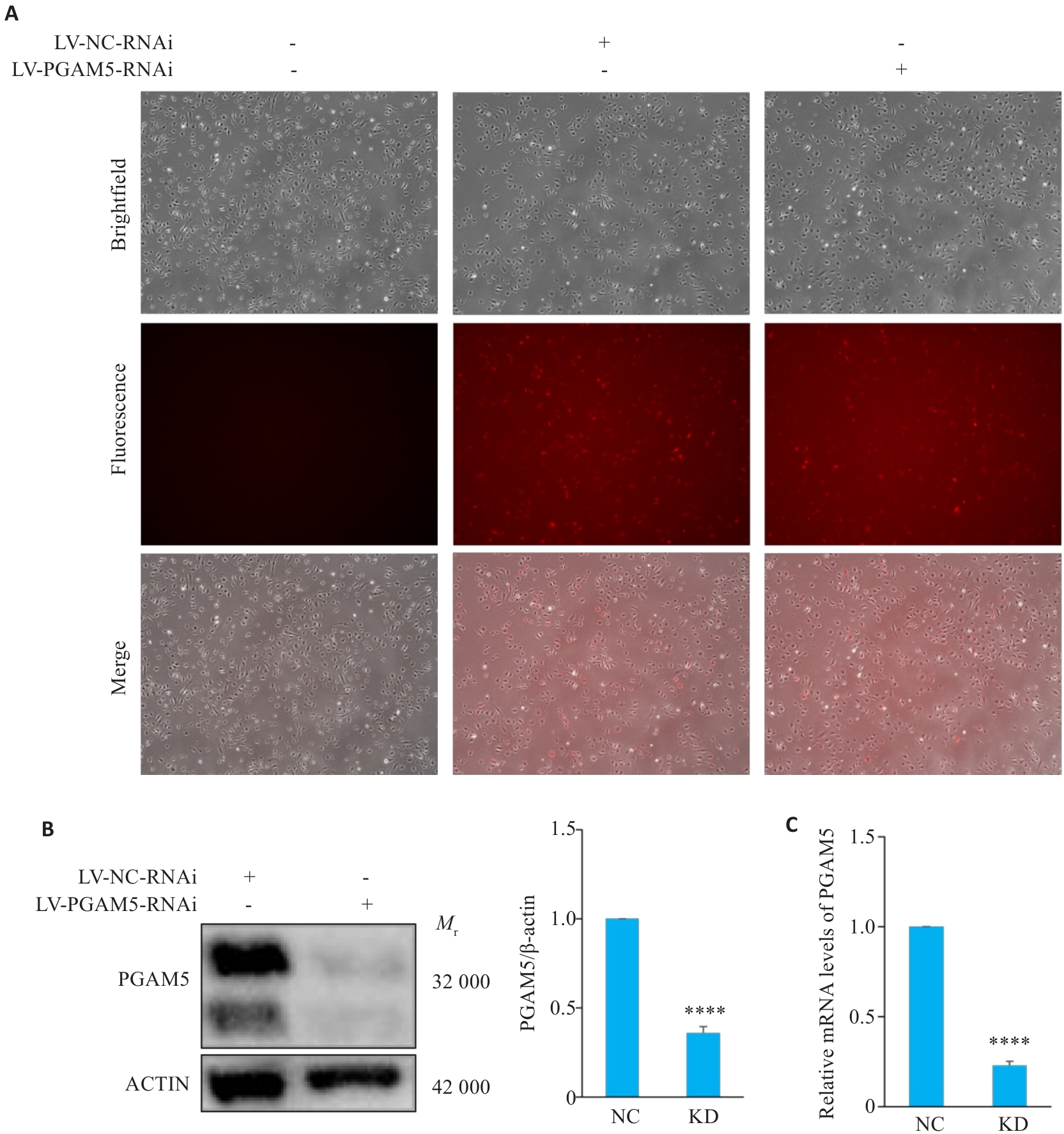
图12 HLSECs中PGAM5敲降效果验证
Fig.12 Validation of PGAM5 knockdown in HLSECs. A: Representative fluorescence images (×100) showing viral transduction efficiency in HLSECs. B: Western blotting of PGAM5 protein and quantitative analysis (n=3). C: Relative PGAM5 mRNA expression levels (n=3). ****P<0.0001 vs NC group.
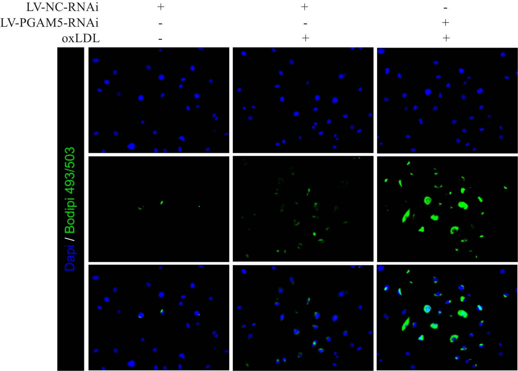
图13 oxLDL处理后HLSECs的Bodipy染色结果
Fig.13 Bodipy staining of HLSECs after oxLDL treatment (×200).
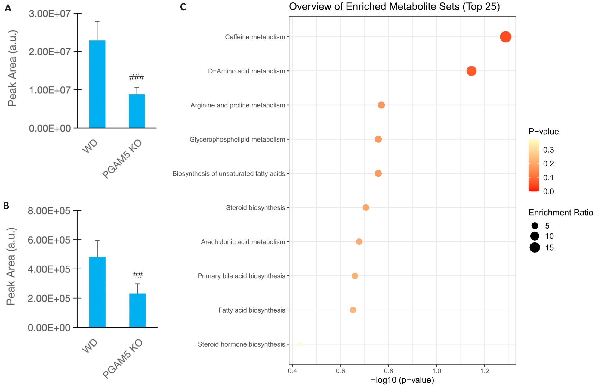
图14 PGAM5缺失显著改变AS小鼠血浆代谢物谱
Fig.14 PGAM5 deficiency reshapes plasma metabolite profile in AS mice. A: Relative abundance of L-palmitoylcarnitine. B: Relative abundance of arachidonic acid. C: Pathway enrichment analysis of top differential metabolites (n=5). ##P<0.01, ###P<0.001 vs WD group.
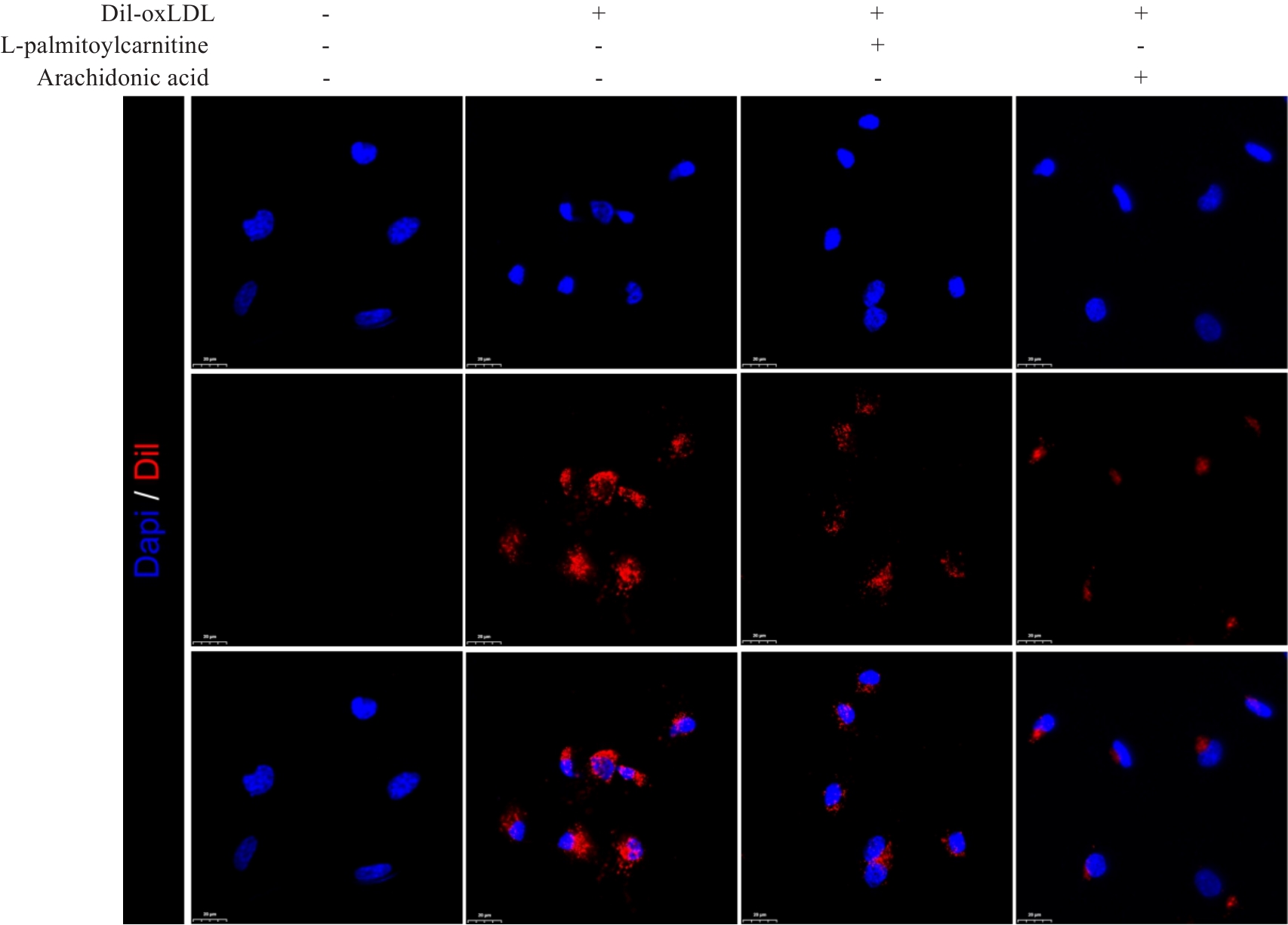
图15 HLSECs对Dil-oxLDL摄取的荧光检测结果
Fig.15 Dil-oxLDL uptake assay in HLSECs (×600).
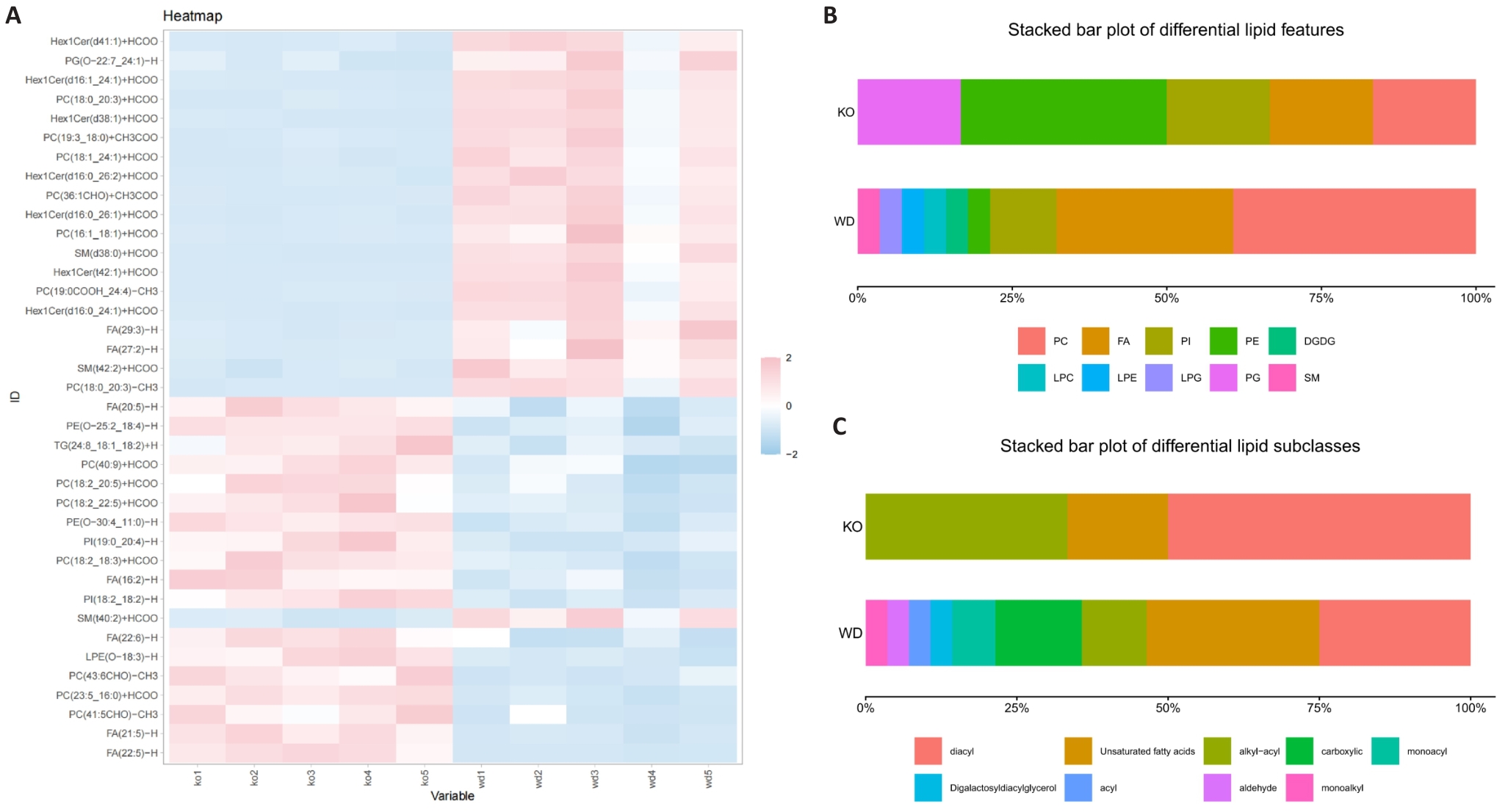
图16 PGAM5缺失显著改变AS小鼠血浆代谢物谱
Fig.16 PGAM5 deficiency significantly alters the plasma metabolite profile in AS mice. A: Heatmap of the representative differential lipids. B: Distribution of differential lipid classes. C: Distribution of differential lipid subclasses.
| [1] | Temtem M, Mendonça MI, Santos M, et al. Validation of the SCORE2 risk prediction algorithm in a Portuguese population: a new model to estimate 10-year cardiovascular disease incidence in Europe[J]. Rev Portuguesa De Cardiol, 2024, 43(8): 437-44. doi:10.1016/j.repc.2023.10.011 |
| [2] | Fu JZ, Deng YH, Ma Y, et al. National and provincial-level prevalence and risk factors of carotid atherosclerosis in Chinese adults[J]. JAMA Netw Open, 2024, 7(1): e2351225. doi:10.1001/jamanetworkopen.2023.51225 |
| [3] | Gusev E, Sarapultsev A. Atherosclerosis and inflammation: insights from the theory of general pathological processes[J]. Int J Mol Sci, 2023, 24(9): 7910. doi:10.3390/ijms24097910 |
| [4] | Miano JM, Fisher EA, Majesky MW. Fate and state of vascular smooth muscle cells in atherosclerosis[J]. Circulation, 2021, 143(21): 2110-6. doi:10.1161/circulationaha.120.049922 |
| [5] | Liu MH, Chen RM, Zheng ZW, et al. Mechanisms of inflammatory microenvironment formation in cardiometabolic diseases: molecular and cellular perspectives[J]. Front Cardiovasc Med, 2025, 11: 1529903. doi:10.3389/fcvm.2024.1529903 |
| [6] | Mortensen MB, Dzaye O, Bøtker HE, et al. Low-density lipoprotein cholesterol is predominantly associated with atherosclerotic cardiovascular disease events in patients with evidence of coronary atherosclerosis: the western Denmark heart registry[J]. Circulation, 2023, 147(14): 1053-63. doi:10.1161/circulationaha.122.061010 |
| [7] | Zhang HL, Long XY, Niu GN, et al. Genetic insights into lipid traits and atherosclerosis risk: a Mendelian randomization and polygenic risk score analysis[J]. Int J Surg, 2025, 111(10): 6802-15. doi:10.1097/js9.0000000000002869 |
| [8] | Ference BA, Ginsberg HN, Graham I, et al. Low-density lipoproteins cause atherosclerotic cardiovascular disease. 1. Evidence from genetic, epidemiologic, and clinical studies. A consensus statement from the European Atherosclerosis Society Consensus Panel[J]. Eur Heart J, 2017, 38(32): 2459-72. doi:10.1093/eurheartj/ehx144 |
| [9] | Josefs T, Barrett TJ, Brown EJ, et al. Neutrophil extracellular traps promote macrophage inflammation and impair atherosclerosis resolution in diabetic mice[J]. JCI Insight, 2020, 5(7): e134796. doi:10.1172/jci.insight.134796 |
| [10] | Badmus OO, Hillhouse SA, Anderson CD, et al. Molecular mechanisms of metabolic associated fatty liver disease (MAFLD): functional analysis of lipid metabolism pathways[J]. Clin Sci, 2022, 136(18): 1347-66. doi:10.1042/cs20220572 |
| [11] | Zhang S, Hong FF, Ma C, et al. Hepatic lipid metabolism disorder and atherosclerosis[J]. Endocr Metab Immune Disord Drug Targets, 2022, 22(6): 590-600. doi:10.2174/1871530322666211220110810 |
| [12] | Zhu LY, Liu FZ, Shi CY, et al. Hepatic micropeptide modulates mitochondrial RNA processing machinery in hepatocellular carcinoma[J]. Mol Cell, 2025, 85(12): 2303-19.e7. doi:10.1016/j.molcel.2025.05.027 |
| [13] | Heeren J, Scheja L. Metabolic-associated fatty liver disease and lipoprotein metabolism[J]. Mol Metab, 2021, 50: 101238. doi:10.1016/j.molmet.2021.101238 |
| [14] | Rogge MM. The role of impaired mitochondrial lipid oxidation in obesity[J]. Biol Res Nurs, 2009, 10(4): 356-73. doi:10.1177/1099800408329408 |
| [15] | Mayr JA. Lipid metabolism in mitochondrial membranes[J]. J Inherit Metab Dis, 2015, 38(1): 137-44. doi:10.1007/s10545-014-9748-x |
| [16] | Ademowo OS, Dias HKI, Burton DGA, et al. Lipid (per) oxidation in mitochondria: an emerging target in the ageing process?[J]. Biogerontology, 2017, 18(6): 859-79. doi:10.1007/s10522-017-9710-z |
| [17] | Di L, Maiseyeu A. Low-density lipoprotein nanomedicines: mechanisms of targeting, biology, and theranostic potential[J]. Drug Deliv, 2021, 28(1): 408-21. doi:10.1080/10717544.2021.1886199 |
| [18] | Bruckdorfer KR. Antioxidants and CVD: symposium on ‘diet and CVD’[J]. Proc Nutr Soc, 2008, 67(2): 214-22. doi:10.1017/s0029665108007052 |
| [19] | Holte C, Szafranska K, Kruse L, et al. Highly oxidized albumin is cleared by liver sinusoidal endothelial cells via the receptors stabilin-1 and-2[J]. Sci Rep, 2023, 13(1): 19121. doi:10.1038/s41598-023-46462-9 |
| [20] | She L, Tu H, Zhang YZ, et al. Inhibition of phosphoglycerate mutase 5 reduces necroptosis in rat hearts following ischemia/reperfusion through suppression of dynamin-related protein 1[J]. Cardiovasc Drugs Ther, 2019, 33(1): 13-23. doi:10.1007/s10557-018-06848-8 |
| [21] | He WB, He WL, Sun ZC, et al. Physiological and pathological roles of PGAM5: an update and future trend[J]. iScience, 2025, 28(2): 111539. doi:10.1016/j.isci.2024.111539 |
| [22] | Qi YX, Rajbanshi B, Hao RH, et al. The dual role of PGAM5 in inflammation[J]. Exp Mol Med, 2025, 57(2): 298-311. doi:10.1038/s12276-025-01391-7 |
| [23] | Zhang Q, Zheng P, Pan Y, et al. Phosphoglycerate mutase 5 is important mediator for instigating arterial lipid accumulation and aggravating atherosclerosis[J]. JACC Basic Transl Sci, 2025, 10(5): 652-73. doi:10.1016/j.jacbts.2024.12.007 |
| [24] | Xu JS, Guo PC, Hao SJ, et al. A spatiotemporal atlas of mouse liver homeostasis and regeneration[J]. Nat Genet, 2024, 56(5): 953-69. doi:10.1038/s41588-024-01709-7 |
| [25] | Du YY, Wu SS, Xi SY, et al. ASH1L in hepatoma cells and hepatic stellate cells promotes fibrosis-associated hepatocellular carcinoma by modulating tumor-associated macrophages[J]. Adv Sci, 2024, 11(45): 2404756. doi:10.1002/advs.202404756 |
| [26] | Sinha S, Ali Q, Zhang T, et al. Aging disrupts hepatocyte zonation homeostasis in mice and humans[J]. Hepatology, 2026, 83(5): 1143-57. doi:10.1097/hep.0000000000001451 |
| [27] | Dai CY, Tsai YM, Chang CY, et al. Reconstruction of the hepatic microenvironment and pathological changes underlying type II diabetes through single-cell RNA sequencing[J]. Int J Biol Sci, 2024, 20(14): 5531-47. doi:10.7150/ijbs.99176 |
| [28] | Yang W, He H, Wang TT, et al. Single-cell transcriptomic analysis reveals a hepatic stellate cell-activation roadmap and myofibroblast origin during liver fibrosis in mice[J]. Hepatology, 2021, 74(5): 2774-90. doi:10.1002/hep.31987 |
| [29] | Rocha AS, Vidal V, Mertz M, et al. The angiocrine factor Rspondin3 is a key determinant of liver zonation[J]. Cell Rep, 2015, 13(9): 1757-64. doi:10.1016/j.celrep.2015.10.049 |
| [30] | Wang B, Zhao LD, Fish M, et al. Self-renewing diploid Axin2+ cells fuel homeostatic renewal of the liver[J]. Nature, 2015, 524(7564): 180-5. doi:10.1038/nature14863 |
| [31] | Halpern KB, Shenhav R, Massalha H, et al. Paired-cell sequencing enables spatial gene expression mapping of liver endothelial cells[J]. Nat Biotechnol, 2018, 36(10): 962-70. doi:10.1038/nbt.4231 |
| [32] | Su TT, Yang YL, Lai SC, et al. Single-cell transcriptomics reveals zone-specific alterations of liver sinusoidal endothelial cells in cirrhosis[J]. Cell Mol Gastroenterol Hepatol, 2021, 11(4): 1139-61. doi:10.1016/j.jcmgh.2020.12.007 |
| [33] | Lin ZY, Liu F, Shi PL, et al. Fatty acid oxidation promotes reprogramming by enhancing oxidative phosphorylation and inhibiting protein kinase C[J]. Stem Cell Res Ther, 2018, 9(1): 47. doi:10.1186/s13287-018-0792-6 |
| [34] | Berra-Romani R, Faris P, Negri S, et al. Arachidonic acid evokes an increase in intracellular Ca2+ concentration and nitric oxide production in endothelial cells from human brain microcirculation[J]. Cells, 2019, 8(7): 689. doi:10.3390/cells8070689 |
| [35] | Vial G, Dubouchaud H, Couturier K, et al. Effects of a high-fat diet on energy metabolism and ROS production in rat liver[J]. J Hepatol, 2011, 54(2): 348-56. doi:10.1016/j.jhep.2010.06.044 |
| [36] | Patten DA, Wilkinson AL, O'Keeffe A, et al. Scavenger receptors: novel roles in the pathogenesis of liver inflammation and cancer[J]. Semin Liver Dis, 2022, 42(1): 61-76. doi:10.1055/s-0041-1733876 |
| [37] | Shetty S, Lalor PF, Adams DH. Liver sinusoidal endothelial cells: gatekeepers of hepatic immunity[J]. Nat Rev Gastroenterol Hepatol, 2018, 15(9): 555-67. doi:10.1038/s41575-018-0020-y |
| [38] | Wilkinson AL, Qurashi M, Shetty S. The role of sinusoidal endothelial cells in the axis of inflammation and cancer within the liver[J]. Front Physiol, 2020, 11: 990. doi:10.3389/fphys.2020.00990 |
| [39] | Smedsrød B, Melkko J, Araki N, et al. Advanced glycation end products are eliminated by scavenger-receptor-mediated endocytosis in hepatic sinusoidal Kupffer and endothelial cells[J]. Biochem J, 1997, 322(2): 567-73. doi:10.1042/bj3220567 |
| [40] | Lee SJ, Evers S, Roeder D, et al. Mannose receptor-mediated regulation of serum glycoprotein homeostasis[J]. Science, 2002, 295(5561): 1898-901. doi:10.1126/science.1069540 |
| [41] | Lalor PF, Lai WK, Curbishley SM, et al. Human hepatic sinusoidal endothelial cells can be distinguished by expression of phenotypic markers related to their specialised functions in vivo [J]. World J Gastroenterol, 2006, 12(34): 5429-39. doi:10.3748/wjg.v12.i34.5429 |
| [42] | Hu YF, Li W, Cheng X, et al. Emerging roles and therapeutic applications of arachidonic acid pathways in cardiometabolic diseases[J]. Circ Res, 2024, 135(1): 222-60. doi:10.1161/circresaha.124.324383 |
| [43] | Badimon L, Vilahur G, Rocca B, et al. The key contribution of platelet and vascular arachidonic acid metabolism to the pathophysiology of atherothrombosis[J]. Cardiovasc Res, 2021, 117(9): 2001-15. doi:10.1093/cvr/cvab003 |
| [44] | Gong X, Qiu T, Zhou JY, et al. TGF-β1 promotes angiogenesis via endothelial-to-mesenchymal transition in infantile hemangioma[J]. Arterioscler Thromb Vasc Biol, 2025, 45(10): 1912-27. doi:10.1161/atvbaha.125.322793 |
| [45] | Ba HX, Guo YF, Jiang YJ, et al. Unveiling the metabolic landscape of pulmonary hypertension: insights from metabolomics[J]. Respir Res, 2024, 25(1): 221. doi:10.1186/s12931-024-02775-5 |
| [46] | Taiwo A, Merrill RA, Wendt L, et al. Metabolite perturbations in type 1 diabetes associated with metabolic dysfunction-associated steatotic liver disease[J]. Front Endocrinol, 2025, 16: 1500242. doi:10.3389/fendo.2025.1500242 |
| [47] | Chong SY, Wang XY, van Bloois L, et al. Injectable liposomal docosahexaenoic acid alleviates atherosclerosis progression and enhances plaque stability[J]. J Control Release, 2023, 360: 344-64. doi:10.1016/j.atherosclerosis.2023.06.702 |
| [48] | Djuricic I, Calder PC. N-3 fatty acids (EPA and DHA) and cardiovascular health-updated review of mechanisms and clinical outcomes[J]. Curr Atheroscler Rep, 2025, 27(1): 116. doi:10.1007/s11883-025-01363-2 |
| [1] | 胡晨曦, 顾曦, 牛鹏程, 王祎婷, 方廖琼, 王智彪, 白晋. 大肠杆菌外膜囊泡依赖靶细胞内吞抑制大鼠胶质瘤细胞增殖[J]. 南方医科大学学报, 2026, 46(8): 1730-1742. |
| [2] | 王宏杨, 朱文怡, 陈栩燊, 张童, 曹志伟, 王金, 解渤, 刘强, 任雪峰. “气经-血络”截断动脉粥样硬化斑块形成:降脂化斑方调控小鼠肝脏逆向胆固醇转运途径改善动脉粥样硬化的机制[J]. 南方医科大学学报, 2025, 45(9): 1818-1829. |
| [3] | 夏冰, 彭进, 丁九阳, 王杰, 唐国伟, 刘国杰, 王沄, 万昌武, 乐翠云. ATF3通过NF-κB信号通路调控动脉粥样硬化斑块内的炎症反应[J]. 南方医科大学学报, 2025, 45(6): 1131-1142. |
| [4] | 张珊苑, 蔡巧燕, 祁江晗, 殷恺馨, 何晨晨, 高铸烨, 张铃, 褚剑锋. 清心解瘀颗粒抗动脉粥样硬化的药效学及调控机制[J]. 南方医科大学学报, 2024, 44(8): 1518-1528. |
| [5] | 李洪涛, 邓 宇, 王添乐, 黄克勇, 于传沛, 陈朝俊. 丹参新醌乙减轻ox-LDL诱导的内皮细胞损伤:基于抑制NF-κB/NLRP3信号通路介导的细胞焦亡[J]. 南方医科大学学报, 2023, 43(8): 1425-1431. |
| [6] | 陆 琦, 鲍迎春, 陈旭姣, 王春明. 白兔局部动脉粥样硬化不稳定斑块对其余血管斑块形成的影响[J]. 南方医科大学学报, 2023, 43(1): 117-121. |
| [7] | 刘泽鑫, 曹 赛, 陈 清, 符方勇, 程梅容, 黄显莹. MicroRNA-132通过诱导线粒体氧化应激障碍-铁死亡进程促进动脉粥样硬化[J]. 南方医科大学学报, 2022, 42(1): 143-149. |
| [8] | 许名东, 李丽华, 李晚泉, 刘家超, 李小丹, 叶浩彬, 陈健芳, 邹文毕. 半乳糖凝集素-3通过lncARSR影响小鼠冠状动脉粥样硬化易损斑块稳定的机制[J]. 南方医科大学学报, 2021, 41(7): 1067-1072. |
| [9] | 邱 峰, 张 琳, 郑介婷, 操龙斌, 张子康, 邓屹琪. 穗花杉双黄酮激活PPAR-α/γ抑制THP-1源性巨噬细胞向M1型极化[J]. 南方医科大学学报, 2021, 41(3): 344-351. |
| [10] | 徐 俊, 史嘉炜, 蔡欣玲, 黄盛娜, 李 刚, 徐 燕. 富心方通过调控c-Fos-NR4A1-p38通路改善人动脉血管内皮细胞缺氧引起的损伤[J]. 南方医科大学学报, 2021, 41(2): 200-209. |
| [11] | 杨渊博, 李忠豪, 刘 蔷, 谢国喜, 冯衍秋. ApoE-/-小鼠三维DANTE-FLASH颈动脉血管壁成像:基于7.0 T磁共振技术[J]. 南方医科大学学报, 2021, 41(2): 216-222. |
| [12] | 顾毓艳, 蒋 晶, 张雅心, 陈育尧, 陈奕澔, 江伟豪, 黄志勇, 周凤华. 从心论治方改善ApoE-/-小鼠动脉粥样硬化及上调ACE2蛋白表达[J]. 南方医科大学学报, 2021, 41(11): 1623-1630. |
| [13] | 林子祥,刘 丹,薛雅芝,吴同薇,曾晓容,郭志刚,曹世平. 血清膜联蛋白A2在冠心病患者血清中明显升高[J]. 南方医科大学学报, 2020, 40(03): 382-387. |
| [14] | 李 挺,何铭垣,李忠豪,王德奖,徐颖怡,吴 炜,燕 翼. 二甲双胍通过调控血管平滑肌肌动蛋白骨架系统抑制小鼠动脉粥样硬化形成[J]. 南方医科大学学报, 2019, 39(11): 1357-1363. |
| [15] | 张磊,祁冀,景亚军,覃波,李义凯,刘刚,郭晓光,扶世杰. 推桥弓手法对食蟹猴轻度颈动脉粥样硬化模型血流动力学的影响[J]. 南方医科大学学报, 2017, 37(12): 1592-. |
| 阅读次数 | ||||||
|
全文 |
|
|||||
|
摘要 |
|
|||||
