
南方医科大学学报 ›› 2026, Vol. 46 ›› Issue (10): 2309-2324.doi: 10.12122/j.issn.1673-4254.2026.10.03
• • 上一篇
卢晓宇1( ), 庞天霄2, 刘烨2, 何学红2(), 李志明2()
收稿日期:2026-03-17
出版日期:2026-10-20
发布日期:2026-10-26
通讯作者:
何学红,李志明
E-mail:dfs1107@163.com;lnzysnk@163.com;zglzmzg@163.com
作者简介:卢晓宇,在读博士研究生,主治医师,E-mail: dfs1107@163.com
基金资助:
Xiaoyu LU1(), Tianxiao PANG2, Ye LIU2, Xuehong HE2(), Zhiming LI2()
Received:2026-03-17
Online:2026-10-20
Published:2026-10-26
Contact:
Xuehong HE, Zhiming LI
E-mail:dfs1107@163.com;lnzysnk@163.com;zglzmzg@163.com
摘要:
目的 探讨参芪泄浊饮(SQXZD)延缓肾纤维化的作用机制。 方法 采用网络药理学富集SQXZD延缓肾纤维化的靶点及信号通路,分子对接分析SQXZD成分与相关靶点的结合能力。体内实验:将49只SPF级雄性SD大鼠随机分为空白组,假手术组,模型组,氯沙坦组(4.6 mg·kg-1·d-1)及SQXZD低、中、高剂量(9.7、19.4、38.8 g·kg-1·d-1)组,7只/组。除空白组及假手术组,其他组大鼠均以单侧输尿管结扎法复制肾纤维化模型。造模24 h后开始灌胃,14 d后取材,Masson染色观察肾组织胶原分布,并检测纤维化相关指标(E-Cadherin)、自噬相关指标(LC3B、p62/SQSTM1)及ERK1/2-mTOR信号通路表达。体外实验:将大鼠肾正常成纤维细胞(NRK-49F)及肾小管上皮细胞(NRK-52E)各分为对照组(未处理)、模型组(TGF-β1处理)、空白血清组(TGF-β1+空白血清处理)、二甲基亚砜(DMSO)组(TGF-β1+DMSO处理)、含药血清组(TGF-β1+SQXZD含药血清处理)、间双没食子酸(DGA)组(TGF-β1+DGA处理)、含药血清+U0126组(TGF-β1+SQXZD含药血清+U0126处理)、DGA+U0126组(TGF-β1+DGA+U0126处理),免疫荧光确定纤维化诱导条件,CCK-8法确定SQXZD含药血清及DGA处理条件,丹酰尸胺染色及透射电子显微镜观察各组细胞自噬水平,检测纤维化相关指标(α-SMA、Col-Ⅰ、E-Cadherin)、自噬相关指标(LC3B、p62/SQSTM1)及ERK1/2-mTOR信号通路表达。 结果 SQXZD主要活性成分为4-O-甲基芍药苷、DGA等54个,与IGF1、KDR、ALB等靶点结合紧密。体内实验显示,与假手术组比较,模型组大鼠术侧肾组织LC3B-Ⅱ表达上调,E-cadherin、p62/SQSTM1、p-mTOR/mTOR表达下调(P<0.01)。与模型组比较,各给药组大鼠术侧肾组织LC3B-Ⅱ表达下调,E-cadherin、p62/SQSTM1、p-mTOR/mTOR表达上调(P<0.05)。体外实验显示,与对照组比较,10 ng/mL TGF-β1处理NRK-49F细胞72 h与5 ng/mL TGF-β1处理NRK-52E细胞36 h后α-SMA表达上调(P<0.01)。与模型组比较,15%体积SQXZD含药血清分别处理TGF-β1诱导的NRK-49F细胞24 h、NRK-52E细胞12 h可抑制细胞活力(P<0.05);NRK-49F及NRK-52E细胞DGAIC50分别为90.15、610.66 ng/mL。与对照组比较,模型组细胞自噬荧光增强、自噬体数量增多,α-SMA、Col-Ⅰ(NRK-49F)、LC3B-Ⅱ、p-ERK1/2/ERK1/2表达上调,E-cadherin(NRK-52E)、p62/SQSTM1、p-mTOR/mTOR表达下调(P<0.01)。与模型组比较,含药血清组、含药血清+U0126组、DGA组、DGA+U0126组细胞自噬荧光减弱、自噬体数量减少,α-SMA、Col-Ⅰ(NRK-49F)、LC3B-Ⅱ、p-ERK1/2/ERK1/2表达下调,E-cadherin(NRK-52E)、p62/SQSTM1、p-mTOR/mTOR表达上调(P<0.05)。 结论 SQXZD可抑制成纤维细胞活化及上皮-间质转化以延缓肾纤维化,其机制可能是通过调控ERK1/2-mTOR信号通路以抑制过度自噬。
卢晓宇, 庞天霄, 刘烨, 何学红, 李志明. 参芪泄浊饮通过调控ERK1/2-mTOR信号通路抑制过度自噬延缓大鼠肾纤维化[J]. 南方医科大学学报, 2026, 46(10): 2309-2324.
Xiaoyu LU, Tianxiao PANG, Ye LIU, Xuehong HE, Zhiming LI. Shenqi Xiezhuo Decoction ameliorates renal fibrosis in rats by inhibiting excessive autophagy via modulating the ERK1/2-mTOR signaling pathway[J]. Journal of Southern Medical University, 2026, 46(10): 2309-2324.
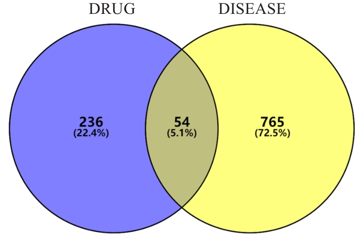
图1 参芪泄浊饮延缓肾纤维化的作用靶点Venn图
Fig.1 Venn diagram of the action targets of SQXZD for delaying progression of renal fibrosis.
| Components | Degree | Components | Degree |
|---|---|---|---|
| benzoyl paeoniflorin | 201 | Digallic acid | 131 |
| Diisooctyl phthalate | 161 | Physciondiglucoside | 129 |
| 4-O-methylpaeoniflorin_qt | 153 | Acanthoside B | 128 |
| Procyanidin B-5,3'-O-gallate | 136 | Poricoic acid B | 128 |
| Poricoic acid A | 135 | Sennoside D_qt | 125 |
表1 参芪泄浊饮延缓肾纤维化的前10个关键成分及Degree值
Tab.1 Top 10 key components of SQXZD for delaying progression of renal fibrosis and their degree values
| Components | Degree | Components | Degree |
|---|---|---|---|
| benzoyl paeoniflorin | 201 | Digallic acid | 131 |
| Diisooctyl phthalate | 161 | Physciondiglucoside | 129 |
| 4-O-methylpaeoniflorin_qt | 153 | Acanthoside B | 128 |
| Procyanidin B-5,3'-O-gallate | 136 | Poricoic acid B | 128 |
| Poricoic acid A | 135 | Sennoside D_qt | 125 |
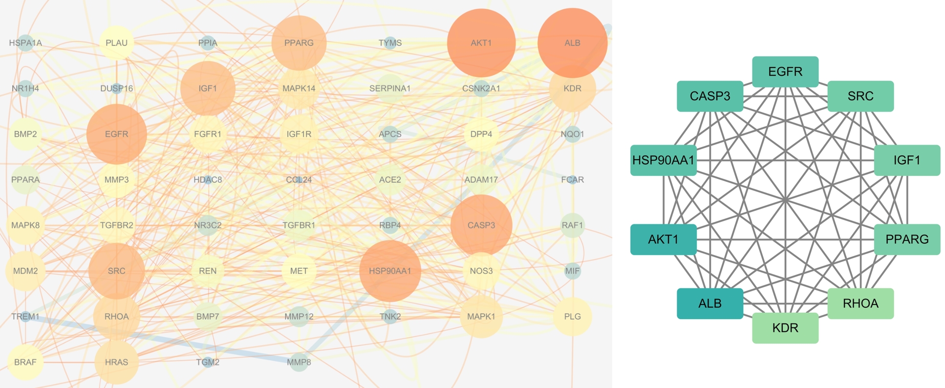
图2 参芪泄浊饮延缓肾纤维化的关键靶点
Fig.2 Key targets of SQXZD for delaying progression of renal fibrosis (Color intensity and node area are based on degree values).
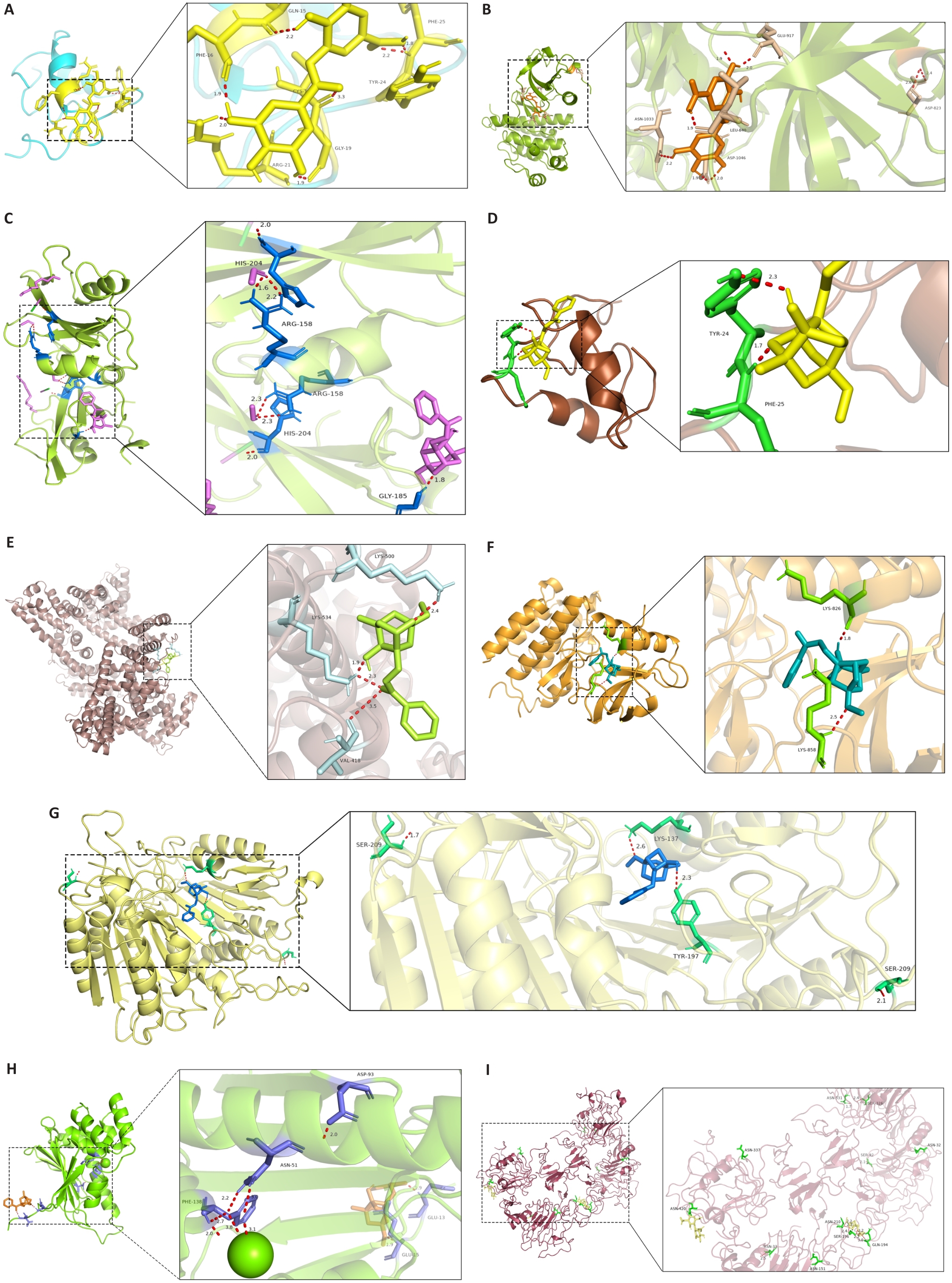
图3 参芪泄浊饮核心活性成分与核心靶点的分子对接模式
Fig.3 Molecular docking of the core active components of SQXZD with their core targets. A: DGA-IGF1. B: DGA-KDR. C: 4-O-methylpaeoniflorin-SRC. D: 4-O-methylpaeoniflorin-IGF1. E: 4-O-methylpaeoniflorin-ALB. F: 4-O-methylpaeoniflorin-KDR. G: 4-O-methylpaeoniflorin-CASP3. H: 4-O-methylpaeoniflorin-HSP90AA1. I: 4-O-methylpaeoniflorin-EGFR. DGA: Digallic acid.
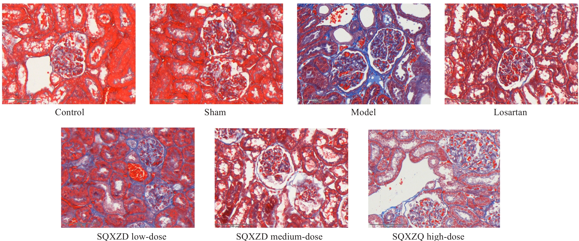
图4 参芪泄浊饮对UUO大鼠术侧肾组织胶原沉积的影响
Fig.4 Effect of SQXZD on collagen deposition in the surgical-side kidney in UUO rats (Original magnification: ×400).
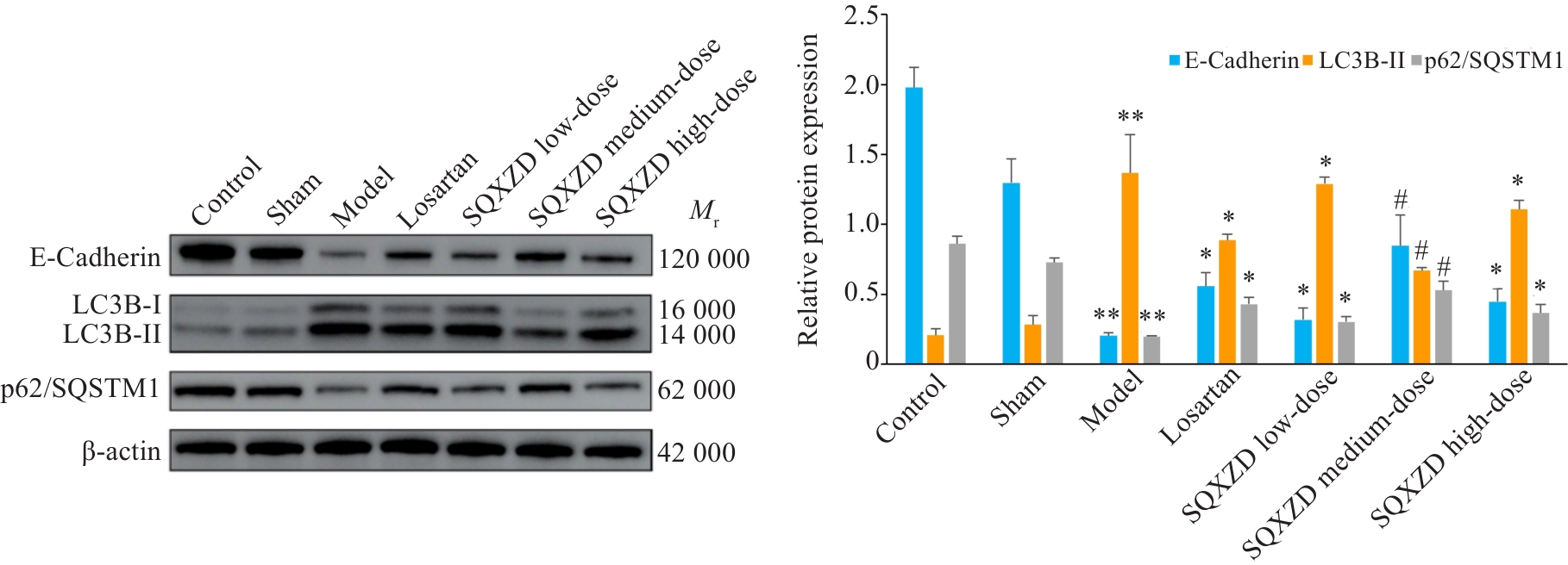
图6 参芪泄浊饮对UUO大鼠术侧肾组织EMT及自噬相关蛋白表达的影响
Fig.6 Effect of SQXZD on expression of EMT and autophagy-related proteins in the surgical-side kidney in UUO rats. **P<0.01 vs Sham group; *P<0.05, #P<0.01 vs Model group, n=3.
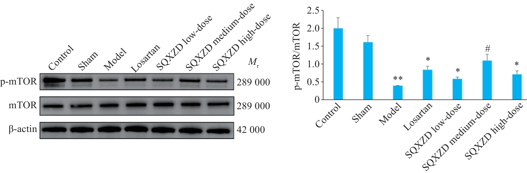
图7 参芪泄浊饮对UUO大鼠术侧肾组织p-mTOR、mTOR表达的影响
Fig.7 Effect of SQXZD on the expression of p-mTOR and mTOR in the surgical-side kidney in UUO rats. **P<0.01 vs Sham group; *P<0.05, #P<0.01 vs Model group, n=3.
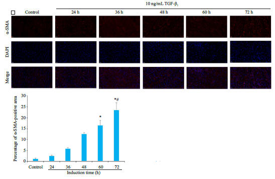
图9 不同浓度TGF-β1处理后对NRK-52E细胞α-SMA表达的影响
Fig.9 Effects of different concentrations of TGF-β1 on α-SMA expression in NRK-52E cells (×400). Induction time: A, 12 h; B, 24 h; C, 36 h; D, 48 h. *P<0.05, #P<0.01 vs Control group; △P<0.01 vs 12 h-induced group by 10 ng/mL TGF-β1; ▲P<0.05 vs 12 h-induced group by 12.5 ng/mL TGF-β1 and 24 h-induced group by 10 ng/mL TGF-β1. n=3.
| Item | Blank group | Control group | Dilutions of SQXZD-medicated serum groups (%) | |||||||
|---|---|---|---|---|---|---|---|---|---|---|
| 2.5 | 5 | 7.5 | 10 | 12.5 | 15 | 17.5 | 20 | |||
| NRK-49F | 0.189±0.006 | 2.062±0.116 | 1.831±0.291 | 2.300±0.120 | 2.257±0.123 | 2.282±0.088 | 2.294±0.095 | 2.332±0.112* | 2.164±0.180 | 2.117±0.087 |
| NRK-52E | 0.207±0.001 | 1.256±0.051 | 1.862±0.424 | 1.702±0.151 | 1.992±0.271 | 1.937±0.224 | 2.017±0.050* | 2.284±0.146# | 2.238±0.153* | 0.826±0.065 |
表2 不同体积的参芪泄浊饮含药血清对NRK-49F及NRK-52E细胞活力的影响
Tab.2 Effect of different Dilutions of SQXZD-medicated serum on viability of NRK-49F and NRK-52E cells (Mean±SD, n=3)
| Item | Blank group | Control group | Dilutions of SQXZD-medicated serum groups (%) | |||||||
|---|---|---|---|---|---|---|---|---|---|---|
| 2.5 | 5 | 7.5 | 10 | 12.5 | 15 | 17.5 | 20 | |||
| NRK-49F | 0.189±0.006 | 2.062±0.116 | 1.831±0.291 | 2.300±0.120 | 2.257±0.123 | 2.282±0.088 | 2.294±0.095 | 2.332±0.112* | 2.164±0.180 | 2.117±0.087 |
| NRK-52E | 0.207±0.001 | 1.256±0.051 | 1.862±0.424 | 1.702±0.151 | 1.992±0.271 | 1.937±0.224 | 2.017±0.050* | 2.284±0.146# | 2.238±0.153* | 0.826±0.065 |
| Group | Induction time NRK-49F | Induction time NRK-52E | ||
|---|---|---|---|---|
| Blank | 1.711±0.053 | 1.830±0.018 | ||
| Control | 2.041±0.061 | 2.612±0.427 | ||
| SQXZD-medicated serum | 0 h | 1.999±0.029 | 0 h | 2.052±0.044 |
| 24 h | 2.014±0.104 | 12 h | 2.093±0.039 | |
| 48 h | 1.915±0.059* | 24 h | 2.049±0.038* | |
表3 参芪泄浊饮含药血清不同处理时间对TGF-β1诱导的NRK-49F及NRK-52E细胞活力的影响
Tab.3 Effect of different treatment durations with SQXZD-medicated serum on viability of TGF-β1-induced NRK-49F and NRK-52E cells (Mean±SD, n=3)
| Group | Induction time NRK-49F | Induction time NRK-52E | ||
|---|---|---|---|---|
| Blank | 1.711±0.053 | 1.830±0.018 | ||
| Control | 2.041±0.061 | 2.612±0.427 | ||
| SQXZD-medicated serum | 0 h | 1.999±0.029 | 0 h | 2.052±0.044 |
| 24 h | 2.014±0.104 | 12 h | 2.093±0.039 | |
| 48 h | 1.915±0.059* | 24 h | 2.049±0.038* | |
| Cell | Blank group | Control group | Concentration of DGA (ng/mL) | ||||
|---|---|---|---|---|---|---|---|
| 44.5 | 89 | 178 | 356 | 712 | |||
| NRK-49F | 1.927±0.046 | 1.808±0.015 | 2.259±0.150 | 0.980±0.080 | 0.273±0.009 | 0.611±0.019 | 0.921±0.016 |
| NRK-52E | 1.256±0.051 | 1.267±0.110 | 1.818±0.172 | 1.018±0.090 | 0.848±0.066 | 0.634±0.150 | 0.862±0.122 |
表4 不同浓度的DGA对NRK-49F及NRK-52E细胞活力的影响
Tab.4 Effect of different concentrations of DGA on viability of NRK-49F and NRK-52E cells (Mean±SD, n=3)
| Cell | Blank group | Control group | Concentration of DGA (ng/mL) | ||||
|---|---|---|---|---|---|---|---|
| 44.5 | 89 | 178 | 356 | 712 | |||
| NRK-49F | 1.927±0.046 | 1.808±0.015 | 2.259±0.150 | 0.980±0.080 | 0.273±0.009 | 0.611±0.019 | 0.921±0.016 |
| NRK-52E | 1.256±0.051 | 1.267±0.110 | 1.818±0.172 | 1.018±0.090 | 0.848±0.066 | 0.634±0.150 | 0.862±0.122 |
| Group | Induction time NRK-49F | Induction time NRK-52E | ||
|---|---|---|---|---|
| Blank | 1.711±0.053 | 1.819±0.031 | ||
| Control | 2.093±0.063 | 2.219±0.041 | ||
| DGA | 0 h | 2.101±0.032 | 0 h | 2.108±0.027* |
| 24 h | 2.082±0.113 | 12 h | 2.052±0.084* | |
| 48 h | 1.974±0.049* | 24 h | 1.943±0.039# | |
表5 DGA不同处理时间对TGF-β1诱导的NRK-49F及NRK-52E细胞活力的影响
Tab.5 Effect of different treatment durations with DGA on viability of TGF-β1-induced NRK-49F and NRK-52E (Mean±SD, n=3)
| Group | Induction time NRK-49F | Induction time NRK-52E | ||
|---|---|---|---|---|
| Blank | 1.711±0.053 | 1.819±0.031 | ||
| Control | 2.093±0.063 | 2.219±0.041 | ||
| DGA | 0 h | 2.101±0.032 | 0 h | 2.108±0.027* |
| 24 h | 2.082±0.113 | 12 h | 2.052±0.084* | |
| 48 h | 1.974±0.049* | 24 h | 1.943±0.039# | |
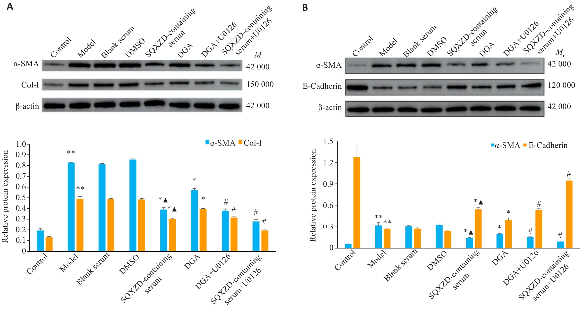
图10 参芪泄浊饮含药血清及DGA对TGF-β1诱导的NRK-49F及NRK-52E细胞α-SMA、Col-I、E-Cadherin表达的影响
Fig.10 Effects of SQXZD-medicated serum and DGA on expression of α-SMA, Col-I, and E-cadherin in TGF-β1-induced NRK-49F (A) and NRK-52E cells (B). **P<0.01 vs Control group; *P<0.05, #P<0.01 vs Model group; ▲P<0.05 vs DGA group. n=3.
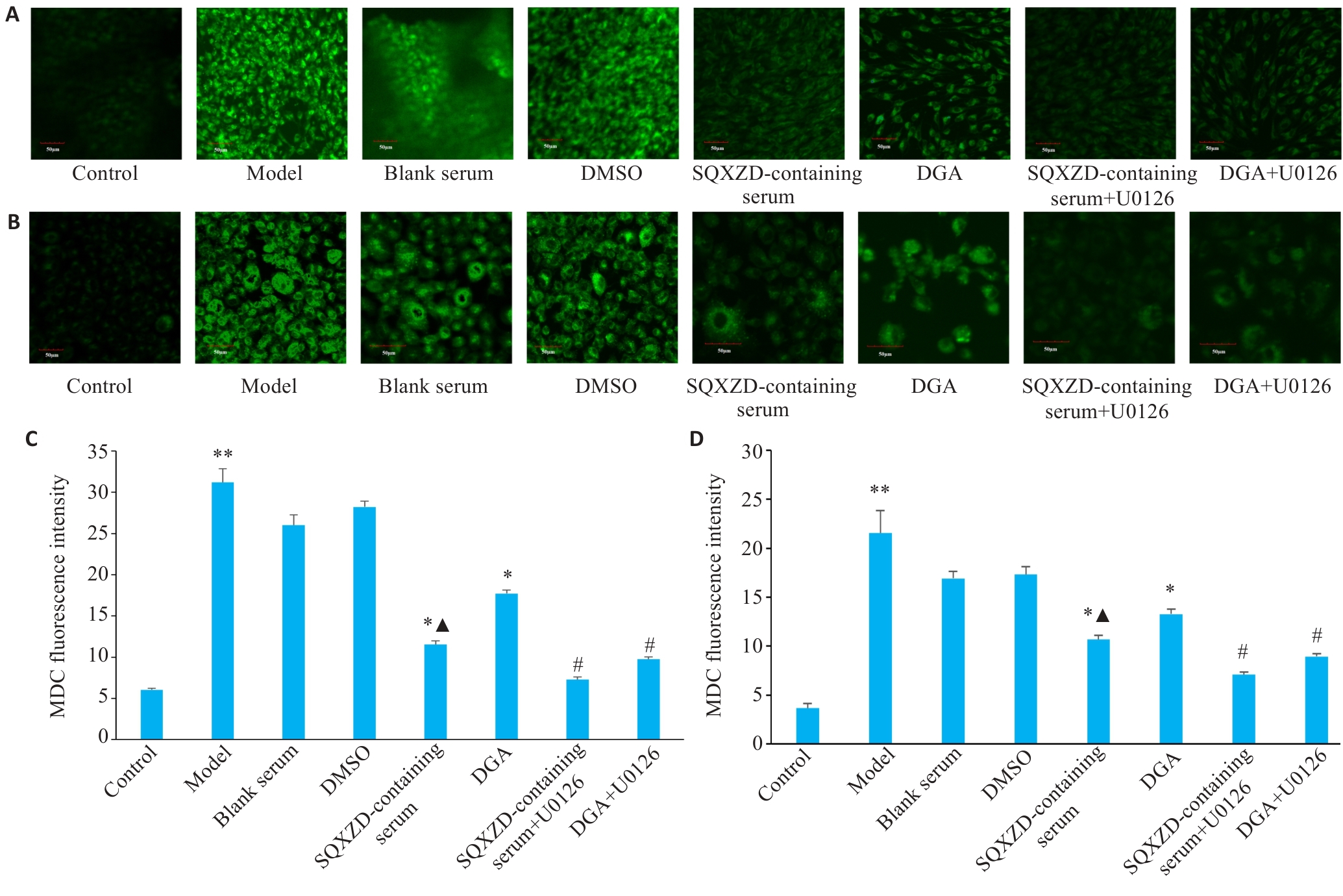
图11 参芪泄浊饮含药血清及DGA对TGF-β1诱导的NRK-49F及NRK-52E细胞自噬荧光的影响
Fig.11 Effects of SQXZD-medicated serum and DGA on autophagic fluorescence in TGF-β1-induced NRK-49F cells (A, C) and NRK-52E cells (B, D) (×400). **P<0.01 vs Control group; *P<0.05, #P<0.01 vs Model group; ▲P<0.05 vs DGA group. n=3.
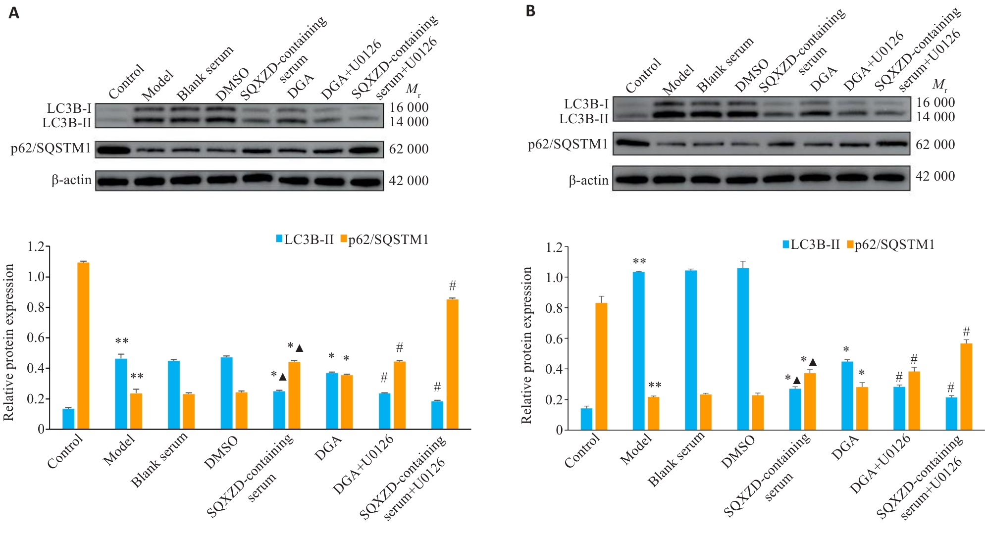
图12 参芪泄浊饮含药血清及DGA对TGF-β1诱导的NRK-49F及NRK-52E细胞LC3B-II、p62/SQSTM1表达的影响
Fig.12 Effects of SQXZD-medicated serum and DGA on the expression of LC3B-II and p62/SQSTM1 in TGF-β1-induced NRK-49F (A) and NRK-52E cells (B). **P<0.01 vs Control group; *P<0.05, #P<0.01 vs Model group; ▲P<0.05 vs DGA group. n=3.
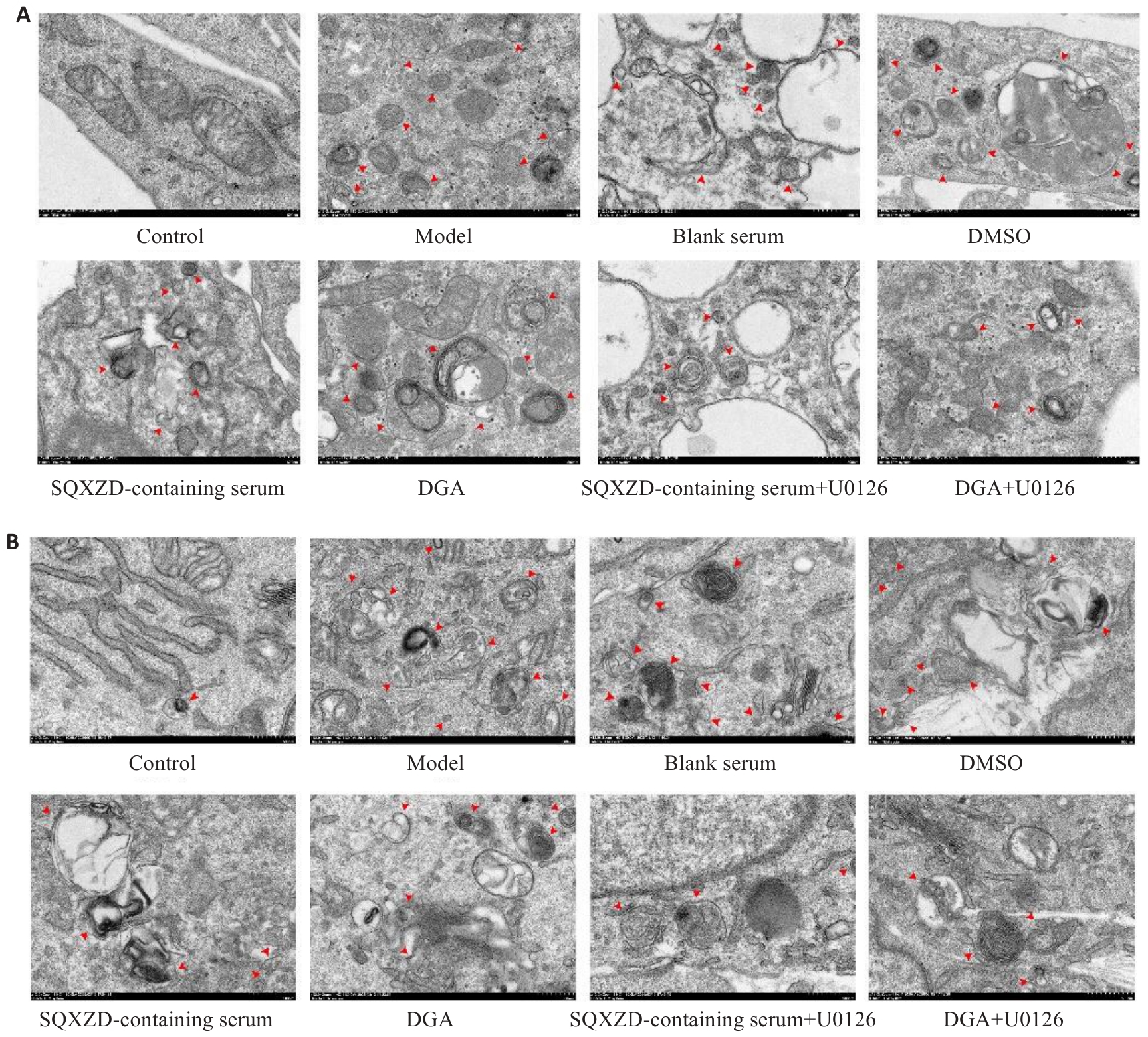
图13 参芪泄浊饮含药血清及DGA对TGF-β1诱导的NRK-49F及NRK-52E细胞超微结构的影响
Fig.13 Effects of SQXZD-medicated serum and DGA on ultrastructure of TGF-β1-induced NRK-49F (A) and NRK-52E cells (B) (Transmission electron microscopy, ×12 000), red arrows indicate autophagosomes.
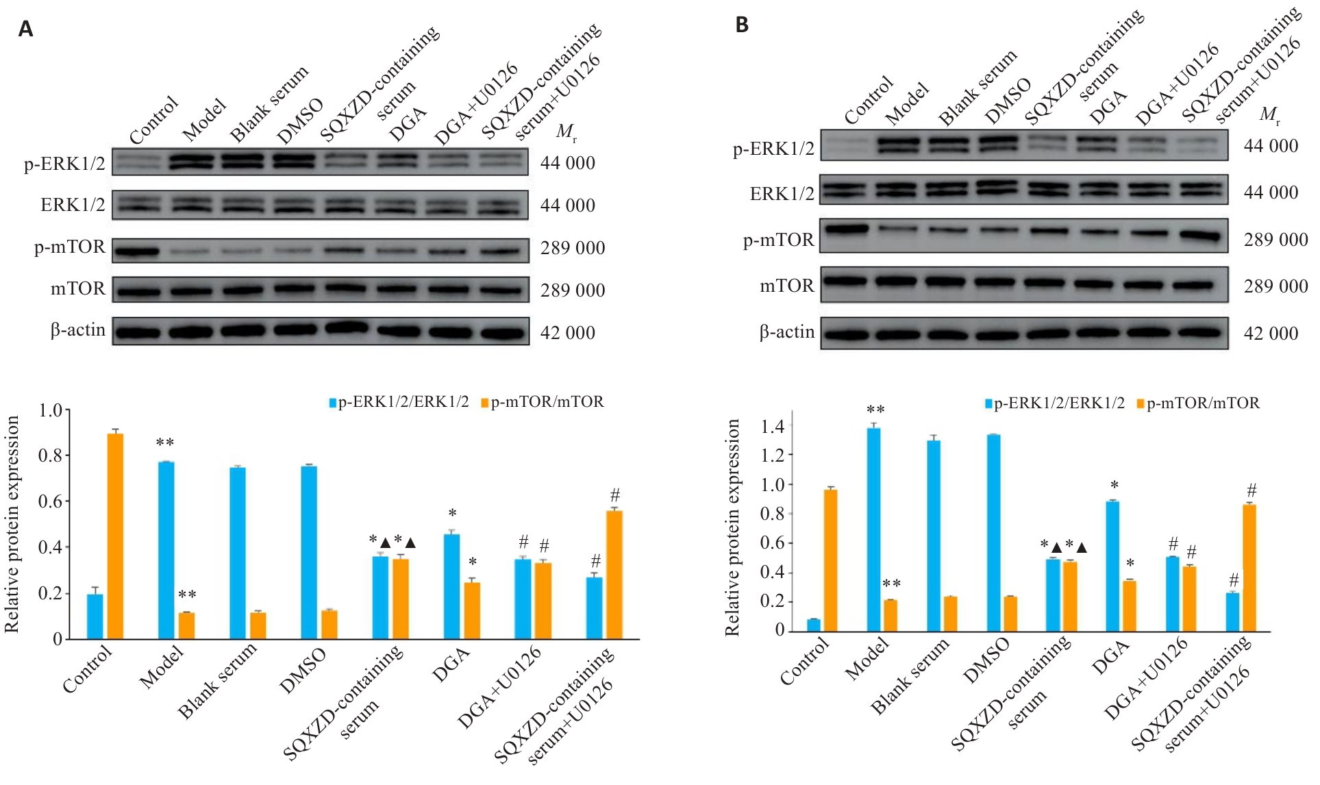
图14 参芪泄浊饮含药血清及DGA对TGF-β1诱导的NRK-49F及NRK-52E细胞p-ERK1/2/ERK1/2、p-mTOR/mTOR表达的影响
Fig.14 Effects of SQXZD-medicated serum and DGA on expression of p-ERK1/2/ERK1/2 and p-mTOR/mTOR in TGF-β1-induced NRK-49F (A) and NRK-52E cells (B). **P<0.01 vs Control group; *P<0.05, #P<0.01 vs Model group; ▲P<0.05 vs DGA group. n=3.
| [1] | Liu YH. Cellular and molecular mechanisms of renal fibrosis[J]. Nat Rev Nephrol, 2011, 7(12): 684-96. doi:10.1038/nrneph.2011.149 |
| [2] | Zeisberg M, Neilson EG. Mechanisms of tubulointerstitial fibrosis[J]. J Am Soc Nephrol, 2010, 21(11): 1819-34. doi:10.1681/asn.2010080793 |
| [3] | GBD 2023 Chronic Kidney Disease Collaborators. Global, regional, and national burden of chronic kidney disease in adults, 1990-2023, and its attributable risk factors: a systematic analysis for the Global Burden of Disease Study 2023[J]. Lancet, 2025,406(10518):2461-82. |
| [4] | Lovisa S, Zeisberg M, Kalluri R. Partial epithelial-to-mesenchymal transition and other new mechanisms of kidney fibrosis[J]. Trends Endocrinol Metab, 2016, 27(10): 681-95. doi:10.1016/j.tem.2016.06.004 |
| [5] | Dai R, Zhang L, Jin H, et al. Autophagy in renal fibrosis: Protection or promotion[J]? Front Pharmacol, 2022, 13: 963920. doi:10.3389/fphar.2022.963920 |
| [6] | Mizushima N, Komatsu M. Autophagy: renovation of cells and tissues[J]. Cell, 2011, 147(4): 728-41. doi:10.1016/j.cell.2011.10.026 |
| [7] | Kaushal GP, Chandrashekar K, Juncos LA, et al. Autophagy function and regulation in kidney disease[J]. Biomolecules, 2020, 10(1): 100. doi:10.3390/biom10010100 |
| [8] | Takemura K, Nishi H, Inagi R. Mitochondrial dysfunction in kidney disease and uremic sarcopenia[J]. Front Physiol, 2020, 11: 565023. doi:10.3389/fphys.2020.565023 |
| [9] | Bai L, Wu Q, Zhang X, Z, et al. Autosis as a selective type of cell death[J]. Front Cell Dev Biol, 2023, 11:1164681-93. doi:10.3389/fcell.2023.1164681 |
| [10] | Inoki K. mTOR signaling in autophagy regulation in the kidney[J]. Semin Nephrol, 2014, 34(1): 2-8. doi:10.1016/j.semnephrol.2013.11.002 |
| [11] | Xue X, Ren JF, Sun XL, et al. Protein kinase Cα drives fibroblast activation and kidney fibrosis by stimulating autophagic flux[J]. J Biol Chem, 2018, 293(28): 11119-30. doi:10.1074/jbc.ra118.002191 |
| [12] | Nam SA, Kim WY, Kim JW, et al. Autophagy attenuates tubulointerstital fibrosis through regulating transforming growth factor‑β and NLRP3 inflammasome signaling pathway[J]. Cell Death Dis, 2019, 10(2): 78. doi:10.1038/s41419-019-1356-0 |
| [13] | Wu YR, Wang LY, Deng D, et al. Renalase protects against renal fibrosis by inhibiting the activation of the ERK signaling pathways[J]. Int J Mol Sci, 2017, 18(5): 855. doi:10.3390/ijms18050855 |
| [14] | Zhu LN, Yang T, Li LJ, et al. TSC1 controls macrophage polarization to prevent inflammatory disease[J]. Nat Commun, 2014, 5: 4696. doi:10.1038/ncomms5696 |
| [15] | Bermudez O, Marchetti S, Pagès G, et al. Post-translational regulation of the ERK phosphatase DUSP6/MKP3 by the mTOR pathway[J]. Oncogene, 2008, 27(26): 3685-91. doi:10.1038/sj.onc.1211040 |
| [16] | Gao Y, Li H, Han Q, et al. Overexpression of DUSP6 enhances chemotherapy-resistance of ovarian epithelial cancer by regulating the ERK signaling pathway[J]. J Cancer, 2020, 11(11): 3151-64. doi:10.7150/jca.37267 |
| [17] | Ponnusamy M, Liu N, Sellamuthu R, et al. Autophagy protects against necrotic renal epithelial cell-induced death of renal interstitial fibroblasts[J]. Am J Physiol Ren Physiol, 2012, 303(1): F83-91. doi:10.1152/ajprenal.00027.2012 |
| [18] | 卢晓宇, 刘智慧, 刘 烨, 等. 参芪泄浊饮通过调控Rap1/MAPK/FoxO3a信号通路改善氧化应激及炎症反应延缓大鼠肾纤维化[J].南方医科大学学报, 2025, 45(12): 2585-97. |
| [19] | 中华中医药学会. T/ ,中药汤剂煎煮规范[S]. 北京: 中国标准出版社, 2021. |
| [20] | 马园园, 刘成海, 陶艳艳. 肾纤维化动物模型特点与研究进展[J]. 中国实验动物学报, 2018, 26(3): 398-403. |
| [21] | Wang SY, Sun ZQ, Yang SX, et al. CTRP6 inhibits cell proliferation and ECM expression in rat mesangial cells cultured under TGF-β1[J]. Biomed Pharmacother, 2018, 97: 280-5. doi:10.1016/j.biopha.2017.10.091 |
| [22] | Bhouri W, Derbel S, Skandrani I, et al. Study of genotoxic, antigenotoxic and antioxidant activities of the digallic acid isolated from Pistacia lentiscus fruits[J]. Toxicol Vitro, 2010, 24(2): 509-15. doi:10.1016/j.tiv.2009.06.024 |
| [23] | 李建省, 王英明, 闫燕顺, 等. “肾虚络瘀,肾络癥瘕”病机观与肾间质纤维化中自噬不足的相关性[J]. 中国实验方剂学杂志, 2023, 29(2):186-94. |
| [24] | Papaconstantinou J. The role of signaling pathways of inflammation and oxidative stress in development of senescence and aging phenotypes in cardiovascular disease[J]. Cells, 2019, 8(11): 1383. doi:10.3390/cells8111383 |
| [25] | Savill J. Apoptosis and the kidney[J]. J Am Soc Nephrol, 1994, 5(1): 12-21. doi:10.1681/asn.v5112 |
| [26] | Bi XX, Han L, Qu TG, et al. Anti-inflammatory effects, SAR, and action mechanism of monoterpenoids from Radix Paeoniae alba on LPS-stimulated RAW 264.7 cells[J]. Molecules, 2017, 22(5): 715. doi:10.3390/molecules22050715 |
| [27] | Kopp JB, Factor VM, Mozes M, et al. Transgenic mice with increased plasma levels of TGF-beta 1 develop progressive renal disease[J]. Lab Invest, 1996, 74(6):991-1003. |
| [28] | Li YS, Yuan Y, Huang ZX, et al. GSDME-mediated pyroptosis promotes inflammation and fibrosis in obstructive nephropathy[J]. Cell Death Differ, 2021, 28(8): 2333-50. doi:10.1038/s41418-021-00755-6 |
| [29] | Wang Z, Zhou ZK, Zhao ZH, et al. A network toxicology and machine learning approach to investigate the mechanism of kidney injury from melamine and cyanuric acid co-exposure[J]. Ecotoxicol Environ Saf, 2025, 293: 118029. doi:10.1016/j.ecoenv.2025.118029 |
| [30] | Yang SB, Zhong S, Deng ZJ, et al. Hyperforin regulates renal fibrosis via targeting the PI3K-AKT/ICAM1 axis[J]. Cell Signal, 2023, 108: 110691. doi:10.1016/j.cellsig.2023.110691 |
| [31] | Yao WJ, Liu MH, Li ZH, et al. CD38hi macrophages promote fibrotic transition following acute kidney injury by modulating NAD+ metabolism[J]. Mol Ther, 2025, 33(7): 3434-52. doi:10.1016/j.ymthe.2025.04.039 |
| [32] | Chen JC, Peng FF, Jiang MD, et al. EGF receptor activation stimulates SOX9 expression in injured renal proximal tubule epithelial cells[J]. J Am Soc Nephrol, 2026, 37(2): 232-42. doi:10.1681/asn.0000000830 |
| [33] | 周 琪, 钱光荣, 汪凤英, 等. 雷帕霉素对阿霉素肾损害大鼠肾组织的保护作用及机制[J]. 山东医药, 2013, 53(41): 23-6. doi:10.3969/j.issn.1002-266X.2013.41.008 |
| [34] | Li XZ, Pan J, Li HL, et al. DsbA-L mediated renal tubulointerstitial fibrosis in UUO mice[J]. Nat Commun, 2020, 11: 4467. doi:10.1038/s41467-020-18304-z |
| [35] | Wang J, Zhuang SG. Src family kinases in chronic kidney disease[J]. Am J Physiol Ren Physiol, 2017, 313(3): F721-8. doi:10.1152/ajprenal.00141.2017 |
| [36] | Genovese F, Manresa AA, Leeming DJ, et al. The extracellular matrix in the kidney: a source of novel non-invasive biomarkers of kidney fibrosis [J]. Fibrogenesis Tissue Repair, 2014, 7(1): 4. doi:10.1186/1755-1536-7-4 |
| [37] | Ong CH, Tham CL, Harith HH, et al. TGF‑β‑induced fibrosis: a review on the underlying mechanism and potential therapeutic strategies[J]. Eur J Pharmacol, 2021, 911: 174510. doi:10.1016/j.ejphar.2021.174510 |
| [38] | Sharma AK, Mauer SM, Kim Y, et al. Interstitial fibrosis in obstructive nephropathy[J]. Kidney Int, 1993, 44(4): 774-88. doi:10.1038/ki.1993.312 |
| [39] | Onikanni SA, Yang CY, Noriega L, et al. U0126 compound triggers thermogenic differentiation in preadipocytes via ERK-AMPK signaling axis[J]. Int J Mol Sci, 2023, 24(9): 7987. doi:10.3390/ijms24097987 |
| [40] | Mateus A, Gordon LJ, Wayne GJ, et al. Prediction of intracellular exposure bridges the gap between target- and cell-based drug discovery[J]. Proc Natl Acad Sci U S A, 2017, 114(30): 6231-9. doi:10.1073/pnas.1701848114 |
| [41] | Liu XR, Wright M, Hop CECA. Rational use of plasma protein and tissue binding data in drug design: miniperspective[J]. J Med Chem, 2014, 57(20): 8238-48. doi:10.1021/jm5007935 |
| [42] | Tian XY, Teng JL, Chen JG. New insights regarding SNARE proteins in autophagosome-lysosome fusion[J]. Autophagy, 2021, 17(10): 2680-8. doi:10.1080/15548627.2020.1823124 |
| [43] | Li JX, Yan Q, Liu N, et al. The prognostic value of autophagy-related markers bclin-1 and LC-3 in colorectal cancers: a systematic review and meta-analysis[J]. Evid Based Complementary Altern Med, 2020, 2020: 8475840. doi:10.1155/2020/8475840 |
| [44] | Lamark T, Svenning S, Johansen T. Regulation of selective autophagy: the p62/SQSTM1 paradigm[J]. Essays Biochem, 2017, 61(6): 609-24. doi:10.1042/ebc20170035 |
| [45] | Zhao YG, Codogno P, Zhang H. Machinery, regulation and pathophysiological implications of autophagosome maturation[J]. Nat Rev Mol Cell Biol, 2021, 22(11): 733-50. doi:10.1038/s41580-021-00392-4 |
| [46] | 魏 升, 钟光辉, 曹晓丹, 等. 温阳消癥方通过ERK2/TFEB信号通路调控肾小管上皮细胞自噬抑制肾间质纤维化的机制研究[J]. 中药材, 2025, (9): 2346-51. |
| [47] | Liu DD, Wang H, Li JL, et al. Non-lethal sonodynamic therapy mitigates hypertensive renal fibrosis through the PI3K/AKT/mTORC1-autophagy pathway[J]. Sci Rep, 2025, 15: 4534. doi:10.1038/s41598-025-86973-1 |
| [48] | Livingston MJ, Ding HF, Huang S, et al. Persistent activation of autophagy in kidney tubular cells promotes renal interstitial fibrosis during unilateral ureteral obstruction[J]. Autophagy, 2016, 12(6): 976-98. doi:10.1080/15548627.2016.1166317 |
| [49] | Li HY, Peng X, Wang YT, et al. Atg5-mediated autophagy deficiency in proximal tubules promotes cell cycle G2/M arrest and renal fibrosis[J]. Autophagy, 2016, 12(9): 1472-86. doi:10.1080/15548627.2016.1190071 |
| [50] | Ganley IG, Lam DH, Wang JR, et al. ULK1·ATG13·FIP200 complex mediates mTOR signaling and is essential for autophagy[J]. J Biol Chem, 2009, 284(18): 12297-305. doi:10.1074/jbc.m900573200 |
| [51] | Yuan HX, Russell RC, Guan KL. Regulation of PIK3C3/VPS34 complexes by MTOR in nutrient stress-induced autophagy[J]. Autophagy, 2013, 9(12): 1983-95. doi:10.4161/auto.26058 |
| [52] | Martina JA, Chen Y, Gucek M, et al. MTORC1 functions as a transcriptional regulator of autophagy by preventing nuclear transport of TFEB[J]. Autophagy, 2012, 8(6): 903-14. doi:10.4161/auto.19653 |
| [53] | Kim J, Kundu M, Viollet B, et al. AMPK and mTOR regulate autophagy through direct phosphorylation of Ulk1[J]. Nat Cell Biol, 2011, 13(2): 132-41. doi:10.1038/ncb2152 |
| [54] | Joshi JN, Lerner AD, Scallo F, et al. mTORC1 activity oscillates throughout the cell cycle, promoting mitotic entry and differentially influencing autophagy induction[J]. Cell Rep, 2024, 43(8): 114543. doi:10.1016/j.celrep.2024.114543 |
| [55] | Wang XY, Zhang Y, Ding ZH, et al. Cross-talk between the RAS-ERK and mTOR signalings-associated autophagy contributes to Tripterygium glycosides tablet-induced liver injury[J]. Biomed Pharmacother, 2023, 160: 114325. doi:10.1016/j.biopha.2023.114325 |
| [56] | 赵 阳, 郭献炳, 许藏藏, 等. 基于PI3K/Akt/mTOR和ERK/mTOR信号通路探究肾消通络方对糖尿病肾病大鼠自噬的调节作用[J]. 中华中医药杂志, 2025, 40(8): 4255-9. |
| [1] | 徐展发, 陈贞月, 谢丽萍, 李馨怡, 秦耿耿, 郜洁. 减味寿胎丸通过抑制Exo-miR-410-3p摄取促进滋养细胞上皮间质转化、迁移及侵袭减少胚胎流产[J]. 南方医科大学学报, 2026, 46(9): 2006-2022. |
| [2] | 陈元欢, 岳斌, 武权生, 毛海燕, 吉秀家, 褚翠君, 陈琳, 张雨知, 李冰霞, 张小花, 冯鹏. CaMKKβ/AMPK/HIF-1α信号通路通过调控线粒体自噬诱导子宫内膜异位症血管生成[J]. 南方医科大学学报, 2026, 46(9): 2046-2055. |
| [3] | 黄丽萍, 邵玉, 周婉冰, 何景萍. 肝细胞癌源性乳酸介导组蛋白H3K18乳酸化修饰调控S100A2转录促进肝癌细胞的增殖、迁移、侵袭及上皮间质转化[J]. 南方医科大学学报, 2026, 46(9): 2056-2068. |
| [4] | 李百超, 王炳哲, 梅向辉, 田野, 韩亮, 张九娜. 磷脂酰肌醇聚糖锚定生物合成T类经由 NF-κB 通路调控上皮间质转化并促进结直肠癌恶性进展[J]. 南方医科大学学报, 2026, 46(9): 2173-2184. |
| [5] | 郭晓喻, 吴家羽, 韩冰, 杨婷, 殷欢欢, 熊津怡, 谢汝佳. C/EBP环磷酸腺苷反应元件结合转录因子同源蛋白通过抑制内质网自噬并增强内质网-线粒体互作促进肝细胞焦亡[J]. 南方医科大学学报, 2026, 46(7): 1648-1659. |
| [6] | 王伟丽, 高怡琳, 洪馨, 陈义珍, 程梦, 张磊, 戴荣, 王亿平. 清肾颗粒通过调控Akt3介导的M1型巨噬细胞极化改善单侧输尿管梗阻小鼠肾纤维化[J]. 南方医科大学学报, 2026, 46(6): 1216-1227. |
| [7] | 杨丹, 赵方毓, 何一多, 朱红, 刘昕, 陈显兵. 头顶一颗珠通过调节内质网自噬保护卒中后认知障碍大鼠的神经功能[J]. 南方医科大学学报, 2026, 46(6): 1365-1373. |
| [8] | 宋明朗, 苏海, 夏洪兴, 陈诗雨, 邵丽丽, 蒋志滨. 痛泻要方通过调控SREBP-1/SCD1通路改善5-HT诱导的BRL 3A细胞氧化应激损伤[J]. 南方医科大学学报, 2026, 46(6): 1385-1394. |
| [9] | 雷艳萍, 宋嘉晟, 徐乐吾, 刘睿, 赵岳. 麦冬皂苷D通过激活β-catenin/FUNDC1/线粒体自噬轴减轻阿霉素诱导的小鼠心肌肥厚[J]. 南方医科大学学报, 2026, 46(4): 803-815. |
| [10] | 李梓源, 苏小宇, 田智涵, 晁兴亮, 王咏, 马度芳. 谷氨酰胺诱导的自噬加剧恶病质裸鼠的肌肉萎缩:基于多组学分析[J]. 南方医科大学学报, 2026, 46(4): 816-824. |
| [11] | 李彩霞, 崔立华, 曹婕, 樊宇星, 周雪颖, 张淑坤, 左艳洁. 活血清解灵通过调控Sirt1-自噬信号通路减轻胆管结扎诱导的大鼠肝纤维化[J]. 南方医科大学学报, 2026, 46(3): 513-522. |
| [12] | 陈义珍, 王伟丽, 程梦, 张威, 高怡琳, 洪馨, 张磊, 戴荣, 王亿平. 清肾颗粒通过调控糖酵解重编程及组蛋白 H3K18 乳酸化减轻小鼠肾纤维化[J]. 南方医科大学学报, 2026, 46(3): 582-591. |
| [13] | 张语洋, 申颖, 佟欣雨, 段宇魁, 罗云娜, 郭文奇. 水飞蓟宾通过诱导细胞自噬及凋亡双重机制抑制兔青光眼术后纤维化[J]. 南方医科大学学报, 2026, 46(3): 666-674. |
| [14] | 乔通, 尹林, 张可妮, 牛民主, 黄菊, 耿志军, 李静, 胡建国. 茯苓新酸A通过调节AMPK/mTOR介导的自噬来减轻葡聚糖硫酸钠诱导的小鼠结肠炎[J]. 南方医科大学学报, 2026, 46(1): 131-140. |
| [15] | 徐嘉艺, 杨迪, 臧开来, 褚孟恩, 赵庆瑶, 李晴, 鲁森, 陈修丽, 李宁. EVA1A过表达通过调节脂质代谢和促进脂滴自噬改善非酒精性脂肪肝[J]. 南方医科大学学报, 2026, 46(1): 150-158. |
| 阅读次数 | ||||||
|
全文 |
|
|||||
|
摘要 |
|
|||||
