南方医科大学学报 ›› 2025, Vol. 45 ›› Issue (5): 1047-1055.doi: 10.12122/j.issn.1673-4254.2025.05.18
收稿日期:2024-10-25
出版日期:2025-05-20
发布日期:2025-05-23
通讯作者:
范应方
E-mail:zs2022088@smu.edu.cn;3070013808@qq.com;fanxifan@smu.edu.cn
作者简介:郑 俊,在读硕士研究生,E-mail: zs2022088@smu.edu.cn基金资助:
Jun ZHENG( ), Zhihua WANG(
 ), Xiaojun HU, Xiang HE, Yingfang FAN(
 )
)
Received:2024-10-25
Online:2025-05-20
Published:2025-05-23
Contact:
Yingfang FAN
E-mail:zs2022088@smu.edu.cn;3070013808@qq.com;fanxifan@smu.edu.cn
摘要:
目的 运用三维可视化技术探索左半肝的门静脉及肝静脉系统的空间解剖,并进行可视化3D分型,为左肝病灶的解剖性肝段、亚肝段切除提供解剖学依据。 方法 筛选244例肝脏增强CT的薄层数据,采用MI-3DV Works软件对左肝内门静脉系统及肝静脉系统进行分割及三维重建,统计左肝门静脉及肝静脉分支的起始位置、数目、空间走行及相对位置关系,分别进行3D分型和肝段划分,指导临床左肝段及亚肝段切除术。 结果 244例肝脏增强CT薄层数据均可清晰重建出左肝门静脉3~4级分支和肝静脉3~4级属支,据其空间分布规律进行左肝内血管3D分型如下:左肝门静脉系统3D特点:242例(99.1%)左肝门静脉单独发出,2例(0.9%)门静脉右前支起自门静脉左支主干,据此将左肝门静脉分为两型。左肝门静脉进一步3D分型如下:II段门静脉据分支数量分为两型;III段门静脉根据分支分布的空间位置细分为三型;而左外叶门静脉根据Ⅱ、Ⅲ段门静脉分支起始点间距离长短进一步分为紧密型和离散型;IV段门静脉根据其分支数量及起始部位分为三型;而第四肝门血管分支(门静脉左支横部血管)据其供应肝段不同分为三型。左肝静脉系统3D分型如下:244例左肝静脉依据其汇入下腔静脉的情况可分为两型;脐裂静脉依据其汇入静脉及其汇入点与该静脉根部的距离可分为三型。基于上述门静脉和肝静脉的分型和肝段划分,分别制定相应部位的不同手术规划。 结论 基于三维可视化技术进行的左肝门静脉和左肝静脉的3D分型,对以肝段或亚肝段为解剖单位的左肝病灶的精准肝切除具有较好的临床指导意义。
郑俊, 王志华, 胡小军, 何翔, 范应方. 左肝内血管可视化3D分型及其在精准肝切除术的临床应用[J]. 南方医科大学学报, 2025, 45(5): 1047-1055.
Jun ZHENG, Zhihua WANG, Xiaojun HU, Xiang HE, Yingfang FAN. 3D visualization-based classification of left intrahepatic vessels and its application in precision hepatectomy[J]. Journal of Southern Medical University, 2025, 45(5): 1047-1055.

图1 血管3D走向示意图
Fig.1 Schematic diagram of 3D directions of blood vessels. A: Schematic diagram of 3D space. B: Schematic diagram of the anatomical position. C: Schematic diagram of the vascular spatial orientation in the anatomical position.
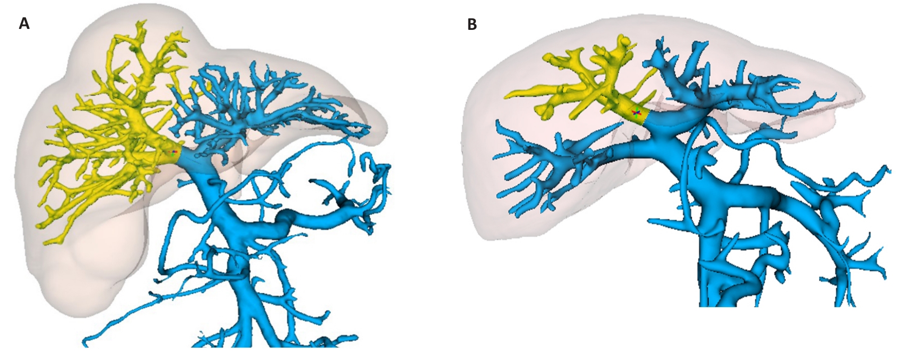
图2 门静脉主干3D分型
Fig.2 3D classification of the main portal vein. A: The left hepatic portal vein has independent origin, and the yellow vessel is the right branch of the portal vein. B: The right anterior branch of the portal vein and the left branch of the portal vein have a common trunk, and the yellow vessel is the right anterior branch of the portal vein.

图3 II段门静脉3D分型
Fig.3 3D classification of portal vein segment II. The yellow vessel is the branch of the portal vein segment II. A: 1 branch. B, C: 2-3 branches.

图4 III段门静脉3D分型
Fig.4 3D classification of segment III of the portal vein. The yellow vessel is the branch of the segment III of the portal vein. A: Branched are distributed in the cephalad and ventral left quadrant. B: Branches are distributed in the foot and ventral left quadrant. C: Branches in both quadrants at the same time.
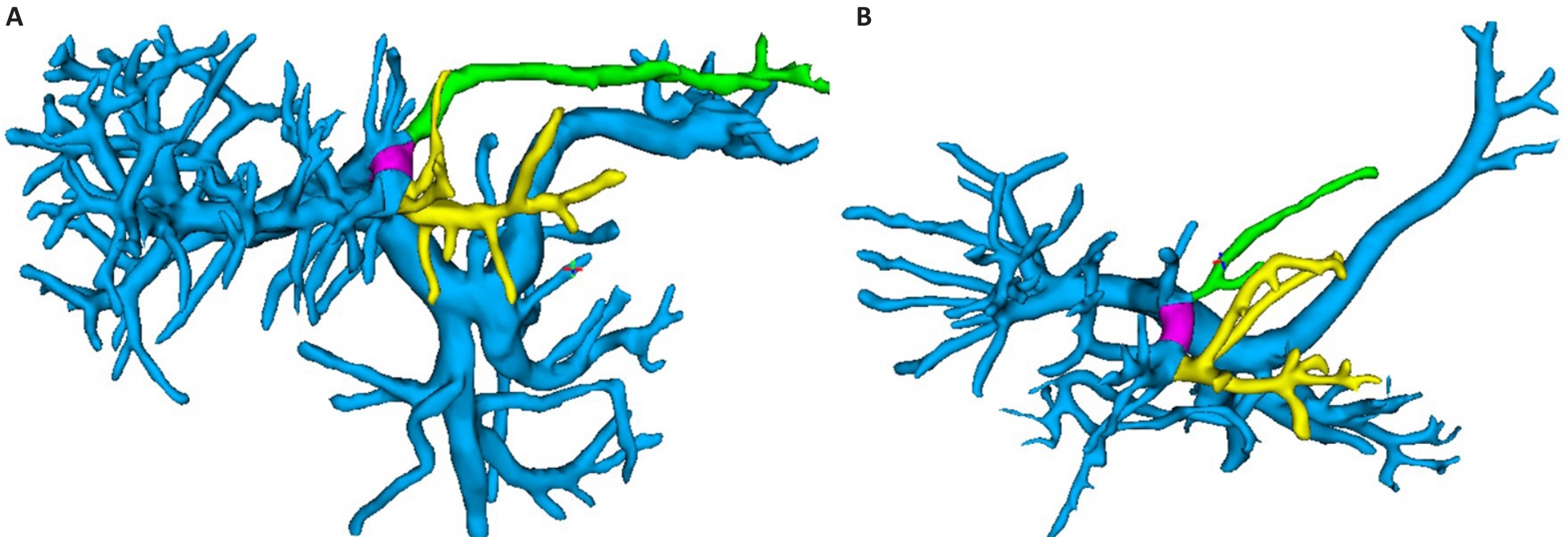
图5 左外叶门静脉3D分型
Fig.5 3D classification of the left lateral lobe of the portal vein. The yellow vessel is the branch of portal vein segment III, the green vessel is the branch of portal vein segment II, and the purple vessel has the shortest distance between them. A: The distance between the origins of the portal vein branch in segments II and III is less than 2 cm. B: The distance is more than 2 cm.

图6 IV段门静脉3D分型
Fig.6 3D classification of portal vein of segment IV. The yellow vessel is from the capsular part of the branch of segment IV, the green vessels is from the sagittal part of the branch of segment IV, and the purple vessel is from the transverse part of the branch of segment IV. A: The branch of the capsular part is clustered and there is no branch in the sagittal part. B: Capsular partial branch + sagittal partial branch. C: Capsular partial branch + sagittal partial branch + transverse partial branch.
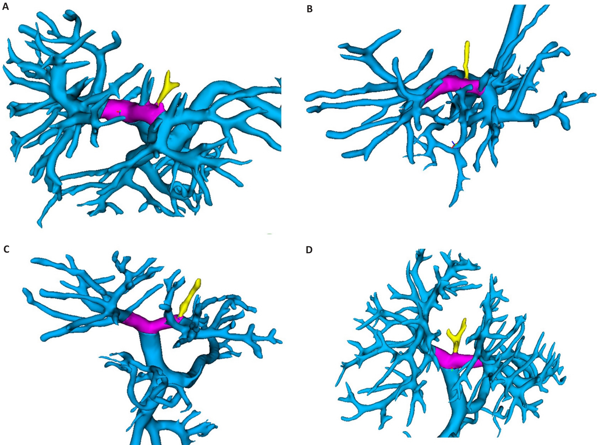
图7 第四肝门血管3D分型
Fig.7 3D classification of the fourth hilar vessel. The yellow vessel is the short portal vessel, and the purple vessel is the transverse portal vein. A: The branches supplying the Spiegel lobes. B: The branches supplying caudate lobulocaval part. C: The branch supplying segment II. D: The branches supplying segment IVa.
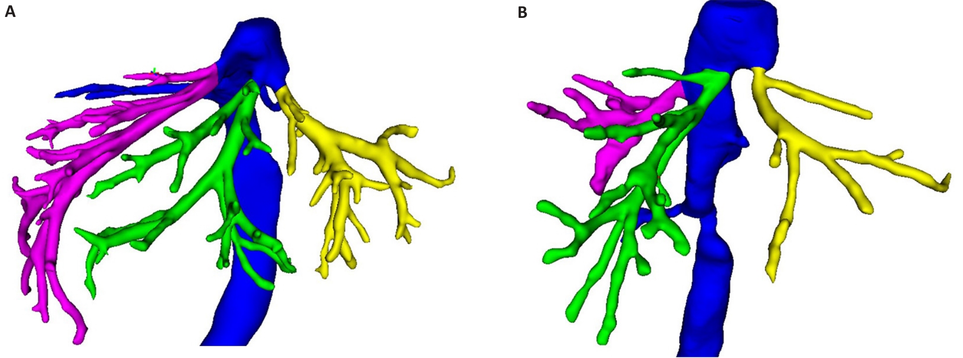
图8 肝静脉3D分型
Fig.8 3D classification of the liver vein. The yellow vessel is the left liver vein, the green vessel is the middle liver vein, and the purple vessel is the right liver vein. A: The left liver vein and the middle liver vein drained into the inferior vena cava. B: The left, middle, and right hepatic veins join the inferior vena cava alone.
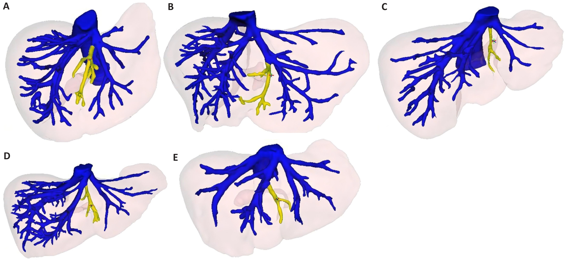
图9 脐裂静脉3D分型
Fig.9 3D classification of the umbilical fissure vein. The yellow vessel is the umbilical fissure vein. A: The umbilical fissure vein merges into the root of the left liver vein within 2 cm. B: The umbilical fissure vein merges 2 cm outside the root of the left hepatic vein. C: Umbilical fissure vein merges into the inferior vena cava. D: The umbilical fissure vein merges into the root of the middle liver vein within 2 cm. E: The umbilical fissure vein merges 2 cm from the root of the middle hepatic vein.
| 1 | Takamoto T, Makuuchi M. Precision surgery for primary liver cancer[J]. Cancer Biol Med, 2019, 16(3): 475-85. |
| 2 | Liao KX, Yang KJ, Cao L, et al. Laparoscopic anatomical versus non-anatomical hepatectomy in the treatment of hepatocellular carcinoma: a randomised controlled trial[J]. Int J Surg, 2022, 102: 106652. |
| 3 | Sugioka A, Kato Y, Tanahashi Y. Systematic extrahepatic Glissonean pedicle isolation for anatomical liver resection based on Laennec's capsule: proposal of a novel comprehensive surgical anatomy of the liver[J]. J Hepatobiliary Pancreat Sci, 2017, 24(1): 17-23. |
| 4 | Zeng N, Tao HS, Fang CH, et al. Individualized preoperative planning using three-dimensional modeling for Bismuth and Corlette type III hilar cholangiocarcinoma[J]. World J Surg Oncol, 2016, 14(1): 44. |
| 5 | Jian Y, Haisu T, Chihua F, et al. Clinical applications of three-dimensional visualization model of arteries supplying the extrahepatic bile duct for patients with biliary obstruction[J]. Am Surg, 2017, 83(1): 8-15. |
| 6 | Cai W, Fan YF, Hu HY, et al. Postoperative liver volume was accurately predicted by a medical image three dimensional visualization system in hepatectomy for liver cancer[J]. Surg Oncol, 2017, 26(2): 188-94. |
| 7 | Lim JSH, Shelat VG. 3D laparoscopy and fluorescence imaging can improve surgical precision for hepatectomy[J]. Hepatobiliary Surg Nutr, 2024, 13(3): 544-7. |
| 8 | Chen H, Wang Y, Xie Z, et al. Application effect of ICG fluorescence real-time imaging technology in laparoscopic hepatectomy[J]. Front Oncol, 2022, 12: 819960. |
| 9 | Zhang P, Luo HL, Zhu W, et al. Real-time navigation for laparoscopic hepatectomy using image fusion of preoperative 3D surgical plan and intraoperative indocyanine green fluorescence imaging[J]. Surg Endosc, 2020, 34(8): 3449-59. |
| 10 | Fujiyama Y, Wakabayashi T, Mishima K, et al. Latest findings on minimally invasive anatomical liver resection[J]. Cancers: Basel, 2023, 15(8): 2218. |
| 11 | 夏穗生, 曾祥熙, 屠頤珠, 等. 肝门外科解剖[J]. 骨科, 1964(2): 81-7. |
| 12 | Yan PN, Tan WF, Yang XW, et al. Applied anatomy of small branches of the portal vein in transverse groove of hepatic hilum[J]. Surg Radiol Anat, 2014, 36(10): 1071-7. |
| 13 | 范应方, 向 飞, 蔡 伟, 等. 基于三维可视化技术的右半肝门静脉3D分型及分段[J]. 南方医科大学学报, 2016, 36(1): 26-31. DOI: 10.3969/j.issn.1673-4254.2016.01.05 |
| 14 | Zhang J, Guo X, Qiao Q, et al. Anatomical study of the hepatic veins in segment 4 of the liver using three-dimensional visualization[J]. Front Surg, 2021, 8: 702280. |
| 15 | Liu ZY, Shen TN, Xia KX, et al. Classification of variant portal vein anatomy based on three-dimensional CT: surgical implications[J]. Surg Radiol Anat, 2024, 46(8): 1177-84. |
| 16 | 禚孝颖, 董 蒨, 吴琳琳, 等. 基于Hisense计算机辅助手术系统的门静脉主干分型的研究[J]. 精准医学杂志, 2019, 34(5): 416-20. DOI: 10.13362/j.jpmed.201905010 |
| 17 | Wang BY, Ou CF, Yu JG, et al. Three-dimensional visual technique based on CT lymphography data combined with methylene blue in endoscopic sentinel lymph node biopsy for breast cancer[J]. Eur J Med Res, 2022, 27(1): 274. |
| 18 | 关明山, 周 波, 韩 娜, 等. 空间封闭点云的八象限三角剖分算法[J]. 哈尔滨理工大学学报, 2009, 14(3): 20-4. DOI: 10.3969/j.issn.1007-2683.2009.03.006 |
| 19 | Cheng YF, Huang TL, Lee TY, et al. Variation of the intrahepatic portal vein; angiographic demonstration and application in living-related hepatic transplantation[J]. Transplant Proc, 1996, 28(3): 1667-8. |
| 20 | Atri M, Bret PM, Fraser-Hill MA. Intrahepatic portal venous variations: prevalence with US[J]. Radiology, 1992, 184(1): 157-8. |
| 21 | 杨陈凤麟, 李 尧, 王 鑫, 等.“门理论”在微创解剖性肝切除术中的应用与展望 [J].中国实用外科杂志, 2024, 44(3): 341-5. |
| 22 | Cazauran JB, Pâris L, Rousset P, et al. Anatomy of the right anterior sector of the liver and its clinical implications in surgery[J]. J Gastrointest Surg, 2018, 22(10): 1819-31. |
| 23 | Zhang K, Zhang SG, Jiang Y, et al. Laparoscopic hepatic left lateral lobectomy combined with fiber choledochoscopic exploration of the common bile duct and traditional open operation[J]. World J Gastroenterol, 2008, 14(7): 1133-6. |
| 24 | 李 斌, 姜小清. 重视“门短静脉” 解剖在围肝门手术中的意义[J]. 中国实用外科杂志, 2019, 39(2): 145-8. |
| 25 | 李 斌, 邱智泉, 闫培宁, 等. “第四肝门” 在围肝门外科的临床意义[J]. 中国普外基础与临床杂志, 2016, 23(11): 1308-10. |
| 26 | Goldsmith NA, Woodburne RT. The surgical anatomy pertaining to liver resection[J]. Surg Gynecol Obstet, 1957, 105(3): 310-8. |
| 27 | Nakamura S, Tsuzuki T. Surgical anatomy of the hepatic veins and the inferior vena Cava [J]. Surg Gynecol Obstet, 1981, 152(1): 43-50. |
| 28 | Tani K, Shindoh J, Akamatsu N, et al. Venous drainage map of the liver for complex hepatobiliary surgery and liver transplantation[J]. HPB, 2016, 18(12): 1031-8. |
| 29 | Kawasaki S, Makuuchi M, Miyagawa S, et al. Extended lateral segmentectomy using intraoperative ultrasound to obtain a partial liver graft[J]. Am J Surg, 1996, 171(2): 286-8. |
| 30 | 董家鸿, 叶 晟. 开启精准肝胆外科的新时代[J]. 中华普外科手术学杂志: 电子版, 2016, 10(3): 181-4. DOI: 10.3877/cma.j.issn.1674-3946.2016.03.001 |
| 31 | Kobayashi K, Hasegawa K, Kokudo T, et al. Extended segmentectomy II to left hepatic vein: importance of preserving umbilical fissure vein to avoid congestion of segment III[J]. J Am Coll Surg, 2017, 225(3): e5-11. |
| [1] | 曾 宁,杨 剑 ,项 楠,文 赛,曾思略,齐 硕,祝 文,胡浩宇,方驰华. 三维可视化联合3D打印在Bismuth-Corlette III、IV型肝门部胆管癌个体化精准外科治疗中的应用[J]. 南方医科大学学报, 2020, 40(08): 1172-1177. |
| [2] | 中华医学会数字医学分会;中国医师协会肝癌专业委员会;中国医师协会临床精准医学专业委员会;中国研究型医院学会数字智能化外科专业委员会. 复杂性肝脏肿瘤切除三维可视化精准诊治指南(2019版)[J]. 南方医科大学学报, 2020, 40(03): 297-307. |
| [3] | 方驰华. 计算机辅助联合吲哚菁绿分子荧光影像技术在肝脏肿瘤诊断和手术导航中的应用指南(2019版)[J]. 南方医科大学学报, 2019, 39(10): 1127-. |
| [4] | 方驰华,方兆山,范应方,李鉴轶,向飞,陶海粟. 三维可视化、3D打印及3D腹腔镜在肝肿瘤外科诊治中的应用[J]. 南方医科大学学报, 2015, 35(05): 639-. |
| [5] | 范应方,方驰华,陈建新,项楠,杨剑,李克晓. 三维可视化技术在精准肝胆管结石诊治中的应用[J]. 南方医科大学学报, 2011, 31(06): 949-. |
| 阅读次数 | ||||||
|
全文 |
|
|||||
|
摘要 |
|
|||||