南方医科大学学报 ›› 2025, Vol. 45 ›› Issue (11): 2483-2495.doi: 10.12122/j.issn.1673-4254.2025.11.21
陶露1,2( ), 陈悦2, 黄林林2, 郑旺2, 宋雪1,2, 项平1,2, 胡建国2()
收稿日期:2025-04-28
出版日期:2025-11-20
发布日期:2025-11-28
通讯作者:
胡建国
E-mail:bytaolu@bbmu.edu.cn;jghu9200@bbmu.edu.cn
作者简介:陶 露,硕士,研究实习员,E-mail: bytaolu@bbmu.edu.cn
基金资助:
Lu TAO1,2(), Yue CHEN2, Linlin HUANG2, Wang ZHENG2, Xue SONG1,2, Ping XIANG1,2, Jianguo HU2()
Received:2025-04-28
Online:2025-11-20
Published:2025-11-28
Contact:
Jianguo HU
E-mail:bytaolu@bbmu.edu.cn;jghu9200@bbmu.edu.cn
摘要:
目的 探讨天然化合物珠子草素(NIR)对克罗恩病样结肠炎的作用及其分子机制。 方法 采用2,4,6-三硝基苯磺酸(TNBS)诱导小鼠建立结肠炎模型,随机分为4组:WT组注射生理盐水;WT+NIR组腹腔注射NIR(10 mg/kg,1次/d,注射7 d),TNBS组用2.5% TNBS造模并给予等体积的生理盐水;TNBS+NIR组用2.5% TNBS造模并腹腔注射NIR(10 mg/kg,1次/d,注射7 d),6只/组。用体质量变化、疾病活动指数(DAI)和结肠长度评估NIR的治疗效果。ELISA法和实时定量PCR(qRT-PCR)检测肠黏膜组织炎症因子(IL-6、IL-1β、TNF-α、IL-17A和IL-10)水平。TUNEL染色和Western blotting检测肠上皮细胞凋亡情况及相关蛋白(Bcl-2/Bax)的表达。Western blotting评估紧密连接蛋白(TJ)(ZO-1、Claudin-1)和p38/JNK通路的活化水平,并通过Diprovocim干预实验验证NIR的调控分子机制。 结果 NIR干预后TNBS小鼠体质量增加,DAI和组织学炎症评分减低,结肠长度增加(P<0.05);ELISA和qRT-PCR结果表明NIR可降低促炎因子(IL-6,IL-1β、IL-17A和TNF-α)的蛋白和mRNA水平,上调抗炎因子IL-10表达水平(P<0.05);TUNEL和Western blotting检测显示NIR可抑制肠上皮细胞凋亡,激活抗凋亡通路(P<0.05);Western blotting结果证实NIR可上调ZO-1和Claudin-1的表达水平,并下调p38和JNK的磷酸化水平(P<0.05);Diprovocim干预可衰减NIR对p38/JNK通路的失活作用。 结论 NIR可通过调控p38/JNK信号的活化抑制肠上皮细胞凋亡,从而改善小鼠CD样肠炎。
陶露, 陈悦, 黄林林, 郑旺, 宋雪, 项平, 胡建国. 珠子草素通过调控p38/JNK信号通路抑制肠上皮细胞凋亡保护肠屏障改善克罗恩病样肠炎[J]. 南方医科大学学报, 2025, 45(11): 2483-2495.
Lu TAO, Yue CHEN, Linlin HUANG, Wang ZHENG, Xue SONG, Ping XIANG, Jianguo HU. Niranthin ameliorates Crohn's disease-like enteritis in mice by inhibiting intestinal epithelial cell apoptosis and protecting intestinal barrier via modulating p38/JNK signaling[J]. Journal of Southern Medical University, 2025, 45(11): 2483-2495.
| Gene | Primer sequences (5'-3') |
|---|---|
| TNF-α | F: CACGCTCTTCTGTCTACTGAACTTC |
| R: CTTGGTGGTTTGTGAGTGTGAGG | |
| IL-1β | F: AATCTCGCAGCAGCACATCAAC |
| R: AGGTCCACGGGAAAGACACAG | |
| IL-6 | F: GAGAGGAGACTTCACAGAGGATACC |
| R: TCATTTCCACGATTTCCCAGAGAAC | |
| IL-10 | F: GGACAACATACTGCTAACCGACTC |
| R: GGGCATCACTTCTACCAGGTAAAAC | |
| IL-17A | F: TGGCGGCTACAGTGAAGGC |
| R: AGGGAGTTAAAGACTTTGAGGTTGAC | |
| GAPDH | F: AACTCCCACTCTTCCACCTTCG |
| R: TCCACCACCCTGTTGCTGTAG |
表1 引物序列
Tab.1 Primer sequences for qRT-PCR in this study
| Gene | Primer sequences (5'-3') |
|---|---|
| TNF-α | F: CACGCTCTTCTGTCTACTGAACTTC |
| R: CTTGGTGGTTTGTGAGTGTGAGG | |
| IL-1β | F: AATCTCGCAGCAGCACATCAAC |
| R: AGGTCCACGGGAAAGACACAG | |
| IL-6 | F: GAGAGGAGACTTCACAGAGGATACC |
| R: TCATTTCCACGATTTCCCAGAGAAC | |
| IL-10 | F: GGACAACATACTGCTAACCGACTC |
| R: GGGCATCACTTCTACCAGGTAAAAC | |
| IL-17A | F: TGGCGGCTACAGTGAAGGC |
| R: AGGGAGTTAAAGACTTTGAGGTTGAC | |
| GAPDH | F: AACTCCCACTCTTCCACCTTCG |
| R: TCCACCACCCTGTTGCTGTAG |
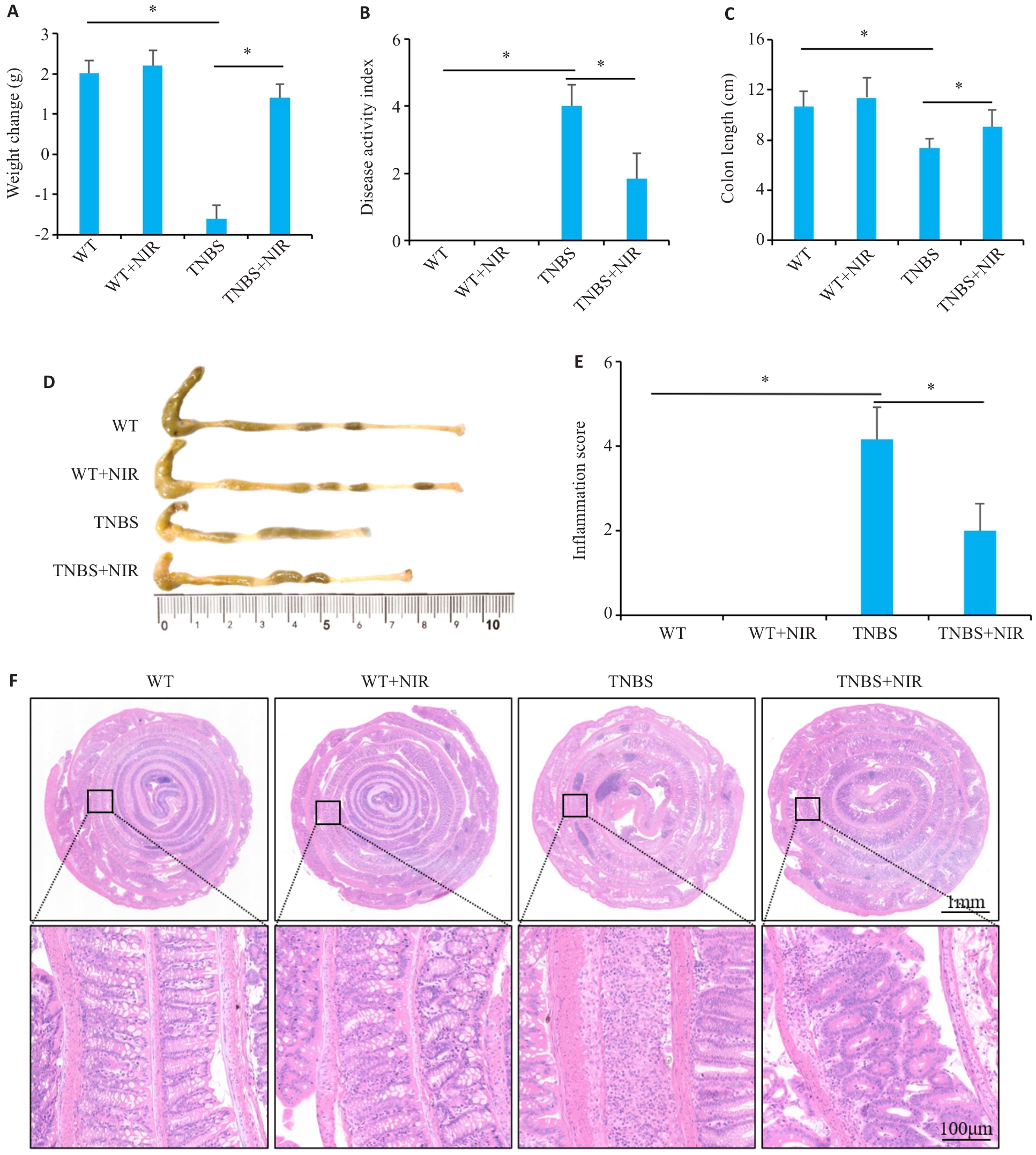
图1 NIR干预对TNBS诱导小鼠结肠炎症状的影响
Fig.1 Effect of niranthin (NIR) treatment on symptoms of TNBS-induced colitis in mice. A: Body weight changes of the mice. B: DAI score. C: Colon length. D: Comparison of colon length of the mice among the 4 groups. E: Colon inflammation score of the mice. F: HE staining of the colon tissues of the mice in the 4 groups.*P<0.05.
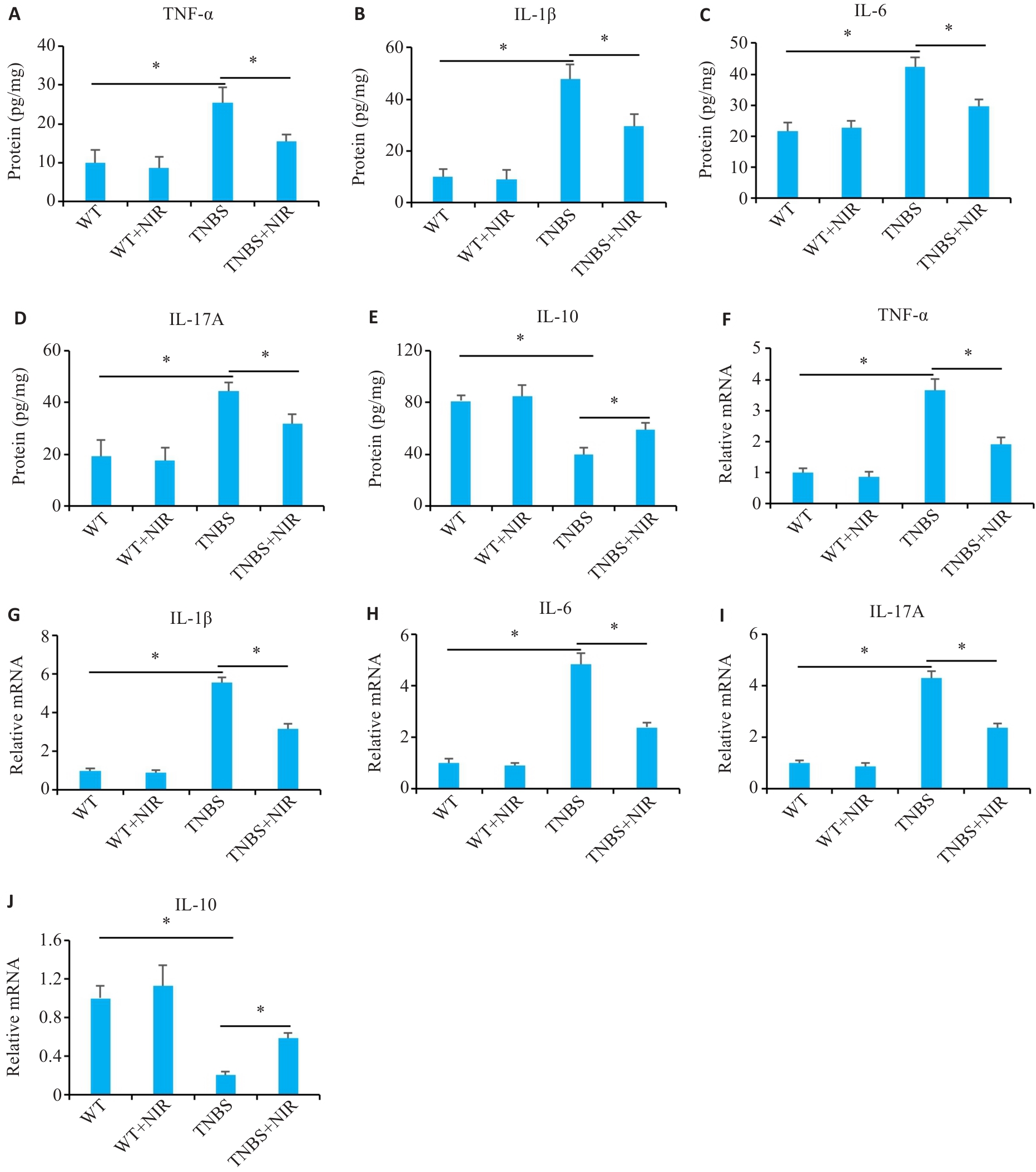
图2 NIR干预对TNBS诱导小鼠结肠炎炎症因子水平的影响
Fig.2 Effect of NIR treatment on inflammatory factor levels in mice with TNBS-induced colitis. A-E: Results of ELISA for detecting the levels of IL-6, IL-1β, IL-17A, TNF-α and IL-10 in the colon tissue of TNBS-induced mice. F-J: qRT-PCR for detecting IL-6, IL-1β, IL-17A, TNF‑α and IL-10 mRNA expressions in the colon tissues of TNBS-induced mice. *P<0.05.
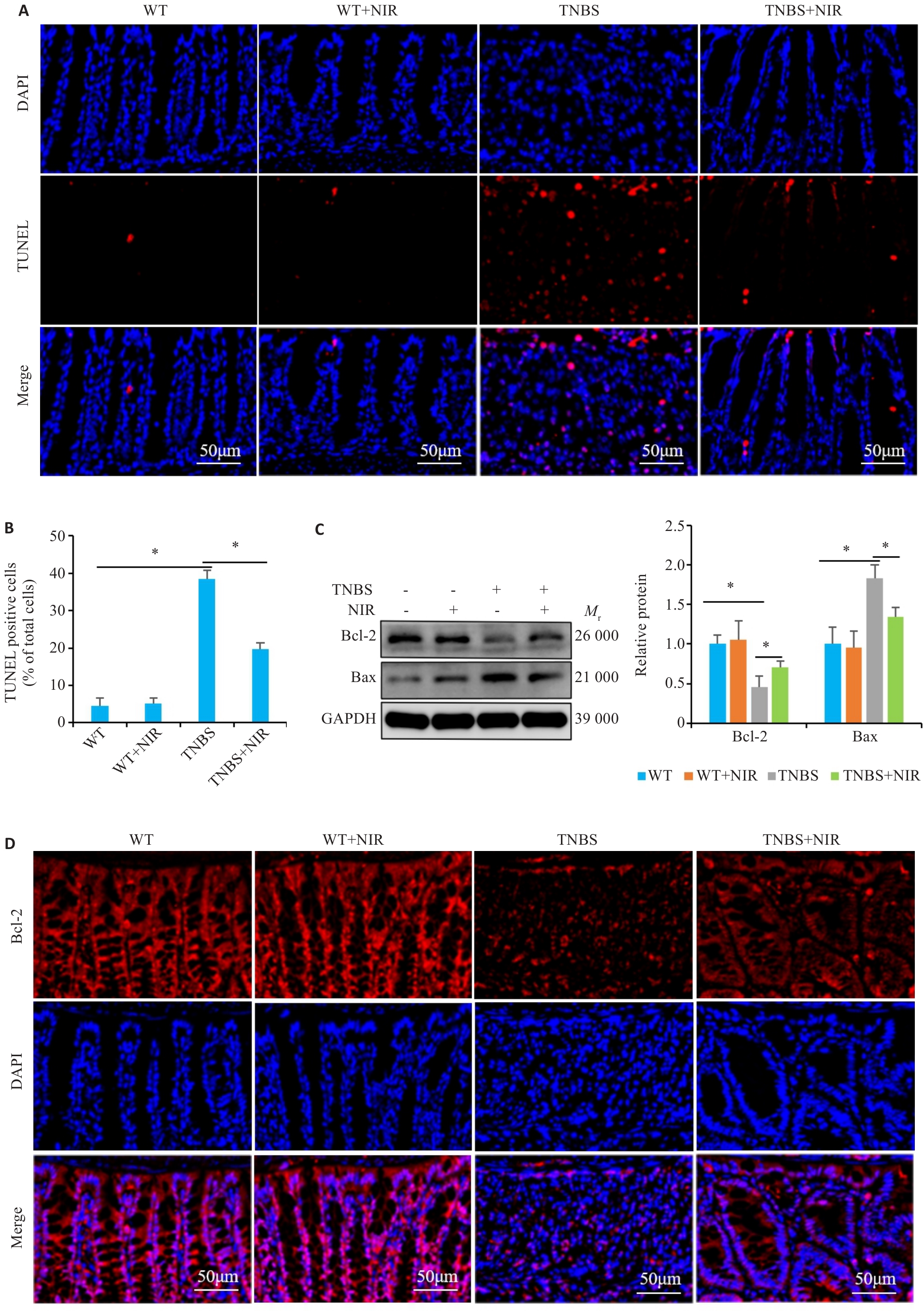
图3 NIR干预对TNBS诱导小鼠结肠炎肠上皮细胞凋亡的影响
Fig.3 Effect of NIR treatment on TNBS-induced apoptosis of intestinal epithelial cells in the mouse models of colitis. A, B: TUNEL staining for detecting apoptosis in TNBS-induced mice with NIR treatment. C: Western blotting for detecting the expression of Bcl-2 and Bax proteins in TNBS-induced mice with NIR treatment. D: Immunofluorescence staining for detecting Bcl-2 expression in TNBS-induced mice with NIR treatment. *P<0.05.

图4 NIR干预对TNBS诱导小鼠结肠炎肠屏障的影响
Fig.4 Effect of NIR treatment on intestinal barrier function in mice with TNBS-induced colitis. A: Immunofluorescence staining for detecting ZO-1 and Claudin-1 expression in TNBS-induced mice with NIR treatment. B: Western blotting for detecting ZO-1 and claudin-1 protein expressions in TNBS-induced mice with NIR treatment. *P<0.05.
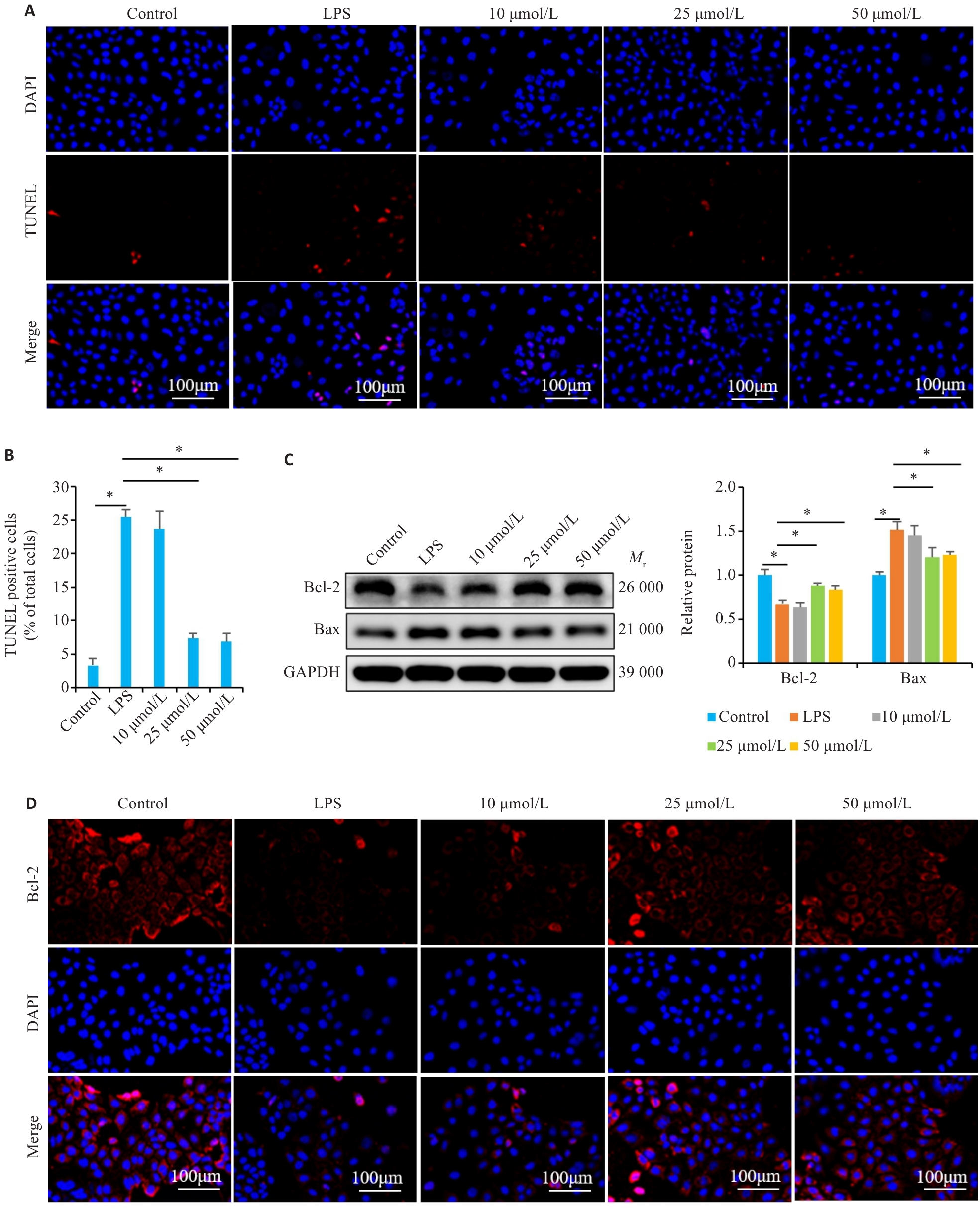
图5 NIR干预对LPS诱导Caco-2细胞凋亡的影响
Fig.5 Effect of NIR treatment on LPS-induced apoptosis in Caco-2 cells. A, B: TUNEL staining for detecting LPS-induced apoptosis of Caco-2 cells with NIR treatment. C: Western blotting for detecting the expression of Bcl-2 and Bax proteins in LPS-induced Caco-2 cells with NIR treatment. D: Immunofluorescence staining for detecting Bcl-2 expression in LPS-induced Caco-2 cells with NIR treatment. *P<0.05.
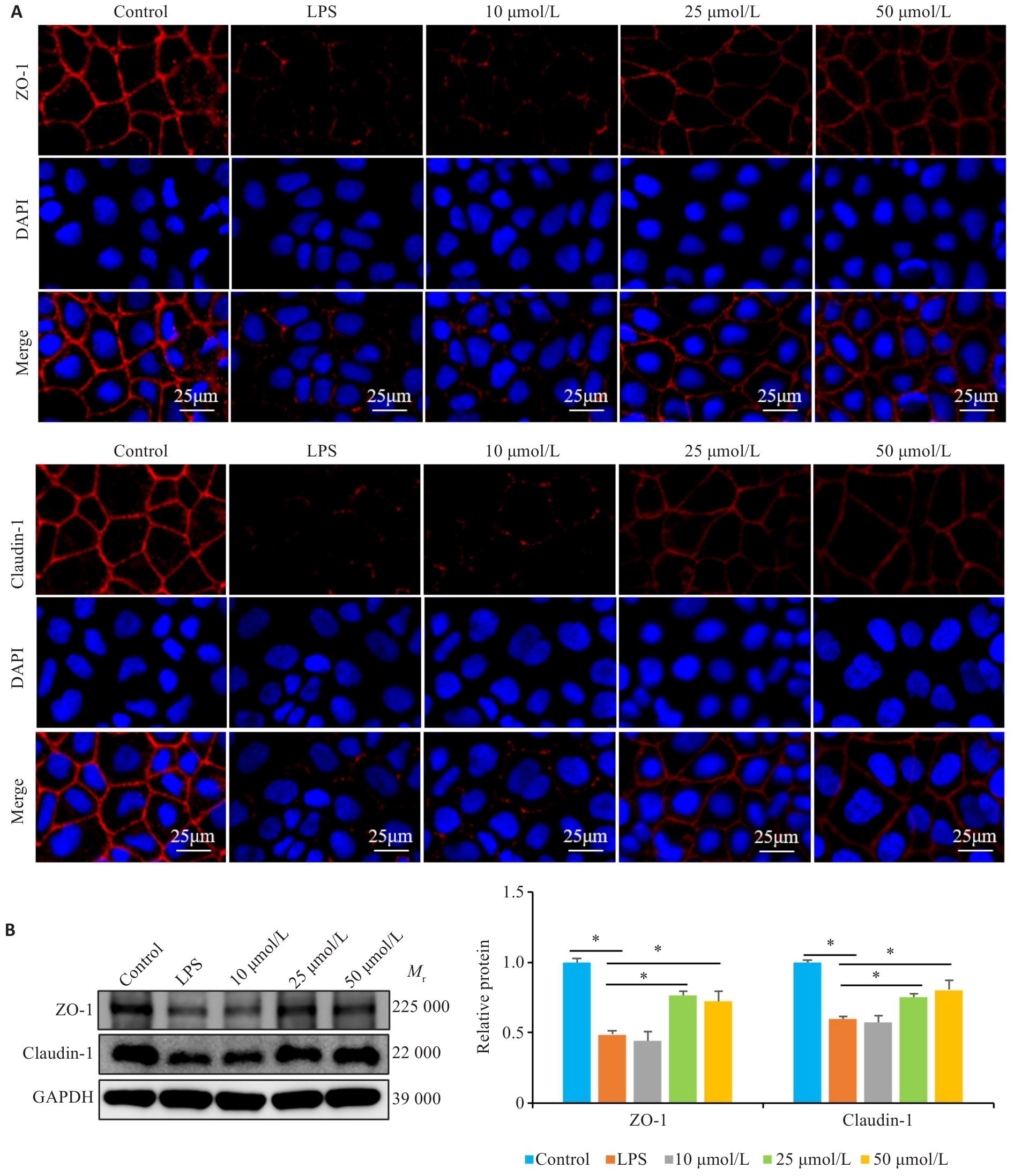
图6 NIR干预对LPS诱导Caco-2细胞TJ蛋白表达的影响
Fig. 6 Effect of NIR treatment on expressions of tight junction proteins in LPS-induced Caco-2 cells. A: Immunofluorescence staining showing ZO-1 and claudin-1 expressions in LPS-induced Caco-2 cells with NIR treatment. B: Western blotting for detecting ZO-1 and claudin-1 protein expressions in LPS-induced Caco-2 cells with NIR treatment. *P<0.05.
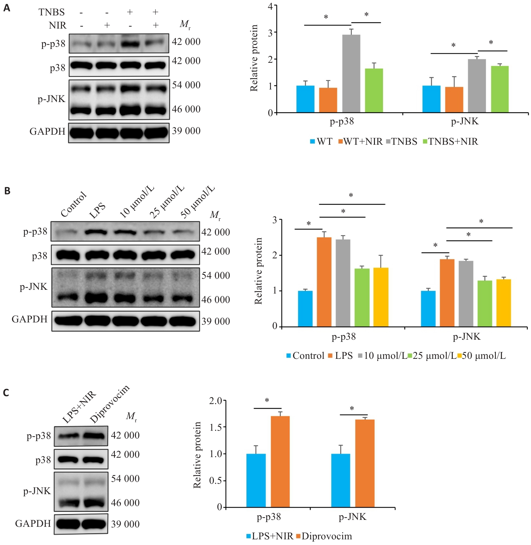
图7 NIR干预可调控p38/JNK信号
Fig. 7 NIR treatment modulates p38/JNK signaling. A: Western blotting for detecting p-p38 and p-JNK protein expressions in TNBS-induced mice with NIR treatment. B: Western blotting for detecting p-p38 and p-JNK protein expressions in LPS-induced Caco-2 cells with NIR treatment. C: Western blotting for detecting p-p38 and p-JNK protein expressions in the colon tissues of the mice with NIR and diprovocim treatment. *P<0.05.
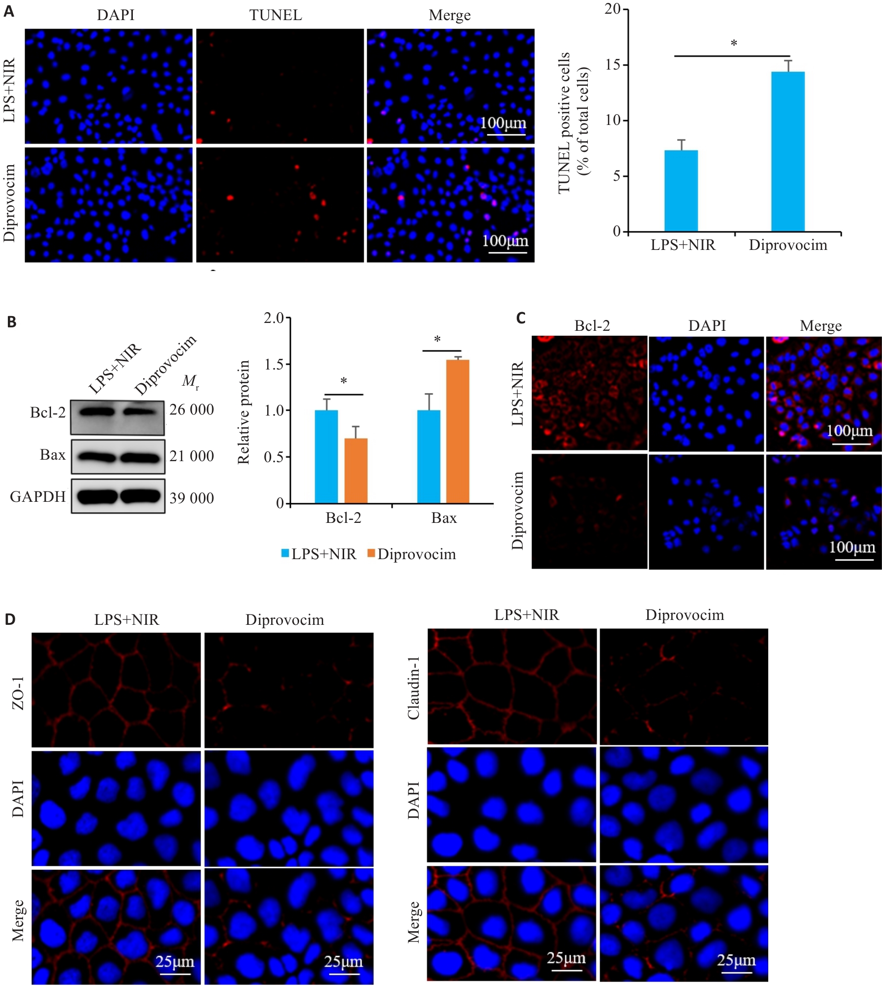
图8 Diprovocim干预对NIR治疗Caco-2细胞中凋亡和TJ蛋白表达的影响
Fig. 8 Effects of diprovocim intervention on apoptosis and expressions of tight junction proteins in NIR-treated Caco-2 cells. A: TUNEL staining for detecting apoptosis in NIR-treated Caco-2 cells by Diprovocim treatment. B: Western blotting for detecting the expression of Bcl-2 and Bax proteins in NIR-treated Caco-2 cells with diprovocim treatment. C, D: Immunofluorescence staining for detecting Bcl-2, ZO-1 and Claudin-1 expression in NIR-treated Caco-2 cells with diprovocim treatment. *P<0.05.
| [1] | Dolinger M, Torres J, Vermeire S. Crohn’s disease[J]. Lancet, 2024, 403(10432): 1177-91. doi:10.1016/s0140-6736(23)02586-2 |
| [2] | Loftus EV Jr, Panés J, Lacerda AP, et al. Upadacitinib induction and maintenance therapy for Crohn’s disease[J]. N Engl J Med, 2023, 388(21): 1966-80. doi:10.1056/nejmoa2212728 |
| [3] | Honap S, Jairath V, Danese S, et al. Navigating the complexities of drug development for inflammatory bowel disease[J]. Nat Rev Drug Discov, 2024, 23(7): 546-62. doi:10.1038/s41573-024-00953-0 |
| [4] | Wang L, Song X, Zhou YQ, et al. Sclareol protected against intestinal barrier dysfunction ameliorating Crohn’s disease-like colitis via Nrf2/NF-B/MLCK signalling[J]. Int Immunopharmacol, 2024, 133: 112140. doi:10.1016/j.intimp.2024.112140 |
| [5] | Zhou L, Zhu L, Wu X, et al. Decreased TMIGD1 aggravates colitis and intestinal barrier dysfunction via the BANF1-NF-κB pathway in Crohn’s disease[J]. BMC Med, 2023, 21(1): 287. doi:10.1186/s12916-023-02989-2 |
| [6] | Xu S, Peng YF, Yang K, et al. PROTAC based STING degrader attenuates acute colitis by inhibiting macrophage M1 polarization and intestinal epithelial cells pyroptosis mediated by STING-NLRP3 axis[J]. Int Immunopharmacol, 2024, 141: 112990. doi:10.1016/j.intimp.2024.112990 |
| [7] | Tan G, Huang C, Chen J, et al. HMGB1 released from GSDME-mediated pyroptotic epithelial cells participates in the tumorigenesis of colitis-associated colorectal cancer through the ERK1/2 pathway[J]. J Hematol Oncol, 2020, 13(1): 149. doi:10.1186/s13045-020-00985-0 |
| [8] | Liu J, Di B, Xu LL. Recent advances in the treatment of IBD: targets, mechanisms and related therapies[J]. Cytokine Growth Factor Rev, 2023, 71: 1-12. doi:10.1016/j.cytogfr.2023.07.001 |
| [9] | Veyrard P, Nancey S, Roblin X. Editorial: 5-ASA in IBD patients on biologics-' stop or continue'[J]? Aliment Pharmacol Ther, 2021, 54(6): 843-4. doi:10.1111/apt.16541 |
| [10] | Sridhar A, Bakke I, Gopalakrishnan S, et al. Tofacitinib and budesonide treatment affect stemness and chemokine release in IBD patient-derived colonoids[J]. Sci Rep, 2025, 15(1): 3753. doi:10.1038/s41598-025-86314-2 |
| [11] | Biologic therapy for inflammatory bowel disease: real-world comparative effectiveness and impact of drug sequencing in 13 222 patients within the UK IBD BioResource[J]. J Crohns Colitis, 2024, 18(6): 790-800. doi:10.1093/ecco-jcc/jjad212.1168 |
| [12] | Ota R, Karasawa D, Oshima M, et al. Asymmetric total synthesis of four bioactive lignans using donor–acceptor cyclopropanes and bioassay of (-)- and (+)-niranthin against hepatitis B and influenza viruses[J]. RSC Adv, 2022, 12(8): 4635-9. doi:10.1039/d2ra00499b |
| [13] | Harikrishnan H, Jantan I, Alagan A, et al. Modulation of cell signaling pathways by Phyllanthus amarus and its major constituents: potential role in the prevention and treatment of inflammation and cancer[J]. Inflammopharmacology, 2020, 28(1): 1-18. doi:10.1007/s10787-019-00671-9 |
| [14] | Chowdhury S, Mukherjee T, Mukhopadhyay R, et al. The lignan niranthin poisons Leishmania donovani topoisomerase IB and favours a Th1 immune response in mice[J]. EMBO Mol Med, 2012, 4(10): 1126-43. doi:10.1002/emmm.201201316 |
| [15] | Harikrishnan H, Jantan I, Haque MA, et al. Anti-inflammatory effects of hypophyllanthin and niranthin through downregulation of NF-κB/MAPKs/PI3K-Akt signaling pathways[J]. Inflammation, 2018, 41(3): 984-95. doi:10.1007/s10753-018-0752-4 |
| [16] | Tan G, Huang CY, Chen JY, et al. Gasdermin-E-mediated pyroptosis participates in the pathogenesis of Crohn’s disease by promoting intestinal inflammation[J]. Cell Rep, 2021, 35(11): 109265. doi:10.1016/j.celrep.2021.109265 |
| [17] | Spencer DM, Veldman GM, Banerjee S, et al. Distinct inflammatory mechanisms mediate early versus late colitis in mice[J]. Gastroenterology, 2002, 122(1): 94-105. doi:10.1053/gast.2002.30308 |
| [18] | Rath HC, Herfarth HH, Ikeda JS, et al. Normal luminal bacteria, especially Bacteroides species, mediate chronic colitis, gastritis, and arthritis in HLA-B27/human beta2 microglobulin transgenic rats[J]. J Clin Invest, 1996, 98(4): 945-53. doi:10.1172/jci118878 |
| [19] | Morin MD, Wang Y, Jones BT, et al. Diprovocims: A New and Exceptionally Potent Class of Toll-like Receptor Agonists[J]. J Am Chem Soc, 2018, 140(43):14440-54. doi:10.1021/jacs.8b09223 |
| [20] | Wei ZY, Ni X, Cui H, et al. Engeletin attenuates the inflammatory response via inhibiting TLR4-NFκB signaling pathway in Crohn’s disease-like colitis[J]. J Ethnopharmacol, 2025, 336: 118733. doi:10.1016/j.jep.2024.118733 |
| [21] | Mehandru S, Colombel JF. The intestinal barrier, an arbitrator turned provocateur in IBD[J]. Nat Rev Gastroenterol Hepatol, 2021, 18(2): 83-4. doi:10.1038/s41575-020-00399-w |
| [22] | Odenwald MA, Turner JR. The intestinal epithelial barrier: a therapeutic target[J]? Nat Rev Gastroenterol Hepatol, 2017, 14(1): 9-21. doi:10.1038/nrgastro.2016.169 |
| [23] | Huang S, Xie Z, Han J, et al. Protocadherin 20 maintains intestinal barrier function to protect against Crohn’s disease by targeting ATF6[J]. Genome Biol, 2023, 24(1): 159. doi:10.1186/s13059-023-02991-0 |
| [24] | Safari F, Sharifi M, Talebi A, et al. Alleviation of cholestatic liver injury and intestinal permeability by lubiprostone treatment in bile duct ligated rats: role of intestinal FXR and tight junction proteins claudin-1, claudin-2, and occludin[J]. Naunyn Schmiedeberg’s Arch Pharmacol, 2023, 396(9): 2009-22. doi:10.1007/s00210-023-02455-z |
| [25] | Pan Z, Huang J, Hu T, et al. Protective effects of selenium nanoparticles against bisphenol A-induced toxicity in porcine intestinal epithelial cells[J]. Int J Mol Sci, 2023, 24(8): 7242. doi:10.3390/ijms24087242 |
| [26] | Yin M, Shen Z, Yang L, et al. Protective effects of CXCR3/HO-1 gene-modified BMMSCs on damaged intestinal epithelial cells: Role of the p38-MAPK signaling pathway[J]. Int J Mol Med, 2019, 43(5): 2086-102. |
| [27] | Zeng ZW, Shi YM, Cai YH, et al. PHLDA1 protects intestinal barrier function via restricting intestinal epithelial cells apoptosis in inflammatory bowel disease[J]. Exp Cell Res, 2024, 443(1): 114322. doi:10.1016/j.yexcr.2024.114322 |
| [28] | Geng Z, Zuo L, Li J, et al. Ginkgetin improved experimental colitis by inhibiting intestinal epithelial cell apoptosis through EGFR/PI3K/AKT signaling[J]. FASEB J, 2024, 38(14): e23817. doi:10.1096/fj.202400211rr |
| [29] | Xu Q, Liu M, Chao X, et al. Stevioside improves antioxidant capacity and intestinal barrier function while attenuating inflammation and apoptosis by regulating the NF-κB/MAPK pathways in diquat-induced oxidative stress of IPEC-J2 cells[J]. Antioxidants: Basel, 2023, 12(5): 1070. doi:10.3390/antiox12051070 |
| [30] | Xing Z, Li X, He J, et al. OLFM4 modulates intestinal inflammation by promoting IL-22+ILC3 in the gut[J]. Commun Biol, 2024, 7(1): 914. doi:10.1038/s42003-024-06601-y |
| [31] | Xiao L, Zhang WH, Huang Y, et al. Intestinal ischemia-reperfusion induces the release of IL-17A to regulate cell inflammation, apoptosis and barrier damage[J]. Exp Ther Med, 2022, 23(2): 158. doi:10.3892/etm.2021.11081 |
| [32] | Chu C, Ru H, Chen Y, et al. Gallic acid attenuates LPS-induced inflammation in Caco-2 cells by suppressing the activation of the NF-κB/MAPK signaling pathway[J]. Acta Biochim Biophys Sin: Shanghai, 2024, 56(6): 905-15. |
| [33] | Tao H, Bao Z, Fu Z, et al. Chlorothalonil induces the intestinal epithelial barrier dysfunction in Caco-2 cell-based in vitro monolayer model by activating MAPK pathway[J]. Acta Biochim Biophys Sin: Shanghai, 2021, 53(11): 1459-68. doi:10.1093/abbs/gmab125 |
| [1] | 牛民主, 殷丽霞, 乔通, 尹林, 张可妮, 胡建国, 宋传旺, 耿志军, 李静. 旱莲苷A通过调控JAK2/STAT3通路抑制M1型巨噬细胞极化改善葡聚糖硫酸钠诱导的小鼠结肠炎[J]. 南方医科大学学报, 2025, 45(6): 1297-1306. |
| [2] | 储菲, 陈孝华, 宋博文, 杨晶晶, 左芦根. 苏荠宁黄酮通过抑制PI3K/AKT信号通路拮抗肠上皮细胞凋亡改善小鼠实验性结肠炎[J]. 南方医科大学学报, 2025, 45(4): 819-828. |
| [3] | 殷丽霞, 牛民主, 张可妮, 耿志军, 胡建国, 李江艳, 李静. 升麻素抑制MAPK通路调节辅助性T细胞免疫平衡改善小鼠克罗恩病样结肠炎[J]. 南方医科大学学报, 2025, 45(3): 595-602. |
| [4] | 黄菊, 殷丽霞, 牛民主, 耿志军, 左芦根, 李静, 胡建国. 紫花前胡苷通过抑制肠上皮细胞焦亡改善2,4,6-三硝基苯磺酸诱导的小鼠实验性结肠炎[J]. 南方医科大学学报, 2025, 45(2): 261-268. |
| [5] | 黄晴晴, 杨晶晶, 姜雪凝, 张文静, 汪煜, 左芦根, 王炼, 王月月, 张小凤, 宋雪, 胡建国. 刺桐碱通过抑制肠上皮炎症反应并改善肠屏障功能缓解小鼠克罗恩病样结肠炎[J]. 南方医科大学学报, 2025, 45(11): 2456-2465. |
| [6] | 刘硕, 李静, 吴兴旺. Swertiamarin通过抑制肠上皮细胞细胞凋亡改善TNBS诱导的实验性结肠炎[J]. 南方医科大学学报, 2024, 44(8): 1545-1552. |
| [7] | 席 进, 张 敏, 张永玉, 张 晨, 张雨路, 王 锐, 申 林, 李 静, 宋 雪. 上调KLF11可改善结肠炎模型小鼠的肠道炎症:基于抑制JAK2/STAT3信号通路[J]. 南方医科大学学报, 2024, 44(4): 765-772. |
| [8] | 牛民主, 殷丽霞, 段婷, 黄菊, 李静, 耿志军, 胡建国, 宋传旺. 川续断皂苷VI通过抑制PI3K/AKT/NF-κB通路拮抗肠上皮细胞凋亡缓解TNBS诱导的小鼠克罗恩病样结肠炎[J]. 南方医科大学学报, 2024, 44(12): 2335-2346. |
| [9] | 邵荣瑢, 杨 子, 张文静, 张 诺, 赵雅静, 张小凤, 左芦根, 葛思堂. 茯苓酸缓解小鼠克罗恩病:基于抑制PI3K/AKT信号通路拮抗肠上皮细胞凋亡[J]. 南方医科大学学报, 2023, 43(6): 935-942. |
| [10] | 杨 子, 赵天豪, 程 阳, 周约青, 李岳彤, 王欣茹, 张小凤, 左芦根, 葛思堂. 香叶木素通过调节小鼠的肠道免疫平衡减轻克罗恩病样结肠炎:基于抑制PI3K/AKT通路[J]. 南方医科大学学报, 2023, 43(3): 474-482. |
| [11] | 肖姝喆, 成燕玲, 朱 云, 唐 芮, 古建标, 兰 林, 何志华, 刘丹琼, 耿岚岚, 程 旸, 龚四堂. WNT2b高表达的成纤维细胞破坏肠道黏膜屏障[J]. 南方医科大学学报, 2023, 43(2): 206-212. |
| [12] | 高 倩,董红霞,李 瑾. 52周英夫利西疗程中第14周中性粒细胞与淋巴细胞比值可预测克罗恩患者是否对治疗产生失应答[J]. 南方医科大学学报, 2020, 40(04): 453-458. |
| [13] | 姜从桥,朱平胜,时依,项武军,葛思堂,张宗兵,左芦根. 原花青素B2 对三硝基苯磺酸结肠炎模型小鼠肠炎及肠屏障的保护作用[J]. 南方医科大学学报, 2019, 39(07): 778-. |
| [14] | 朱振浩,邱琛,张明,陈昭,向城,王新颖. 英夫利西单抗治疗的小肠克罗恩病患者不同肠段黏膜愈合情况分析[J]. 南方医科大学学报, 2017, 37(01): 44-. |
| [15] | 戴世学,顾红祥,武钢,钟涛,菅洪健,湛永乐,张旻海,高勇,徐俊,陈东升,廖广捷,封艳玲,刘洪波, 邹颖,迟宏罡. CD8+CD28+/CD8+CD28- T细胞平衡预测炎症性肠病患者并发消化道出血的价值[J]. 南方医科大学学报, 2016, 36(12): 1609-. |
| 阅读次数 | ||||||
|
全文 |
|
|||||
|
摘要 |
|
|||||