2. 南昌市公安局刑事侦查大队,江西 南昌 330000
2. Criminal Investigation Brigade of Nanchang Public Security Bureau, Nanchang 330000, China
死亡时间(PMI)推断是国内外法医学界研究的热点之一。准确的PMI推断对于排查犯罪嫌疑人、划定侦查范围以及降低案件侦破成本等具有重要意义。目前,PMI推断主要根据角膜混浊、尸斑、尸冷、尸僵等尸体现象的发展程度来粗略估计[1-3],对指导法医实践工作效果并不理想。有学者们从分子层面探索客观可量化的PMI推断方法,如利用死后核酸[4]、氨基酸[5]及诸多离子[6]的含量变化规律推断PMI,但上述检测方法技术条件要求较高,离实际应用还有距离。有研究[7-10]引入计算机图像处理技术分析个体死后角膜图像混浊变化推断PMI,以探索简便、客观的PMI推断途径,但尚未建立具体的死后角膜图像颜色特征参数与PMI的相关线性回归模型。本研究利用MATLAB软件分割出角膜瞳孔区域图像,并提取图像中6项颜色特征参数值(RGBHSV),分别建立各参数与PMI的回归方程,同时比较不同死因对死后角膜瞳孔区域图像颜色变化的影响,以期为PMI推断提供一种无损、客观方法。
1 材料和方法 1.1 实验材料 1.1.1 实验动物健康成年新西兰大白兔40只,雌雄不限,体质量2.3~2.5 kg,购买自辽宁中医药大学实验动物中心。饲养于20 ℃室内,给予充足水和饲料。所有实验用兔均未患眼部疾病。
1.1.2 仪器设备单反相机套机(1200万像素,分辨率3984×2656,EOS 750D,佳能,日本),LED台灯(18瓦,白色灯管,色温6400 K,雷士,中国)。
1.2 实验方法 1.2.1 实验分组将家兔随机分为空气栓塞和溺死两组,每组20只。适应性饲养5 d后,分别采用空气栓塞法[11]和溺死法[12]处死。家兔死后置于温度20 ℃、湿度30%的避光室内。除采集角膜图像外,家兔眼睑全程保持完全闭合。
1.2.2 图像采集参考文献[9]的方法,于兔死后72 h内每隔2 h,使用相机于角膜正上方8.0 cm处采集角膜图像。图像采集过程中,由LED灯提供光源,相机白平衡、曝光模式、对焦模式均设置为自动模式。每只兔眼在各时间点分别采集5张图像,以.jpg格式保存于相机中。
1.2.3 图像分析将角膜图像输入计算机,利用MATLAB(R2014b,Math Works,美国)软件设计相关程序代码[13],分割出角膜瞳孔区域图像。再采用颜色矩阵法[14-15],检测角膜瞳孔区域图像中红色(R)、绿色(G)、蓝色(B)、色调(H)、饱和度(S)、明度(V)6项颜色特征参数值。获得兔死后各时间点的5张角膜瞳孔区域图像的6项颜色特征参数值后,计算各参数的平均值。最终每项参数在各时间点的数值取其相应平均值。
1.3 统计学方法通过SPSS 22.0软件分析死后兔角膜瞳孔区域图像6项颜色特征参数值(RGBHSV)随PMI变化规律,分别制作各参数与PMI关系的拟合曲线图及建立相关回归方程,并比较分析两种死因对各参数与PMI关系的影响。
2 结果 2.1 死后兔角膜瞳孔区域图像颜色肉眼变化溺死及空气栓塞死后72 h内兔角膜瞳孔区域图像颜色变化过程类似,死后24 h内均由透明逐渐变为白色云雾状混浊,直至瞳孔不可透视,至死后24 h后,图像颜色变化经肉眼观察难以分辨(表 1)。
| 表 1 死后72 h内兔角膜瞳孔区域图像颜色变化肉眼观察结果 Table 1 Visual observation results of color changes in the pupil region on rabbit corneal images within 72 h after death |
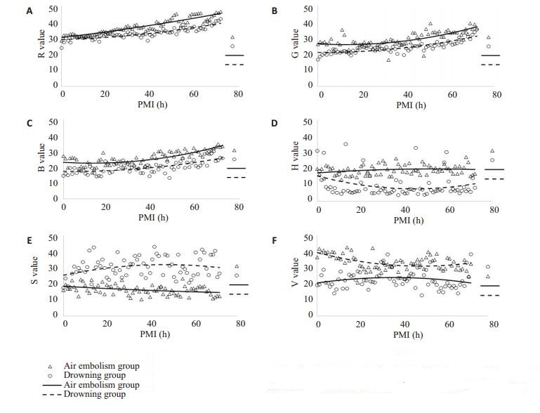
不论空气栓塞或溺死,死后72 h内兔角膜瞳孔区域图像的R、G、B值均随PMI呈上升趋势,死后20 h内变化趋势不明显,20 h后上升趋势加快;经比较发现,空气栓塞组R、G、B实测值总体高于溺死组(图 1A~C)。H、S、V值在死后72 h内没有明显的变化趋势,各时间点测量值无明显分布规律(图 1D~F)。

|
图 1 死后72 h内兔角膜瞳孔区域图像6项颜色特征参数变化趋势图 Figure 1 Variation trends of 6 color feature parameters in the pupil region on rabbit corneal images within 72 h after death. A、B、C、D、E、F: respectively represent the variation trend charts of R、G、B、H、S、V. |
回归分析结果表明,不论空气栓塞或溺死,反映死后72 h内兔角膜瞳孔区域图像颜色变化的R、G、B 3项颜色特征参数均与PMI相关性较好;而H、S、V 3项颜色特征参数与PMI关系均没有统计学意义(表 2)。
| 表 2 死后72 h内兔角膜瞳孔区域图像6项颜色特征参数与PMI回归分析结果 Table 2 Regression analysis of PMI and the 6 color feature parameters in the pupil region on rabbit corneal images within 72 h after deathy represents PMI (h), and x represent different color feature parameters |
研究发现,肉眼观察兔角膜瞳孔区域颜色于死后24 h内由透明逐渐变为白色云雾状混浊,直至瞳孔不可透视。分析与个体死后角膜固有层中粘多糖和水含量发生变化以及角膜基质纤维排列紊乱等因素有关[16-19]。传统法医实践中观察死后角膜混浊推断PMI,主要依据个人主观经验将角膜混浊程度分为轻、中、重度来粗略判断PMI大致范围[18-19]。该方法作为经验法则的应用,致PMI预测误差较大。
为定量分析死后角膜颜色变化,本研究通过MATLAB软件分割出角膜瞳孔区域图像加以分析,这与过去采取分析整个角膜图像的研究方法[7-10]不同。主要考虑在于:完整的角膜图像包括瞳孔区域和虹膜区域,而法医实践中以瞳孔区域能否透视作为角膜混浊程度划分的主要依据[18-19];此外,为提高角膜图像颜色变化的检测灵敏度,要求最大程度减弱原背景颜色对图像分析的干扰,而虹膜本身具有颜色且存在个体差异[20-21]。因此角膜瞳孔区域图像的提取可以保证测量数据结果的准确性和可靠性。
此外,本研究基于MATLAB软件提取死后角膜瞳孔区域图像的6项颜色特征参数值(RGBHSV),并建立各参数与PMI的线性回归模型。以往的研究[7-10]只建立了死后角膜图像灰度值与PMI的回归方程,难以全面反映死后角膜图像颜色变化规律;虽提出死后角膜图像颜色变化与PMI之间存在一定关联,但并未建立具体的颜色特征参数与PMI的回归方程。
计算机图像分析结果显示,死后72 h内,两种不同死的兔角膜瞳孔区域图像的R、G、B值均随PMI呈上升趋势,且与PMI拟合关系较好,而H、S、V值无明显变化,与PMI拟合关系较差。经分析,RGB模型作为图像分析中最常用的颜色模型,可综合反映图像的色彩、深浅、明暗变化[22-24];HSV模型作为一种面向视觉感知的颜色模型,侧重于色彩表示,并不适于反映黑、白、灰类图像的颜色变化[25-27]。由于死后角膜瞳孔区域图像出现灰白色云雾状混浊变化过程,故RGB模型较HSV模型更适于死后角膜瞳孔区域图像混浊变化中的颜色表达。
研究发现,空气栓塞死后兔角膜瞳孔区域图像的R、G、B检测值均较溺死后高。兔空气栓塞死亡模型[11, 28-29]的致死机理是大量气体进入右心腔后,与血液混合形成血性泡沫堵塞肺动脉出口致猝死,死亡过程相对迅速,眼组织缺血缺氧显著;兔溺死模型[12, 30]的死亡过程相对较长,眼组织处于淤血缺氧状态。分析,正是由于上述差异,导致兔空气栓塞死后即刻角膜内皮细胞变性或坏死程度较溺死后严重,反映在角膜瞳孔区域图像上即空气栓塞组R、G、B检测值较高。研究还发现,两种死因死后兔角膜瞳孔区域图像的R、G、B值变化趋势一致,且与PMI相关性相近。这提示死后角膜瞳孔区域图像的颜色特征参数变化趋势受死因影响并不显著。
综上,个体死后角膜瞳孔区域图像颜色变化呈一定时间规律性。本研究建立的角膜图像处理方法及相关回归方程,为PMI推断提供一种无损、客观方法。未来的研究将会考虑外界环境因素差异对死后角膜瞳孔区域图像颜色变化的影响,并积极收集人尸体角膜图像数据资料加以分析。
| [1] |
Hostiuc S, Rusu MC, Mănoiu VS, et al. Usefulness of ultrastructure studies for the estimation of the postmortem interval. A systematic review[J].
Romn J Morphol Embryol, 2017, 58(2): 377-84.
|
| [2] |
Hubig M, Muggenthaler H, Sinicina I, et al. Temperature based forensic death time estimation: The standard model in experimental test[J].
Leg Med (Tokyo), 2015, 17(5): 381-7.
DOI: 10.1016/j.legalmed.2015.05.005. |
| [3] |
Madea B. Methods for determining time of death[J].
Forensic Sci Med Pathol, 2016, 12(4): 451-85.
DOI: 10.1007/s12024-016-9776-y. |
| [4] |
郑吉龙, 张晓东, 安志远, 等. 兔死后角膜内皮细胞核DNA降解随死亡时间变化规律[J].
中国法医学杂志, 2010, 25(4): 220-2.
DOI: 10.3969/j.issn.1001-5728.2010.04.002. |
| [5] |
Wang J, Kelly GC, Tate WJ, et al. Excitatory amino acid transporter expression in the essential tremor dentate nucleus and cerebellar cortex: a postmortem study[J].
Parkinsonism Relat Disord, 2016, 32: 87-93.
DOI: 10.1016/j.parkreldis.2016.09.003. |
| [6] |
Ansari N, Lodha A, Menon SK. Smart platform for the time since death determination from vitreous humor cystine[J].
Biosens Bioelectron, 2016, 86: 115-21.
DOI: 10.1016/j.bios.2016.06.042. |
| [7] |
Zhou L, Liu Y, Liu L, et al. Image analysis on corneal opacity: a novel method to estimate postmortem interval in rabbits[J].
J Huazhong Univ Sci Technolog Med Sci, 2010, 30(2): 235-9.
DOI: 10.1007/s11596-010-0221-2. |
| [8] |
王晓亮, 方超, 罗思敏. 猪眼角膜混浊程度变化与PMI推断[J].
广东公安科技, 2011, 19(2): 14-7.
|
| [9] |
Kawashima W, Hatake K, Kudo R, et al. Estimating the time after death on the basis of corneal opacity[J].
J Forensic Res, 2015, 6(1): 269.
|
| [10] |
Cantürk İ, Çelik S, Şahin MF, et al. Investigation of opacity development in the human eye for estimation of the postmortem interval[J].
Biocyb Biomed Engin, 2017, 37(3): 559-65.
|
| [11] |
Xia J, Zhang LL, Chen XS. The gas change in the different postmortem intervals after rabbit venous air embolism[J].
Chin J Forens Med, 2008, 23(3): 171-4.
|
| [12] |
Chaudhari KM, A n, Shrigiriwar MB, et al. Histopathology findings of asphyxia in lungs of hanging and drowning deaths[J].
MedicoLegal Update, 2016, 16(1): 138-42.
|
| [13] |
张德丰.
数字图像处理: MATLAB版[M]. 北京: 人民邮电出版社, 2015.
|
| [14] |
Zhou H, Shen Y, Zhu X, et al. Digital image modification detection using color information and its histograms[J].
Forensic Sci Int, 2016, 266: 379-88.
DOI: 10.1016/j.forsciint.2016.06.005. |
| [15] |
Karakasis EG, Papakostas GA, Koulouriotis DE, et al. A unified methodology for computing accurate quaternion color moments and moment invariants[J].
IEEE Trans Image Process, 2014, 23(2): 596-611.
|
| [16] |
Aslam TM, Shakir S, Wong J, et al. Use of iris recognition camera technology for the quantification of corneal opacification in mucopolysaccharidoses[J].
Br J Ophthalmol, 2012, 96(12): 1466-8.
DOI: 10.1136/bjophthalmol-2011-300996. |
| [17] |
Torricelli AA, Wilson SE. Cellular and extracellular matrix modulation of corneal stromal opacity[J].
Exp Eye Res, 2014, 129: 151-60.
DOI: 10.1016/j.exer.2014.09.013. |
| [18] |
Napoli PE, Nioi M, D'aloja E, et al. Post-Mortem corneal thickness measurements with a portable optical coherence tomography system: a reliability study[J].
Sci Rep, 2016, 6: 30428.
DOI: 10.1038/srep30428. |
| [19] |
李晓娜, 郑吉龙, 单迪, 等. 兔死后角膜内皮细胞活性率变化的时间规律性研究[J].
解剖科学进展, 2014, 20(2): 115-8.
|
| [20] |
Norton HL, Edwards M, Krithika S, et al. Quantitative assessment of skin, hair, and iris variation in a diverse sample of individuals and associated genetic variation[J].
Am J Phys Anthropol, 2016, 160(4): 570-81.
DOI: 10.1002/ajpa.v160.4. |
| [21] |
Hohl DM, Bezus B, Ratowiecki J, et al. Genetic and phenotypic variability of iris color in Buenos Aires population[J].
Genet Mol Biol, 2018, 41(1): 50-8.
DOI: 10.1590/1678-4685-gmb-2017-0175. |
| [22] |
Tugrul S, Degirmenci N, Eren SB, et al. RGB measurements as a novel objective diagnostic test for otitis media with effusion[J].
Acta Otolaryngol, 2015, 135(4): 342-7.
DOI: 10.3109/00016489.2014.984877. |
| [23] |
Singh MK, Nain N. Unobtrusive silhouette extraction using multivariate analysis and shadow removal in RGB color model[J].
J Rel Intell Envir, 2016, 2(4): 175-86.
DOI: 10.1007/s40860-016-0031-9. |
| [24] |
Boopathi E, Thiagarasu V. Edge detection in color images using RGB color model[J].
Int J Comput Appl, 2018, 180(9): 6-11.
|
| [25] |
Chae S, Jun K. HSV color model based hand contour detector robust to noise[J].
J Korea Multimed Societ, 2015, 18(10): 1149-56.
DOI: 10.9717/kmms.2015.18.10.1149. |
| [26] |
Jae-Hyoung Y, Kim YT, Lee NK, et al. Effective color correction method employing HSV color model[J].
J Measurement Sci Instrument, 2012, 3(1): 39-45.
|
| [27] |
Ma L, Zhang XH. Relationship between Saturation and Brightness Value in HSV Color Space[J].
J Comp Graph, 2014, 26(8): 1272-8.
|
| [28] |
Bernaldo De Quirós Y, González-Díaz O, Møllerløkken A, et al. Differentiation at autopsy between in vivo gas embolism and putrefaction using gas composition analysis[J].
Int J Legal Med, 2013, 127(2): 437-45.
DOI: 10.1007/s00414-012-0783-6. |
| [29] |
Bernaldo De Quirós Y, Saavedra P, Møllerløkken A, et al. Differentiation at necropsy between in vivo gas embolism and putrefaction using a gas score[J].
Res Vet Sci, 2016, 106: 48-55.
DOI: 10.1016/j.rvsc.2016.03.007. |
| [30] |
Hyodoh H, Terashima R, Rokukawa M, et al. Experimental drowning lung images on postmortem CT - Difference between sea water and fresh water[J].
Leg Med (Tokyo), 2016, 19: 11-5.
DOI: 10.1016/j.legalmed.2016.01.006. |
 2018, Vol. 38

