Journal of Southern Medical University ›› 2025, Vol. 45 ›› Issue (2): 422-436.doi: 10.12122/j.issn.1673-4254.2025.02.23
Previous Articles Next Articles
Zhixiong ZENG1( ), Yongbo WANG2, Zongyue LIN2, Zhaoying BIAN1,3, Jianhua MA1()
Received:2024-10-15
Online:2025-02-20
Published:2025-03-03
Contact:
Jianhua MA
E-mail:zxzeng@smu.edu.cn;jhma@smu.edu.cn
Supported by:Zhixiong ZENG, Yongbo WANG, Zongyue LIN, Zhaoying BIAN, Jianhua MA. A segmented backprojection tensor degradation feature encoding model for motion artifacts correction in dental cone beam computed tomography[J]. Journal of Southern Medical University, 2025, 45(2): 422-436.
Add to citation manager EndNote|Ris|BibTeX
URL: https://www.j-smu.com/EN/10.12122/j.issn.1673-4254.2025.02.23
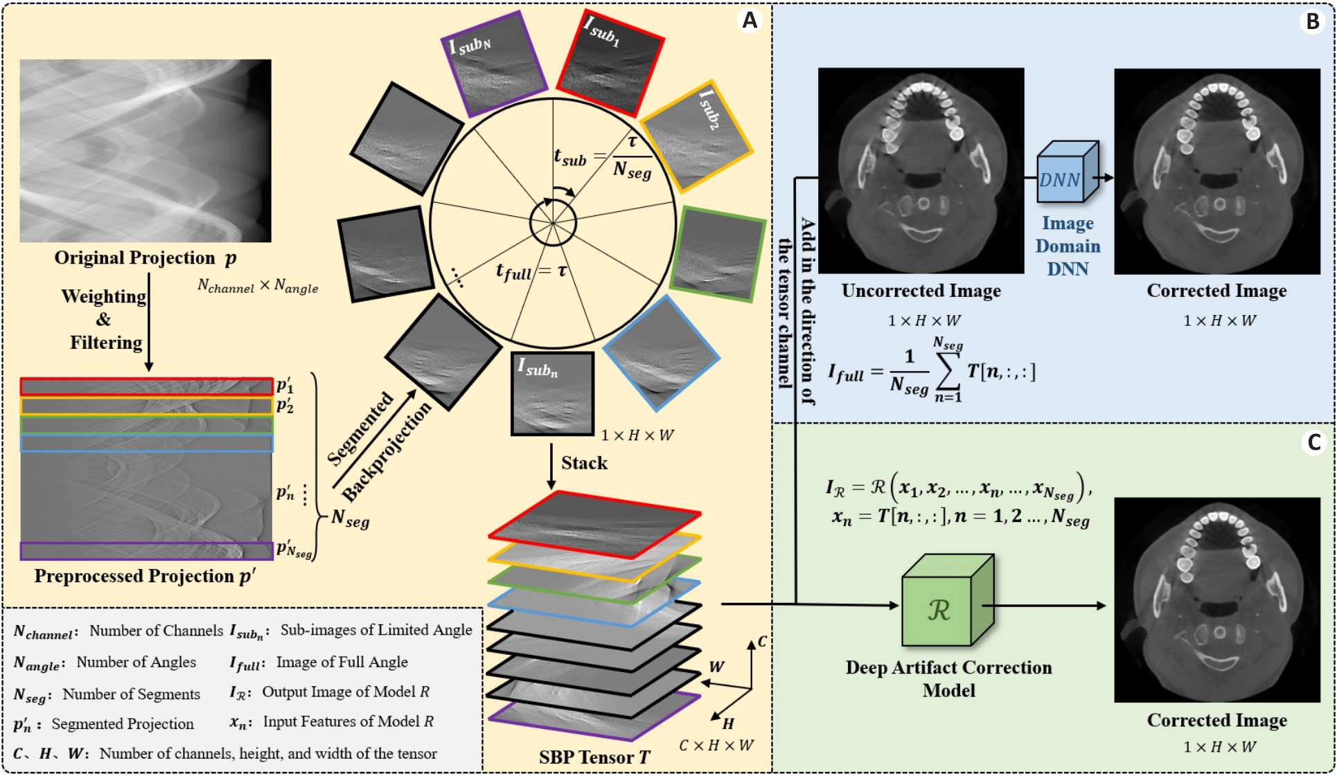
Fig.1 Schematic diagram of the segmented backprojection tensor. A: Origin of the segmented backprojection tensor. B: Training idea of DNN in the traditional image domain. C: Training idea of the proposed model.
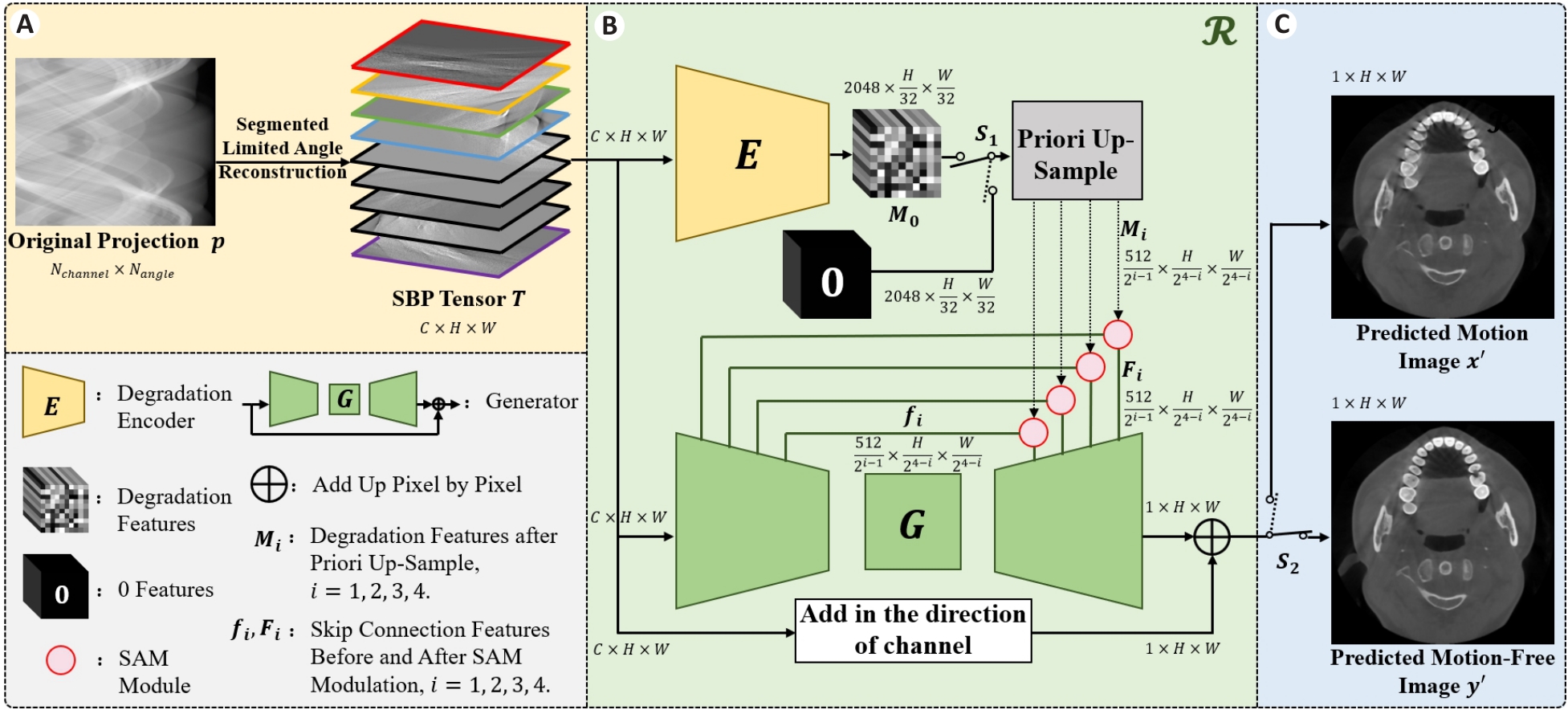
Fig.2 Flow chart of the SBP-MAC model. A: Preparation of the input data of the model. B: Composition of the proposed model. C: Output results of the model.
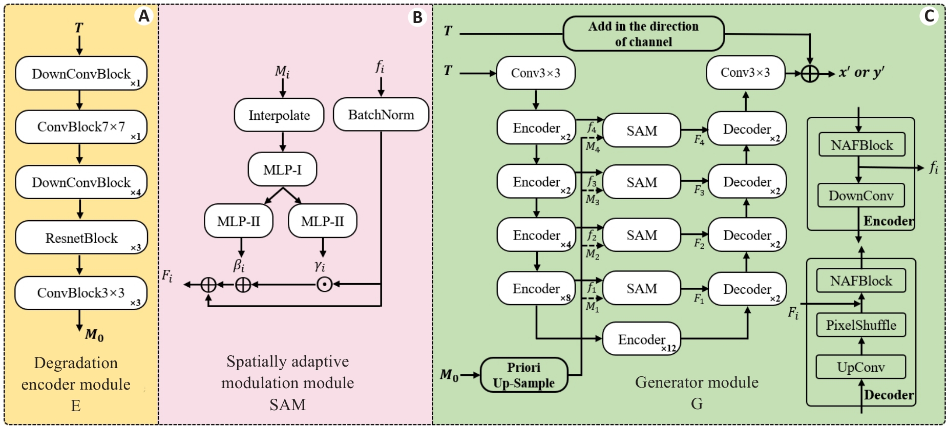
Fig.3 Detailed archetecture of the key modules. A: Degradation Encoder Module (E). B: Spatial Adaptive Modulation Module (SAM). C: Generator Module (G). The number of stacks for each smaller module is indicated at the bottom right corner.
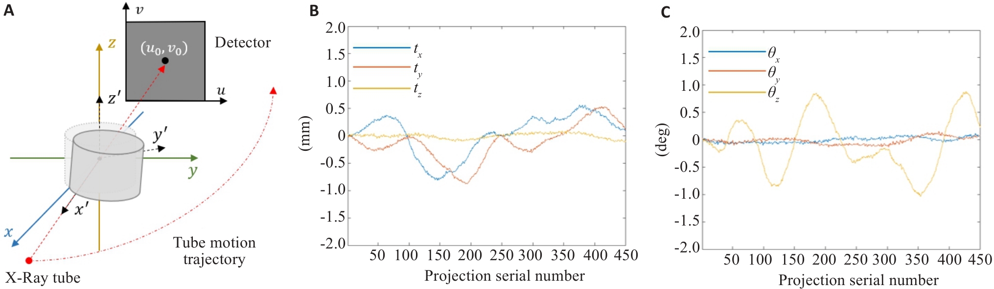
Fig.4 Schematic diagram of CBCT scanning and motion waveforms. A: Schematic diagram of CBCT scanning. B: Translational motion waveform used in the simulation. C: Rotational motion waveform used in the simulation.
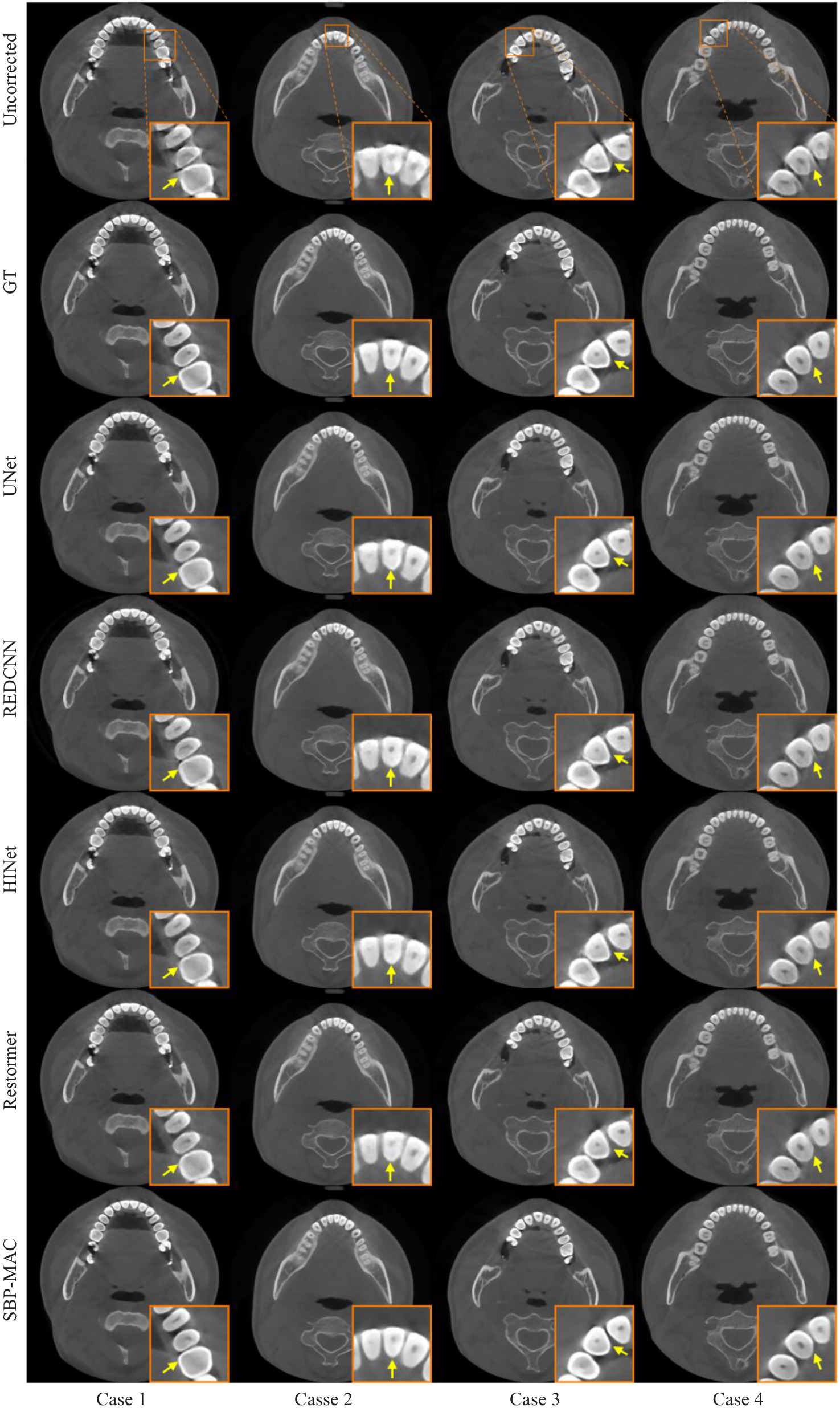
Fig.5 Transverse-section results of different motion artifact correction methods on simulation data.
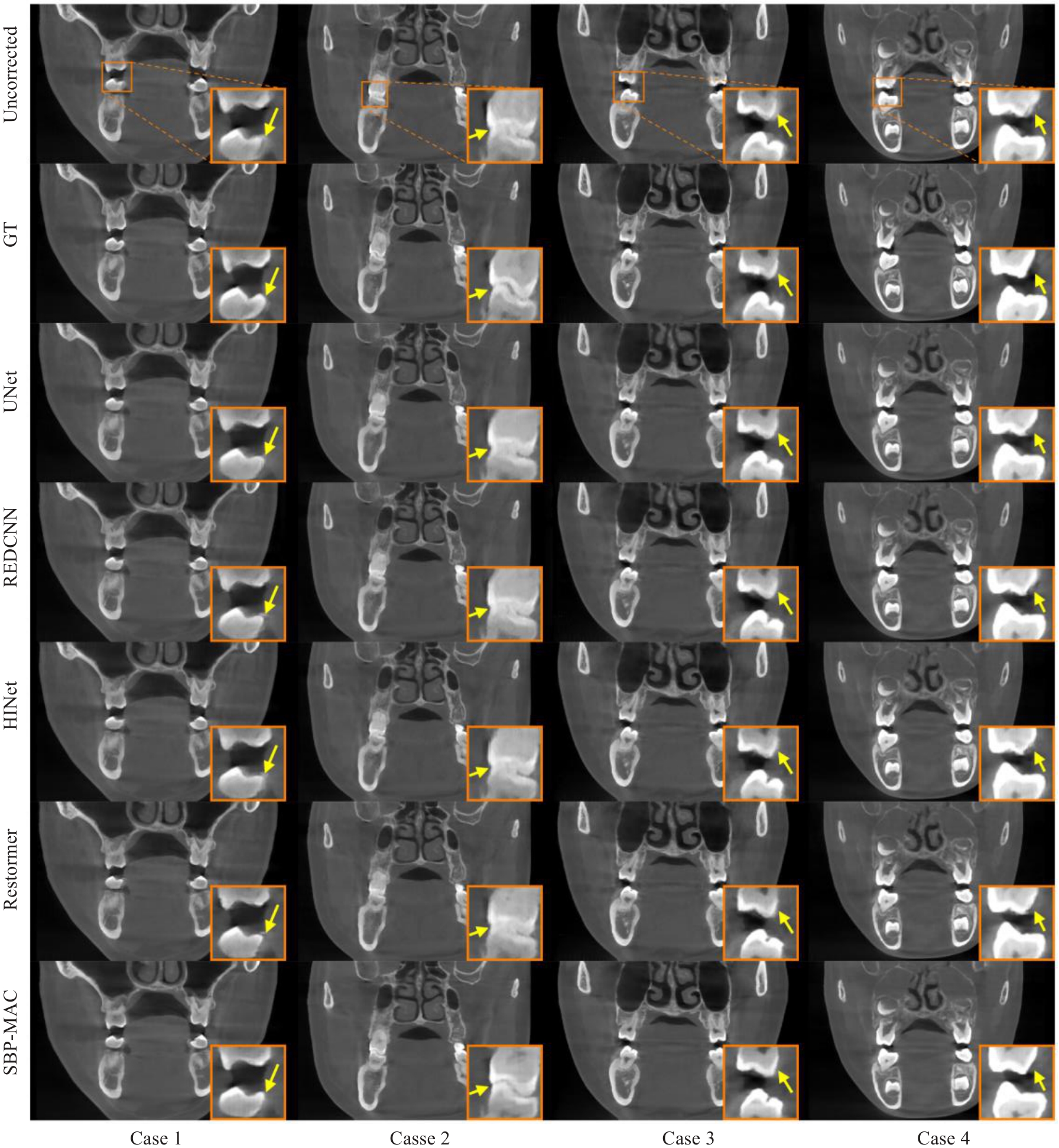
Fig.6 Coronal-section results of different motion artifact correction methods on simulation data.
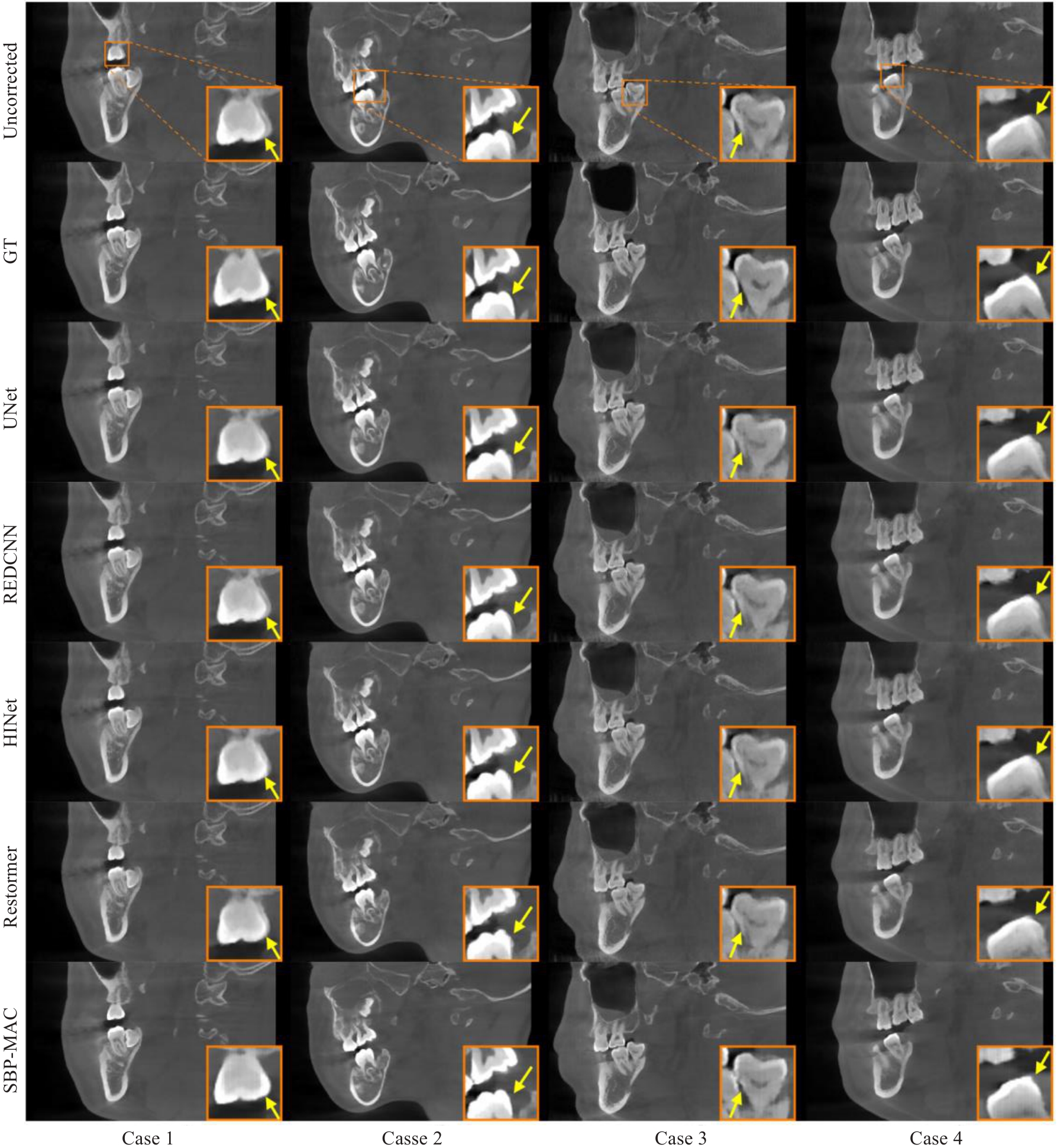
Fig.7 Sagittal-section results of different motion artifact correction methods on simulation data.
| Methods | PSNR | RMSE | SSIM |
|---|---|---|---|
| Uncorrected | 29.5479±1.5036 | 8.6211±1.4713 | 0.9276±0.0191 |
| UNet | 30.2005±1.5400 | 8.0025±1.3922 | 0.9339±0.0191 |
| REDCNN | 30.0276±1.5185 | 8.1599±1.3991 | 0.9318±0.0185 |
| HINet | 30.2865±1.5343 | 7.9228±1.3729 | 0.9346±0.0183 |
| Restormer | 30.3057±1.5646 | 7.9111±1.4128 | 0.9342±0.0191 |
| SBP-MAC | 31.9936±1.9277 | 6.5657±1.4119 | 0.9547±0.0144 |
Tab.1 Quantitative indicators of different motion artifact correction algorithms on simulation data (Mean±SD)
| Methods | PSNR | RMSE | SSIM |
|---|---|---|---|
| Uncorrected | 29.5479±1.5036 | 8.6211±1.4713 | 0.9276±0.0191 |
| UNet | 30.2005±1.5400 | 8.0025±1.3922 | 0.9339±0.0191 |
| REDCNN | 30.0276±1.5185 | 8.1599±1.3991 | 0.9318±0.0185 |
| HINet | 30.2865±1.5343 | 7.9228±1.3729 | 0.9346±0.0183 |
| Restormer | 30.3057±1.5646 | 7.9111±1.4128 | 0.9342±0.0191 |
| SBP-MAC | 31.9936±1.9277 | 6.5657±1.4119 | 0.9547±0.0144 |

Fig.10 Coronal-section results of different motion artifact correction methods on real data.
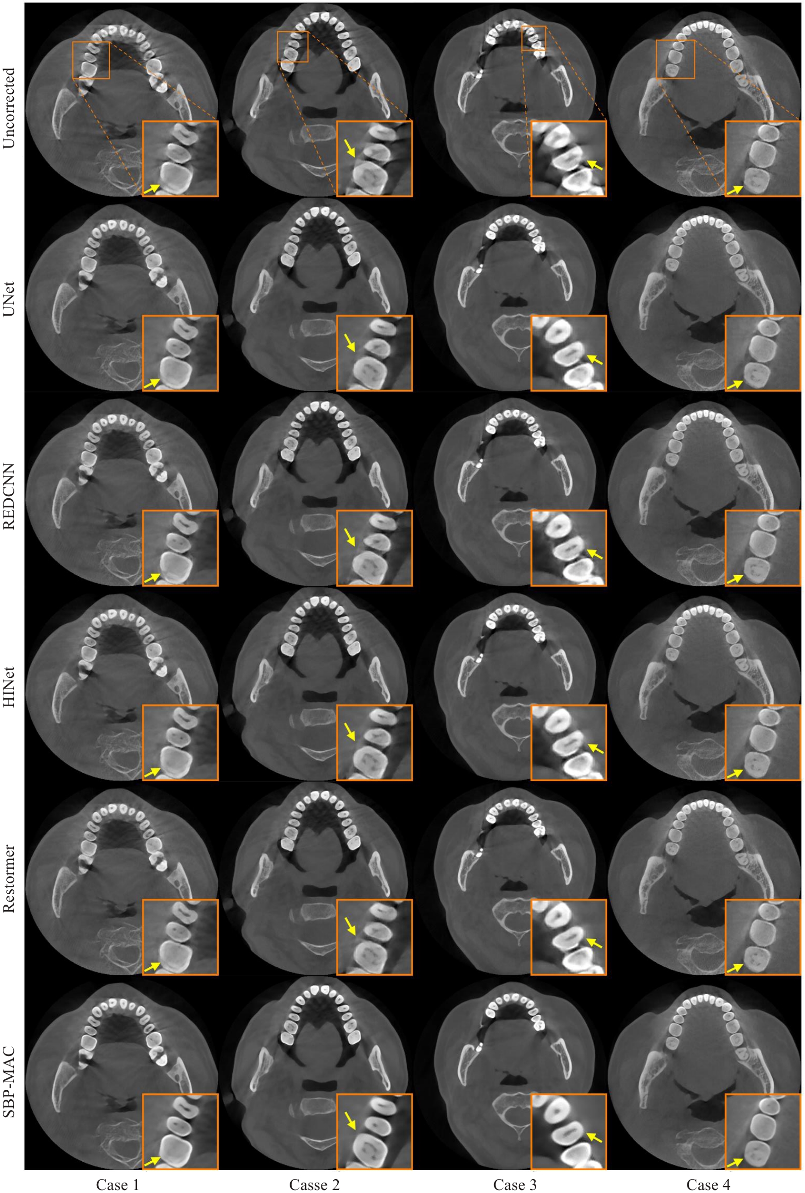
Fig.8 Transverse-section results of different motion artifact correction methods on real data.
| Methods | Scores (Mean±SD) | P |
|---|---|---|
| UNet | 3.7263±0.5915 | 0.0018 |
| REDCNN | 3.8000±0.5416 | 0.0042 |
| HINet | 3.7895±0.4026 | 0.0012 |
| Restormer | 3.9211±0.3084 | 0.0078 |
| SBP-MAC | 4.2421±0.3548 | - |
Tab.2 Image quality expert score statistics on real clinical data
| Methods | Scores (Mean±SD) | P |
|---|---|---|
| UNet | 3.7263±0.5915 | 0.0018 |
| REDCNN | 3.8000±0.5416 | 0.0042 |
| HINet | 3.7895±0.4026 | 0.0012 |
| Restormer | 3.9211±0.3084 | 0.0078 |
| SBP-MAC | 4.2421±0.3548 | - |
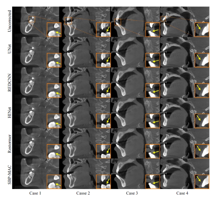
Fig.10 Sagittal-section results of different motion artifact correction methods on real data.
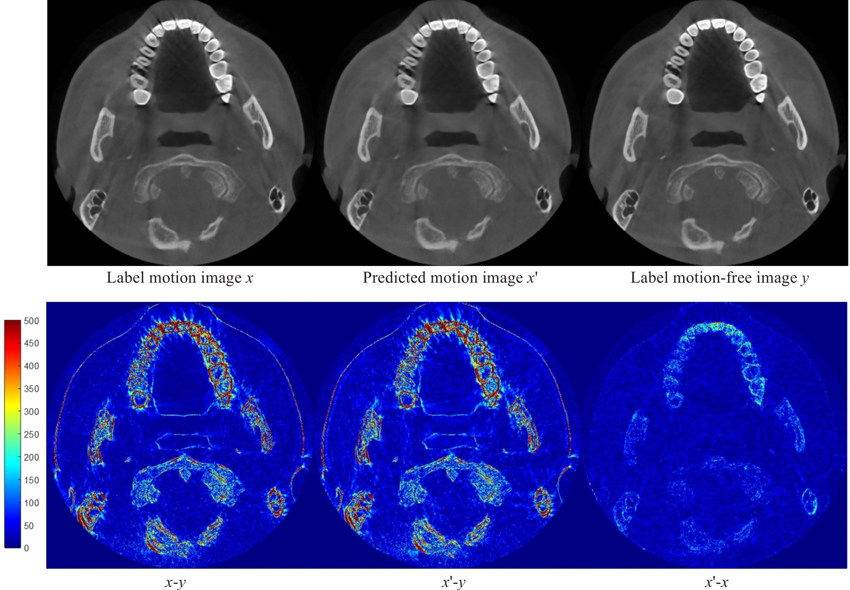
Fig.11 Displacement of motion artifact image generation results.
| PSNR | RMSE | SSIM | |
|---|---|---|---|
| 29.4475±1.5051 | 8.7216±1.4902 | 0.9272±0.0190 | |
| 30.9235±2.0620 | 7.4532±1.7300 | 0.9410±0.0210 | |
| 31.6159±1.7890 | 6.8346±1.3598 | 0.9497±0.0156 | |
| 31.6953±1.8010 | 6.7756±1.3774 | 0.9519±0.0150 | |
| 31.7653±1.9522 | 6.7456±1.4842 | 0.9513±0.0169 | |
| 31.9936±1.9277 | 6.5657±1.4119 | 0.9547±0.0144 | |
| 31.5523±2.0146 | 6.9245±1.5739 | 0.9504±0.0182 | |
| Uncorrected | 29.5479±1.5036 | 8.6211±1.4713 | 0.9276±0.0191 |
Tab.3 Ablation experiments of different hyperparameters λ of the loss function on simulation data (Mean±SD)
| PSNR | RMSE | SSIM | |
|---|---|---|---|
| 29.4475±1.5051 | 8.7216±1.4902 | 0.9272±0.0190 | |
| 30.9235±2.0620 | 7.4532±1.7300 | 0.9410±0.0210 | |
| 31.6159±1.7890 | 6.8346±1.3598 | 0.9497±0.0156 | |
| 31.6953±1.8010 | 6.7756±1.3774 | 0.9519±0.0150 | |
| 31.7653±1.9522 | 6.7456±1.4842 | 0.9513±0.0169 | |
| 31.9936±1.9277 | 6.5657±1.4119 | 0.9547±0.0144 | |
| 31.5523±2.0146 | 6.9245±1.5739 | 0.9504±0.0182 | |
| Uncorrected | 29.5479±1.5036 | 8.6211±1.4713 | 0.9276±0.0191 |
Tensor as input | Degradation encoder guidance | Artifact consistency loss | PSNR | RMSE | SSIM |
|---|---|---|---|---|---|
| -- | -- | -- | 30.3743±1.6067 | 7.8556±1.4400 | 0.9368±0.0180 |
| √ | -- | -- | 30.7192±1.8001 | 7.5822±1.5518 | 0.9414±0.0181 |
| √ | √ | -- | 31.5523±2.0146 | 6.9245±1.5739 | 0.9504±0.0182 |
| √ | √ | √ | 31.9936±1.9277 | 6.5657±1.4119 | 0.9547±0.0144 |
| Uncorrected | 29.5479±1.5036 | 8.6211±1.4713 | 0.9276±0.0191 | ||
Tab.4 Ablation experiments of different key factors on simulation data (Mean±SD)
Tensor as input | Degradation encoder guidance | Artifact consistency loss | PSNR | RMSE | SSIM |
|---|---|---|---|---|---|
| -- | -- | -- | 30.3743±1.6067 | 7.8556±1.4400 | 0.9368±0.0180 |
| √ | -- | -- | 30.7192±1.8001 | 7.5822±1.5518 | 0.9414±0.0181 |
| √ | √ | -- | 31.5523±2.0146 | 6.9245±1.5739 | 0.9504±0.0182 |
| √ | √ | √ | 31.9936±1.9277 | 6.5657±1.4119 | 0.9547±0.0144 |
| Uncorrected | 29.5479±1.5036 | 8.6211±1.4713 | 0.9276±0.0191 | ||
| Fixed hyperparameter | Variable hyperparameter | PSNR | RMSE | SSIM |
|---|---|---|---|---|
| 30.5183±1.9243 | 7.7814±1.6805 | 0.9386±0.0212 | ||
| 30.6998±1.9764 | 7.6334±1.7315 | 0.9400±0.0219 | ||
| 30.7374±2.0913 | 7.6229±1.8165 | 0.9409±0.0226 | ||
| 31.9936±1.9277 | 6.5657±1.4119 | 0.9547±0.0144 | ||
| 31.2309±1.7166 | 7.1332±1.3707 | 0.9445±0.0169 | ||
| 30.9549±1.9805 | 7.4097±1.6358 | 0.9389±0.0219 | ||
| 31.8414±1.5808 | 6.6301±1.1826 | 0.9508±0.0122 | ||
| 31.9936±1.9277 | 6.5657±1.4119 | 0.9547±0.0144 | ||
| 30.4684±1.6176 | 7.7740±1.4537 | 0.9349±0.0190 | ||
| 29.5610±1.4616 | 8.6027±1.4620 | 0.9293±0.0210 | ||
| Uncorrected | 29.5479±1.5036 | 8.6211±1.4713 | 0.9276±0.0191 | |
Tab.5 Exploratory experiment on the impact of α1 and α2 on model performance (Mean±SD)
| Fixed hyperparameter | Variable hyperparameter | PSNR | RMSE | SSIM |
|---|---|---|---|---|
| 30.5183±1.9243 | 7.7814±1.6805 | 0.9386±0.0212 | ||
| 30.6998±1.9764 | 7.6334±1.7315 | 0.9400±0.0219 | ||
| 30.7374±2.0913 | 7.6229±1.8165 | 0.9409±0.0226 | ||
| 31.9936±1.9277 | 6.5657±1.4119 | 0.9547±0.0144 | ||
| 31.2309±1.7166 | 7.1332±1.3707 | 0.9445±0.0169 | ||
| 30.9549±1.9805 | 7.4097±1.6358 | 0.9389±0.0219 | ||
| 31.8414±1.5808 | 6.6301±1.1826 | 0.9508±0.0122 | ||
| 31.9936±1.9277 | 6.5657±1.4119 | 0.9547±0.0144 | ||
| 30.4684±1.6176 | 7.7740±1.4537 | 0.9349±0.0190 | ||
| 29.5610±1.4616 | 8.6027±1.4620 | 0.9293±0.0210 | ||
| Uncorrected | 29.5479±1.5036 | 8.6211±1.4713 | 0.9276±0.0191 | |
| PSNR | RMSE | SSIM | |
|---|---|---|---|
| 1 | 30.2443±1.8714 | 8.0235±1.7334 | 0.9318±0.0192 |
| 3 | 30.9100±2.1304 | 7.4803±1.8122 | 0.9425±0.0217 |
| 6 | 31.8551±1.7499 | 6.6448±1.3210 | 0.9536±0.0130 |
| 9 | 31.9936±1.9277 | 6.5657±1.4119 | 0.9547±0.0144 |
| 15 | 31.8019±1.8482 | 6.7010±1.4063 | 0.9516±0.0148 |
| Uncorrected | 29.5479±1.5036 | 8.6211±1.4713 | 0.9276±0.0191 |
Tab.6 Exploratory experiment on the impact of Nseg on model performance (Mean±SD)
| PSNR | RMSE | SSIM | |
|---|---|---|---|
| 1 | 30.2443±1.8714 | 8.0235±1.7334 | 0.9318±0.0192 |
| 3 | 30.9100±2.1304 | 7.4803±1.8122 | 0.9425±0.0217 |
| 6 | 31.8551±1.7499 | 6.6448±1.3210 | 0.9536±0.0130 |
| 9 | 31.9936±1.9277 | 6.5657±1.4119 | 0.9547±0.0144 |
| 15 | 31.8019±1.8482 | 6.7010±1.4063 | 0.9516±0.0148 |
| Uncorrected | 29.5479±1.5036 | 8.6211±1.4713 | 0.9276±0.0191 |
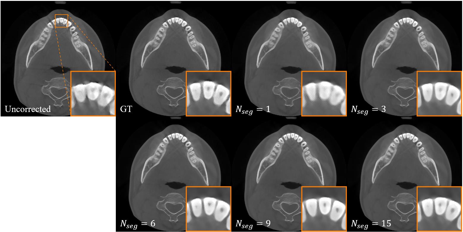
Fig.12 Artifact correction results under different values of the Nseg .
| 1 | Ng SY. Comparison between conventional dental radiography and CBCT[M]// Cone Beam CT in Dentistry. Cham: Springer, 2023: 271-330. |
| 2 | Schulze RKW, Drage NA. Cone-beam computed tomography and its applications in dental and maxillofacial radiology[J]. Clin Radiol, 2020, 75(9): 647-57. |
| 3 | Nemtoi A, Czink C, Haba D, et al. Cone beam CT: a current overview of devices[J]. Dentomaxillofac Radiol, 2013, 42(8): 20120443. |
| 4 | Spin-Neto R, Wenzel A. Patient movement and motion artefacts in cone beam computed tomography of the dentomaxillofacial region: a systematic literature review[J]. Oral Surg Oral Med Oral Pathol Oral Radiol, 2016, 121(4): 425-33. |
| 5 | Keriş EY. Effect of patient anxiety on image motion artefacts in CBCT[J]. BMC Oral Health, 2017, 17(1): 73. |
| 6 | Donaldson K, O'Connor S, Heath N. Dental cone beam CT image quality possibly reduced by patient movement[J]. Dentomaxillofac Radiol, 2013, 42(2): 91866873. |
| 7 | Spin-Neto R, Hauge Matzen L, Hermann L, et al. Head motion and perception of discomfort by young children during simulated CBCT examinations[J]. Dentomaxillofac Radiol, 2021, 50(3): 20200445. |
| 8 | Spin-Neto R, Costa C, Salgado DM, et al. Patient movement characteristics and the impact on CBCT image quality and interpretability[J]. Dentomaxillofac Radiol, 2018, 47(1): 20170216. |
| 9 | Spin-Neto R, Matzen L H, Schropp L, et al. Factors affecting patient movement and re-exposure in cone beam computed tomography examination [J]. Oral Surg Oral Med Oral Pathol Oral Radiol, 2015, 119(5): 572-8. |
| 10 | Nardi C, Taliani GG, Castellani A, et al. Repetition of examination due to motion artifacts in horizontal cone beam CT: comparison among three different kinds of head support[J]. J Int Soc Prev Community Dent, 2017, 7(4): 208-13. |
| 11 | Yildizer Keris E, Demirel O, Ozdede M. Evaluation of motion artifacts in cone-beam computed tomography with three different patient positioning[J]. Oral Radiol, 2021, 37(2): 276-81. |
| 12 | Jacobson MW, Stayman JW. Compensating for head motion in slowly-rotating cone beam CT systems with optimization transfer based motion estimation[C]//2008 IEEE Nuclear Science Symposium Conference Record. Dresden, Germany. IEEE, 2008: 5240-5. |
| 13 | Kyme AZ, Fulton RR. Motion estimation and correction in SPECT, PET and CT[J]. Phys Med Biol, 2021, 66(18): 18TR02. |
| 14 | Ouadah S, Jacobson M, Stayman JW, et al. Correction of patient motion in cone-beam CT using 3D-2D registration[J]. Phys Med Biol, 2017, 62(23): 8813-31. |
| 15 | Sun T, Jacobs R, Pauwels R, et al. A motion correction approach for oral and maxillofacial cone-beam CT imaging[J]. Phys Med Biol, 2021, 66(12): 125008. |
| 16 | Berger M, Müller K, Aichert A, et al. Marker-free motion correction in weight-bearing cone-beam CT of the knee joint[J]. Med Phys, 2016, 43(3): 1235-48. |
| 17 | Niebler S, Schömer E, Tjaden H, et al. Projection-based improvement of 3D reconstructions from motion-impaired dental cone beam CT data[J]. Med Phys, 2019, 46(10): 4470-80. |
| 18 | Hernandez D, Eldib ME, Hegazy MAA, et al. A head motion estimation algorithm for motion artifact correction in dental CT imaging[J]. Phys Med Biol, 2018, 63(6): 065014. |
| 19 | Berger M, Xia Y, Aichinger W, et al. Motion compensation for cone-beam CT using Fourier consistency conditions[J]. Phys Med Biol, 2017, 62(17): 7181-215. |
| 20 | Aichert A, Berger M, Wang J, et al. Epipolar consistency in transmission imaging[J]. IEEE Trans Med Imaging, 2015, 34(11): 2205-19. |
| 21 | A'lvarez-Borrego J. Fast autofocus algorithm for automated microscopes[J]. Opt Eng, 2005, 44(6): 063601. |
| 22 | Wicklein J, Kyriakou Y, Kalender WA, et al. An online motion- and misalignment-correction method for medical flat-detector CT[C]//SPIE Proceedings", "Medical Imaging 2013: Physics of Medical Imaging. Lake Buena Vista (Orlando Area), Florida, USA. SPIE, 2013: 466-72. |
| 23 | Sisniega A, Stayman JW, Yorkston J, et al. Motion compensation in extremity cone-beam CT using a penalized image sharpness criterion[J]. Phys Med Biol, 2017, 62(9): 3712-34. |
| 24 | Zhang Y, Zhang LY. A rigid motion artifact reduction method for CT based on blind deconvolution[J]. Algorithms, 2019, 12(8): 155. |
| 25 | Gao C, Feng AQ, Liu XT, et al. A fully differentiable framework for 2D/3D registration and the projective spatial transformers[J]. IEEE Trans Med Imag, 2024, 43(1): 275-85. |
| 26 | Ali ASRA, Landi C, Sarti C, et al. Non-iterative compensation for patient motion in dental CBCT imaging[C]//2023 IEEE Nuclear Science Symposium, Medical Imaging Conference and International Symposium on Room-Temperature Semiconductor Detectors (NSS MIC RTSD). Vancouver, BC, Canada. IEEE, 2023: 1. |
| 27 | Preuhs A, Manhart M, Roser P, et al. Appearance learning for image-based motion estimation in tomography[J]. IEEE Trans Med Imaging, 2020, 39(11): 3667-78. |
| 28 | Thies M, Wagner F, Maul N, et al. A gradient-based approach to fast and accurate head motion compensation in cone-beam CT[J]. IEEE Trans Med Imaging, 2024, doi: 10.1109/TMI.2024.3474250 |
| 29 | Amirian M, Montoya-Zegarra JA, Herzig I, et al. Mitigation of motion-induced artifacts in cone beam computed tomography using deep convolutional neural networks[J]. Med Phys, 2023, 50(10): 6228-42. |
| 30 | Ko Y, Moon S, Baek J, et al. Rigid and non-rigid motion artifact reduction in X-ray CT using attention module[J]. Med Image Anal, 2021, 67: 101883. |
| 31 | Li DS, Zhang Y, Cheung KC, et al. Learning degradation representations for Image deblurring[M]//Lecture Notes in Computer Science. Cham: Springer Nature Switzerland, 2022: 736-53. |
| 32 | Chen LY, Chu XJ, Zhang XY, et al. Simple baselines for Image restoration[M]//Lecture Notes in Computer Science. Cham: Springer Nature Switzerland, 2022: 17-33. |
| 33 | Park T, Liu MY, Wang TC, et al. Semantic image synthesis with spatially-adaptive normalization[C]//2019 IEEE/CVF Conference on Computer Vision and Pattern Recognition (CVPR). Long Beach, CA, USA. IEEE, 2019: 2332-41. |
| 34 | He KM, Zhang XY, Ren SQ, et al. Deep residual learning for image recognition[C]//2016 IEEE Conference on Computer Vision and Pattern Recognition (CVPR). Las Vegas, NV, USA. IEEE, 2016: 770-8. |
| 35 | Mechrez R, Talmi I, Shama F, et al. Maintaining natural image statistics with the contextual loss[C]//Asian Conference on Computer Vision. Cham: Springer, 2019: 427-443. |
| 36 | Simonyan K, Zisserman A. Very deep convolutional networks for large-scale image recognition[EB/OL]. 2014: arXiv: 1409.1556. |
| 37 | Ronneberger O, Fischer P, Brox T. U-net: Convolutional networks for biomedical image segmentation [C]//2015 Medical Image Computing and Computer Assisted Intervention (MICCAI). Munich, Germany. Springer International Publishing, 2015: 234-41. |
| 38 | Chen H, Zhang Y, Kalra MK, et al. Low-dose CT with a residual encoder-decoder convolutional neural network[J]. IEEE Trans Med Imaging, 2017, 36(12): 2524-35. |
| 39 | Chen LY, Lu X, Zhang J, et al. HINet: half instance normalization network for image restoration[C]//2021 IEEE/CVF Conference on Computer Vision and Pattern Recognition Workshops (CVPRW). Nashville, TN, USA. IEEE, 2021: 182-92. |
| 40 | Zamir SW, Arora A, Khan S, et al. Restormer: efficient transformer for high-resolution image restoration[C]//2022 IEEE/CVF Conference on Computer Vision and Pattern Recognition (CVPR). New Orleans, LA, USA. IEEE, 2022: 5718-29. |
| No related articles found! |
| Viewed | ||||||
|
Full text |
|
|||||
|
Abstract |
|
|||||