Journal of Southern Medical University ›› 2025, Vol. 45 ›› Issue (2): 395-408.doi: 10.12122/j.issn.1673-4254.2025.02.21
Wenwei LI1( ), Zerui MAO1, Yongbo WANG2, Zhaoying BIAN1,3, Jing HUANG1()
Received:2024-10-11
Online:2025-02-20
Published:2025-03-03
Contact:
Jing HUANG
E-mail:l22220309@smu.edu.cn;hjing@smu.edu.cn
Supported by:Wenwei LI, Zerui MAO, Yongbo WANG, Zhaoying BIAN, Jing HUANG. A sparse-view cone-beam CT reconstruction algorithm based on bidirectional flow field- guided projection completion[J]. Journal of Southern Medical University, 2025, 45(2): 395-408.
Add to citation manager EndNote|Ris|BibTeX
URL: https://www.j-smu.com/EN/10.12122/j.issn.1673-4254.2025.02.21
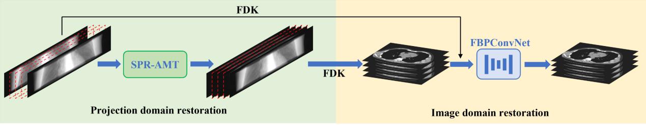
Fig.1 Schematic diagram of the BBC-Recon method.
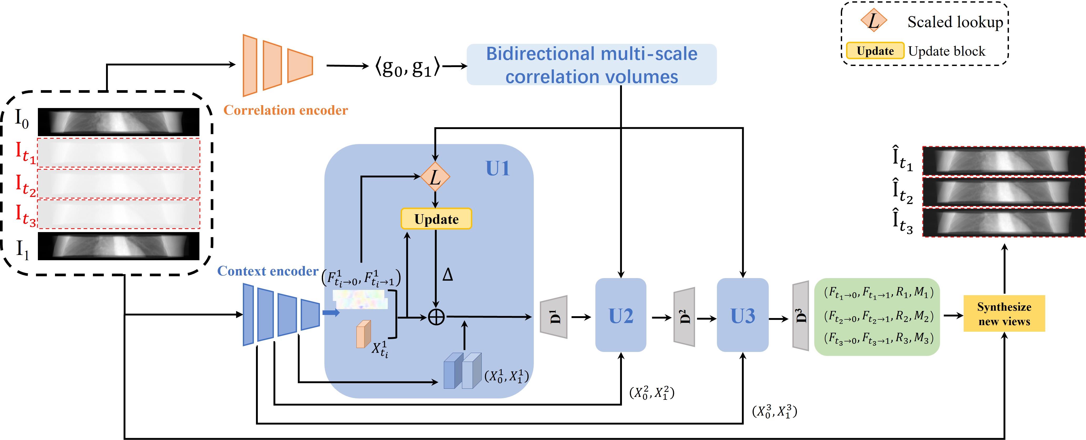
Fig.2 The projection recovery module based on bidirectional flow field guided projection completion.
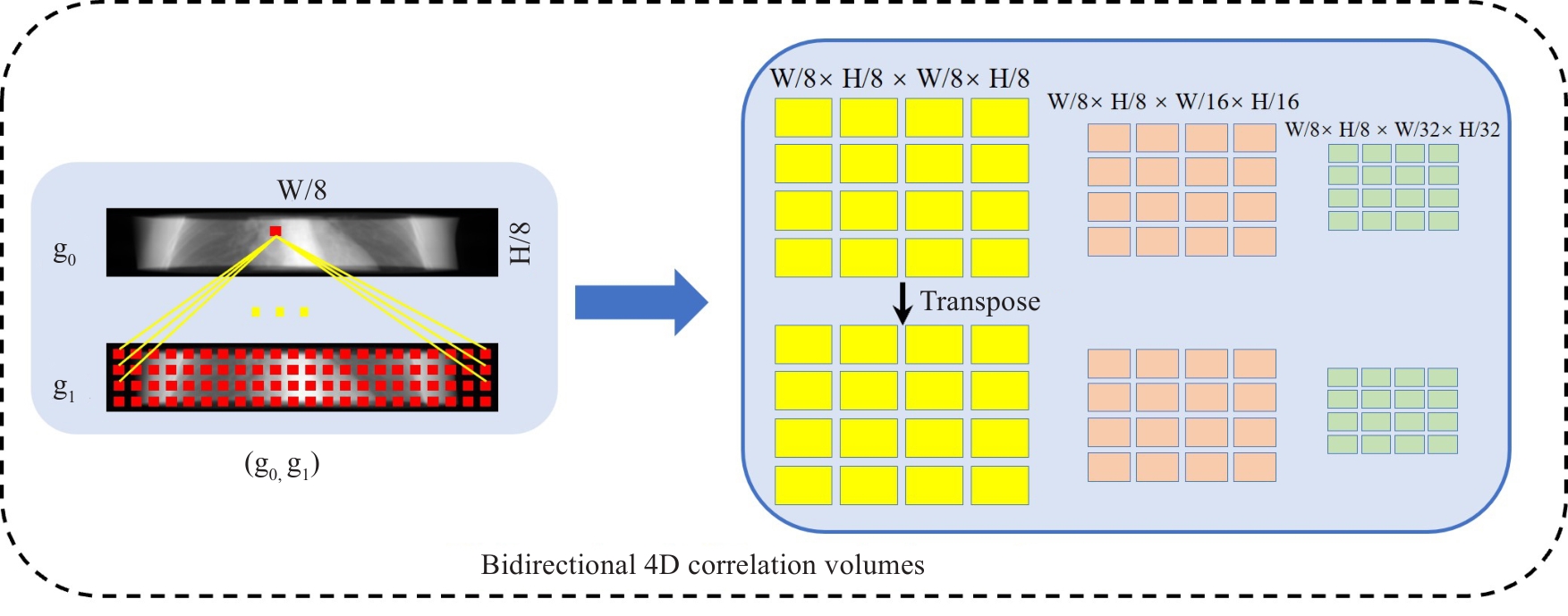
Fig.3 Schematic diagram of bidirectional multi-scale correlation volume calculation.
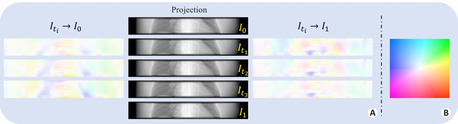
Fig.4 Schematic diagram of bidirectional flow field. A: Estimated bidirectional flow field, B: Color legend.
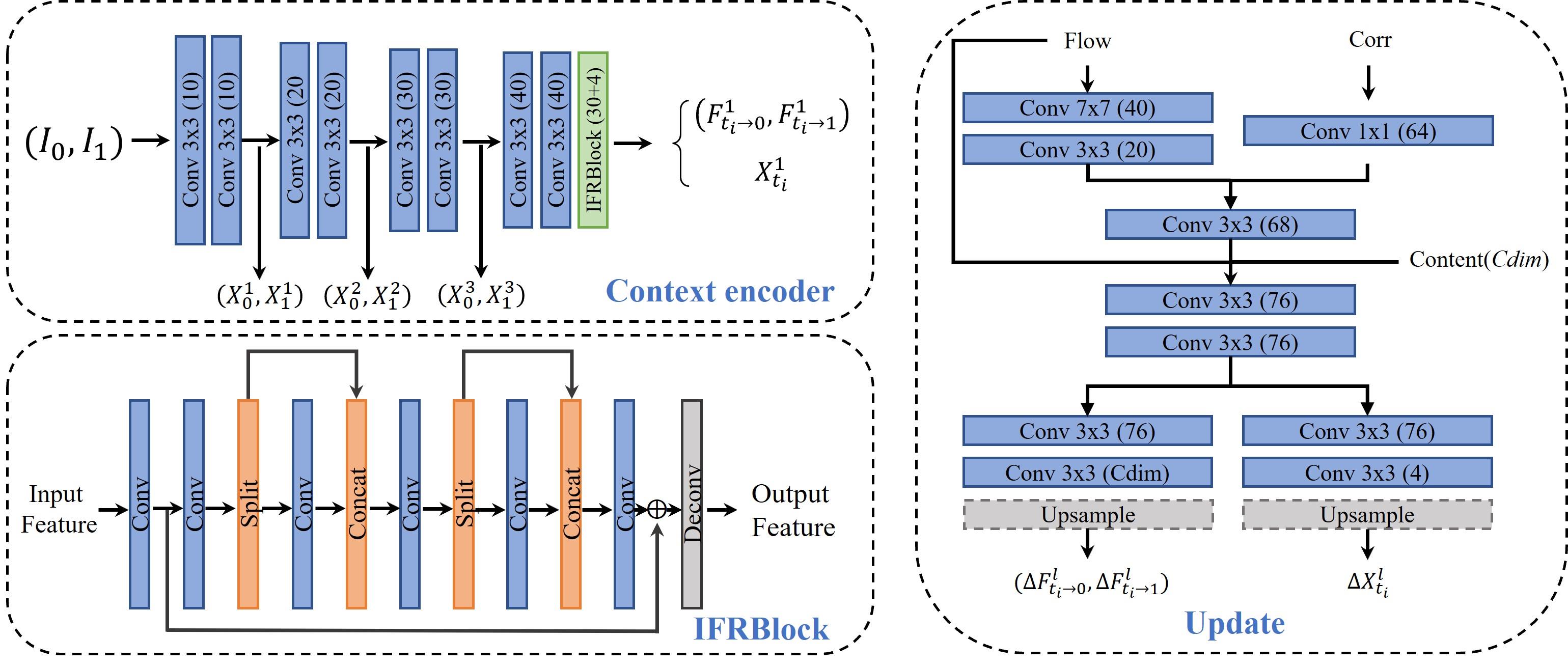
Fig.5 Structural diagram of each functional module for projection recovery.
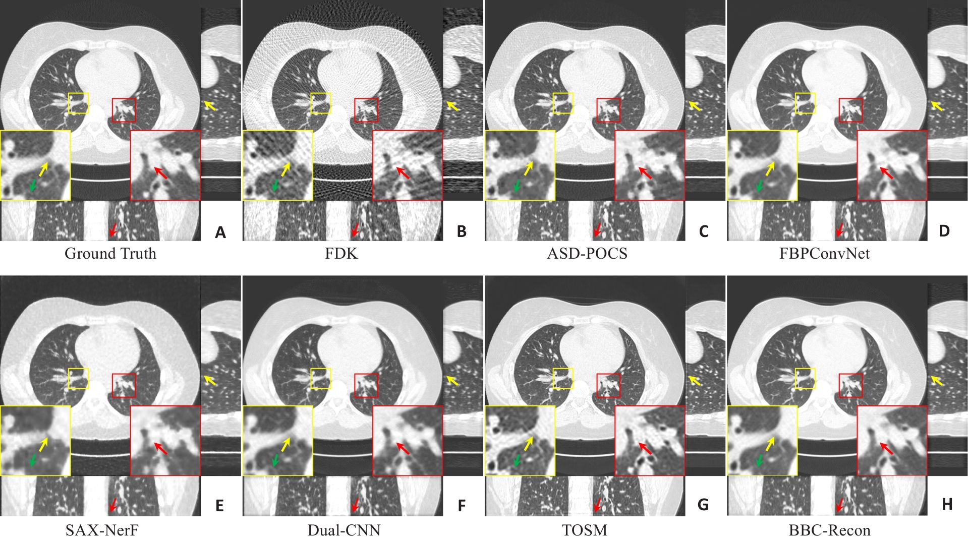
Fig.6 Comparison of 3D imaging results with 150-views of the chest. A: Ground Truth; B: FDK reconstruction; C: ASD-POCS; D: FBPConvNet; E: SAX-NerF; F: Dual-CNN; G: TOSM; H: BBC-Recon (the proposed method). The display window is [-1300,100]HU.
| Method | Axial | Coronal | Sagittal | ||||||
|---|---|---|---|---|---|---|---|---|---|
| PSNR | SSIM | RMSE | PSNR | SSIM | RMSE | PSNR | SSIM | RMSE | |
| FDK | 30.9635 | 0.9677 | 84.0423 | 31.2190 | 0.7775 | 82.9474 | 31.1780 | 0.9634 | 83.1501 |
| ASD-POCS | 37.5809 | 0.9928 | 39.3558 | 38.2010 | 0.8781 | 38.1144 | 37.7018 | 0.9902 | 39.0479 |
| FBPConvNet | |||||||||
| SAX-NeRF | 36.2327 | 0.9902 | 45.9355 | 36.8490 | 0.8478 | 44.5609 | 36.4561 | 0.9880 | 45.4288 |
| Dual-CNN | 35.5758 | 0.9885 | 50.2866 | 36.4565 | 0.8518 | 48.2645 | 35.8967 | 0.9866 | 49.4623 |
| TOSM | 37.8617 | 0.9933 | 38.1402 | 38.4367 | 0.8666 | 36.9161 | 38.0829 | 0.9919 | 37.6555 |
| BBC-Recon | 39.4176 | 0.9953 | 33.3770 | 40.2626 | 0.9252 | 32.1349 | 39.7163 | 0.9945 | 32.9198 |
Tab.1 Comparison of 3D imaging quantitative metrics with 150-views of the chest
| Method | Axial | Coronal | Sagittal | ||||||
|---|---|---|---|---|---|---|---|---|---|
| PSNR | SSIM | RMSE | PSNR | SSIM | RMSE | PSNR | SSIM | RMSE | |
| FDK | 30.9635 | 0.9677 | 84.0423 | 31.2190 | 0.7775 | 82.9474 | 31.1780 | 0.9634 | 83.1501 |
| ASD-POCS | 37.5809 | 0.9928 | 39.3558 | 38.2010 | 0.8781 | 38.1144 | 37.7018 | 0.9902 | 39.0479 |
| FBPConvNet | |||||||||
| SAX-NeRF | 36.2327 | 0.9902 | 45.9355 | 36.8490 | 0.8478 | 44.5609 | 36.4561 | 0.9880 | 45.4288 |
| Dual-CNN | 35.5758 | 0.9885 | 50.2866 | 36.4565 | 0.8518 | 48.2645 | 35.8967 | 0.9866 | 49.4623 |
| TOSM | 37.8617 | 0.9933 | 38.1402 | 38.4367 | 0.8666 | 36.9161 | 38.0829 | 0.9919 | 37.6555 |
| BBC-Recon | 39.4176 | 0.9953 | 33.3770 | 40.2626 | 0.9252 | 32.1349 | 39.7163 | 0.9945 | 32.9198 |
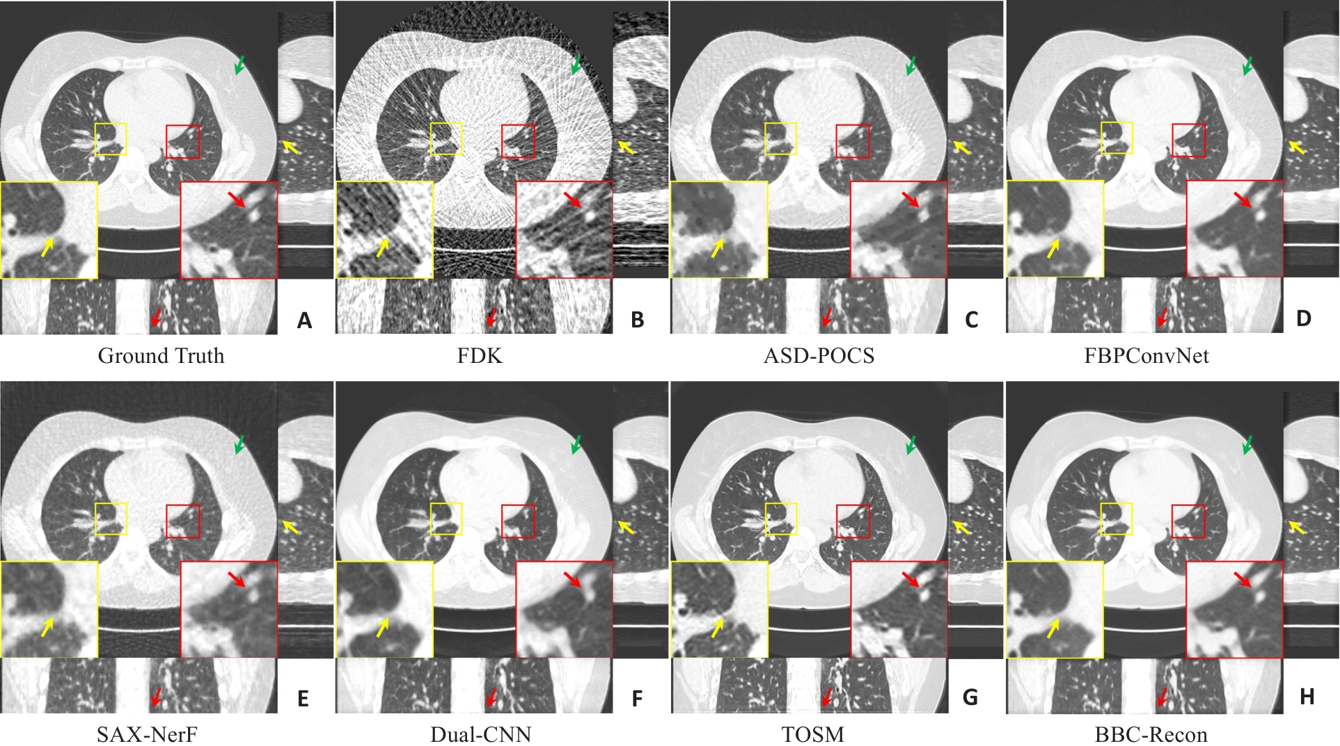
Fig.7 Comparison of 3D imaging results with 75-views of the chest. A: Ground Truth; B: FDK reconstruction; C: ASD-POCS; D: FBPConvNet; E: SAX-NerF; F: Dual-CNN; G: TOSM; H: BBC-Recon (the proposed method). The display window is [-1300,100]HU.
| Method | Axial | Coronal | Sagittal | ||||||
|---|---|---|---|---|---|---|---|---|---|
| PSNR | SSIM | RMSE | PSNR | SSIM | RMSE | PSNR | SSIM | RMSE | |
| FDK | 26.0442 | 0.9056 | 148.0213 | 26.3768 | 0.6830 | 145.5730 | 26.3245 | 0.8951 | 145.9907 |
| ASD-POCS | 34.9296 | 0.9866 | 53.3403 | 35.9176 | 0.8432 | 50.7807 | 35.2250 | 0.9839 | 52.4890 |
| FBPConvNet | 36.2275 | 0.9903 | 46.9397 | 37.3632 | 44.4852 | 36.5693 | 0.9885 | 46.1690 | |
| SAX-NeRF | 34.7707 | 0.9862 | 54.3179 | 35.5614 | 0.8335 | 52.2195 | 35.0749 | 0.9833 | 53.5090 |
| Dual-CNN | 32.9485 | 0.9807 | 67.4798 | 34.3106 | 0.8104 | 64.2221 | 33.4249 | 0.9775 | 66.2729 |
| TOSM | 41.9589 | 0.8602 | |||||||
| BBC-Recon | 37.2343 | 0.9923 | 38.3114 | 0.8956 | 40.0071 | 37.6296 | 0.9910 | 41.2971 | |
Tab.2 Comparison of 3D imaging quantitative metrics with 75-views of the chest
| Method | Axial | Coronal | Sagittal | ||||||
|---|---|---|---|---|---|---|---|---|---|
| PSNR | SSIM | RMSE | PSNR | SSIM | RMSE | PSNR | SSIM | RMSE | |
| FDK | 26.0442 | 0.9056 | 148.0213 | 26.3768 | 0.6830 | 145.5730 | 26.3245 | 0.8951 | 145.9907 |
| ASD-POCS | 34.9296 | 0.9866 | 53.3403 | 35.9176 | 0.8432 | 50.7807 | 35.2250 | 0.9839 | 52.4890 |
| FBPConvNet | 36.2275 | 0.9903 | 46.9397 | 37.3632 | 44.4852 | 36.5693 | 0.9885 | 46.1690 | |
| SAX-NeRF | 34.7707 | 0.9862 | 54.3179 | 35.5614 | 0.8335 | 52.2195 | 35.0749 | 0.9833 | 53.5090 |
| Dual-CNN | 32.9485 | 0.9807 | 67.4798 | 34.3106 | 0.8104 | 64.2221 | 33.4249 | 0.9775 | 66.2729 |
| TOSM | 41.9589 | 0.8602 | |||||||
| BBC-Recon | 37.2343 | 0.9923 | 38.3114 | 0.8956 | 40.0071 | 37.6296 | 0.9910 | 41.2971 | |

Fig.8 Comparison of 3D imaging results with 150-views of the abdomen. A: Ground Truth; B: FDK reconstruction; C: ASD-POCS; D: FBPConvNet; E: SAX-NerF; F: Dual-CNN; G: TOSM; H: BBC-Recon (the proposed method). The display window is [-140,260]HU.
| Method | Axial | Coronal | Sagittal | ||||||
|---|---|---|---|---|---|---|---|---|---|
| PSNR | SSIM | RMSE | PSNR | SSIM | RMSE | PSNR | SSIM | RMSE | |
| FDK | 31.6740 | 0.9933 | 40.2281 | 32.2543 | 0.7547 | 38.8053 | 31.9684 | 0.9102 | 39.6143 |
| ASD-POCS | 39.2508 | 0.9988 | 39.9507 | 0.8566 | 16.5868 | 39.6122 | 0.9492 | ||
| FBPConvNet | 17.6568 | 16.9073 | |||||||
| SAX-NeRF | 35.9638 | 0.9975 | 24.7276 | 36.9414 | 0.8466 | 23.5581 | 36.3126 | 0.9361 | 24.2745 |
| Dual-CNN | 36.3912 | 0.9978 | 24.0718 | 37.2046 | 0.8321 | 23.4742 | 36.8023 | 0.9328 | 23.7924 |
| TOSM | 38.8606 | 0.9987 | 17.9193 | 39.7214 | 0.8671 | 17.1003 | 39.2508 | 0.9471 | 17.5033 |
| BBC-Recon | 41.0348 | 0.9993 | 16.4155 | 43.9115 | 0.9694 | 14.7886 | 42.3120 | 0.9883 | 15.6204 |
Tab.3 Comparison of 3D imaging quantitative metrics with 150-views for the abdomen
| Method | Axial | Coronal | Sagittal | ||||||
|---|---|---|---|---|---|---|---|---|---|
| PSNR | SSIM | RMSE | PSNR | SSIM | RMSE | PSNR | SSIM | RMSE | |
| FDK | 31.6740 | 0.9933 | 40.2281 | 32.2543 | 0.7547 | 38.8053 | 31.9684 | 0.9102 | 39.6143 |
| ASD-POCS | 39.2508 | 0.9988 | 39.9507 | 0.8566 | 16.5868 | 39.6122 | 0.9492 | ||
| FBPConvNet | 17.6568 | 16.9073 | |||||||
| SAX-NeRF | 35.9638 | 0.9975 | 24.7276 | 36.9414 | 0.8466 | 23.5581 | 36.3126 | 0.9361 | 24.2745 |
| Dual-CNN | 36.3912 | 0.9978 | 24.0718 | 37.2046 | 0.8321 | 23.4742 | 36.8023 | 0.9328 | 23.7924 |
| TOSM | 38.8606 | 0.9987 | 17.9193 | 39.7214 | 0.8671 | 17.1003 | 39.2508 | 0.9471 | 17.5033 |
| BBC-Recon | 41.0348 | 0.9993 | 16.4155 | 43.9115 | 0.9694 | 14.7886 | 42.3120 | 0.9883 | 15.6204 |
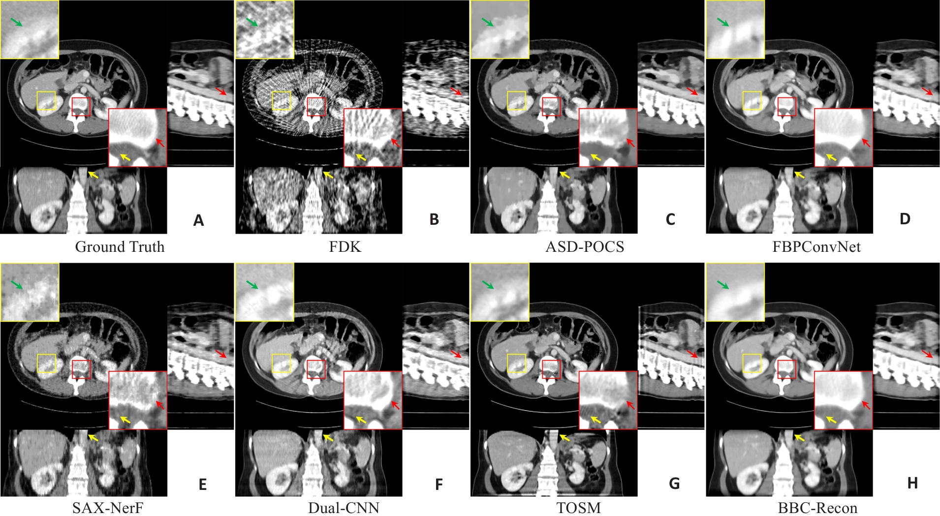
Fig.9 Comparison of 3D imaging results with 75-views of the abdomen. A: Ground Truth; B: FDK reconstruction; C: ASD-POCS; D: FBPConvNet; E: SAX-NerF; F: Dual-CNN; G: TOSM; H: BBC-Recon (the proposed method). The display window is [-140,260]HU.
| Method | Axial | Coronal | Sagittal | ||||||
|---|---|---|---|---|---|---|---|---|---|
| PSNR | SSIM | RMSE | PSNR | SSIM | RMSE | PSNR | SSIM | RMSE | |
| FDK | 25.2239 | 0.9711 | 84.5392 | 25.8403 | 0.6772 | 81.4876 | 25.4900 | 0.8644 | 83.4123 |
| ASD-POCS | 36.9642 | 0.9980 | 22.2091 | 37.9780 | 0.8399 | 20.9628 | 37.6697 | 0.9420 | 21.3747 |
| FBPConvNet | 36.9754 | 0.9981 | 23.5329 | 39.0430 | 21.7162 | 37.9324 | 22.5322 | ||
| SAX-NeRF | 33.8918 | 0.9960 | 31.3303 | 34.6090 | 0.8157 | 30.2131 | 34.3279 | 0.9207 | 30.5888 |
| Dual-CNN | 32.6976 | 0.9951 | 36.4336 | 34.3789 | 0.7750 | 34.6022 | 33.5714 | 0.9221 | 34.8065 |
| TOSM | 38.2694 | 19.1363 | 0.8644 | 18.2285 | 0.9457 | 18.6399 | |||
| BBC-Recon | 0.9986 | 40.7806 | 0.9436 | 39.3995 | 0.9752 | ||||
Tab.4 Comparison of 3D imaging quantitative metrics with 75-views for the abdomen
| Method | Axial | Coronal | Sagittal | ||||||
|---|---|---|---|---|---|---|---|---|---|
| PSNR | SSIM | RMSE | PSNR | SSIM | RMSE | PSNR | SSIM | RMSE | |
| FDK | 25.2239 | 0.9711 | 84.5392 | 25.8403 | 0.6772 | 81.4876 | 25.4900 | 0.8644 | 83.4123 |
| ASD-POCS | 36.9642 | 0.9980 | 22.2091 | 37.9780 | 0.8399 | 20.9628 | 37.6697 | 0.9420 | 21.3747 |
| FBPConvNet | 36.9754 | 0.9981 | 23.5329 | 39.0430 | 21.7162 | 37.9324 | 22.5322 | ||
| SAX-NeRF | 33.8918 | 0.9960 | 31.3303 | 34.6090 | 0.8157 | 30.2131 | 34.3279 | 0.9207 | 30.5888 |
| Dual-CNN | 32.6976 | 0.9951 | 36.4336 | 34.3789 | 0.7750 | 34.6022 | 33.5714 | 0.9221 | 34.8065 |
| TOSM | 38.2694 | 19.1363 | 0.8644 | 18.2285 | 0.9457 | 18.6399 | |||
| BBC-Recon | 0.9986 | 40.7806 | 0.9436 | 39.3995 | 0.9752 | ||||
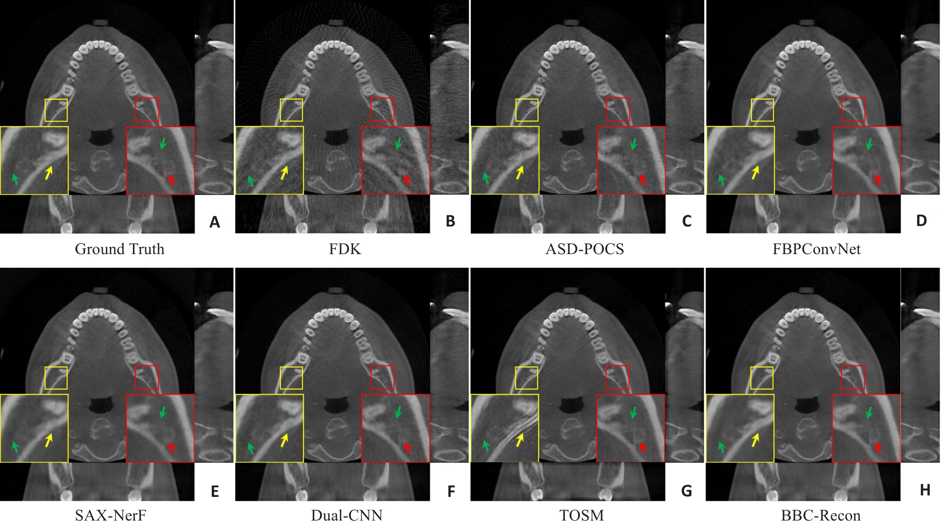
Fig.10 Comparison of 3D imaging results with 75-views of the oral cavity. A: Ground Truth; B: FDK reconstruction; C: ASD-POCS; D: FBPConvNet; E: SAX-NerF; F: Dual-CNN; G: TOSM; H: BBC-Recon (the proposed method). The display window is [-1000,2400]HU.
| Method | Axial | Coronal | Sagittal | ||||||
|---|---|---|---|---|---|---|---|---|---|
| PSNR | SSIM | RMSE | PSNR | SSIM | RMSE | PSNR | SSIM | RMSE | |
| FDK | 31.9519 | 0.9732 | 89.2484 | 32.0827 | 0.8565 | 88.7660 | 32.0100 | 0.8776 | 89.0789 |
| ASD-POCS | 37.2948 | 0.9920 | 48.3739 | 37.6072 | 47.5603 | 37.8578 | 0.9539 | 46.9743 | |
| FBPConvNet | 39.0303 | 0.9946 | 41.0228 | 39.5855 | 0.9437 | 39.9948 | 39.8207 | 39.5142 | |
| SAX-NeRF | 34.0368 | 0.9824 | 71.6770 | 35.2988 | 0.8817 | 66.9498 | 34.8295 | 0.8615 | 69.1129 |
| Dual-CNN | 35.7781 | 0.9816 | 78.9618 | 36.4218 | 0.8724 | 77.1856 | 36.6044 | 0.9081 | 76.4721 |
| TOSM | 0.9647 | 0.9671 | |||||||
| BBC-Recon | 40.0257 | 0.9956 | 36.9586 | 40.5435 | 0.9485 | 36.1054 | 40.7308 | 0.9723 | 35.7568 |
Tab.5 Comparison of 3D imaging quantitative metrics with 150-views for the oral cavity
| Method | Axial | Coronal | Sagittal | ||||||
|---|---|---|---|---|---|---|---|---|---|
| PSNR | SSIM | RMSE | PSNR | SSIM | RMSE | PSNR | SSIM | RMSE | |
| FDK | 31.9519 | 0.9732 | 89.2484 | 32.0827 | 0.8565 | 88.7660 | 32.0100 | 0.8776 | 89.0789 |
| ASD-POCS | 37.2948 | 0.9920 | 48.3739 | 37.6072 | 47.5603 | 37.8578 | 0.9539 | 46.9743 | |
| FBPConvNet | 39.0303 | 0.9946 | 41.0228 | 39.5855 | 0.9437 | 39.9948 | 39.8207 | 39.5142 | |
| SAX-NeRF | 34.0368 | 0.9824 | 71.6770 | 35.2988 | 0.8817 | 66.9498 | 34.8295 | 0.8615 | 69.1129 |
| Dual-CNN | 35.7781 | 0.9816 | 78.9618 | 36.4218 | 0.8724 | 77.1856 | 36.6044 | 0.9081 | 76.4721 |
| TOSM | 0.9647 | 0.9671 | |||||||
| BBC-Recon | 40.0257 | 0.9956 | 36.9586 | 40.5435 | 0.9485 | 36.1054 | 40.7308 | 0.9723 | 35.7568 |
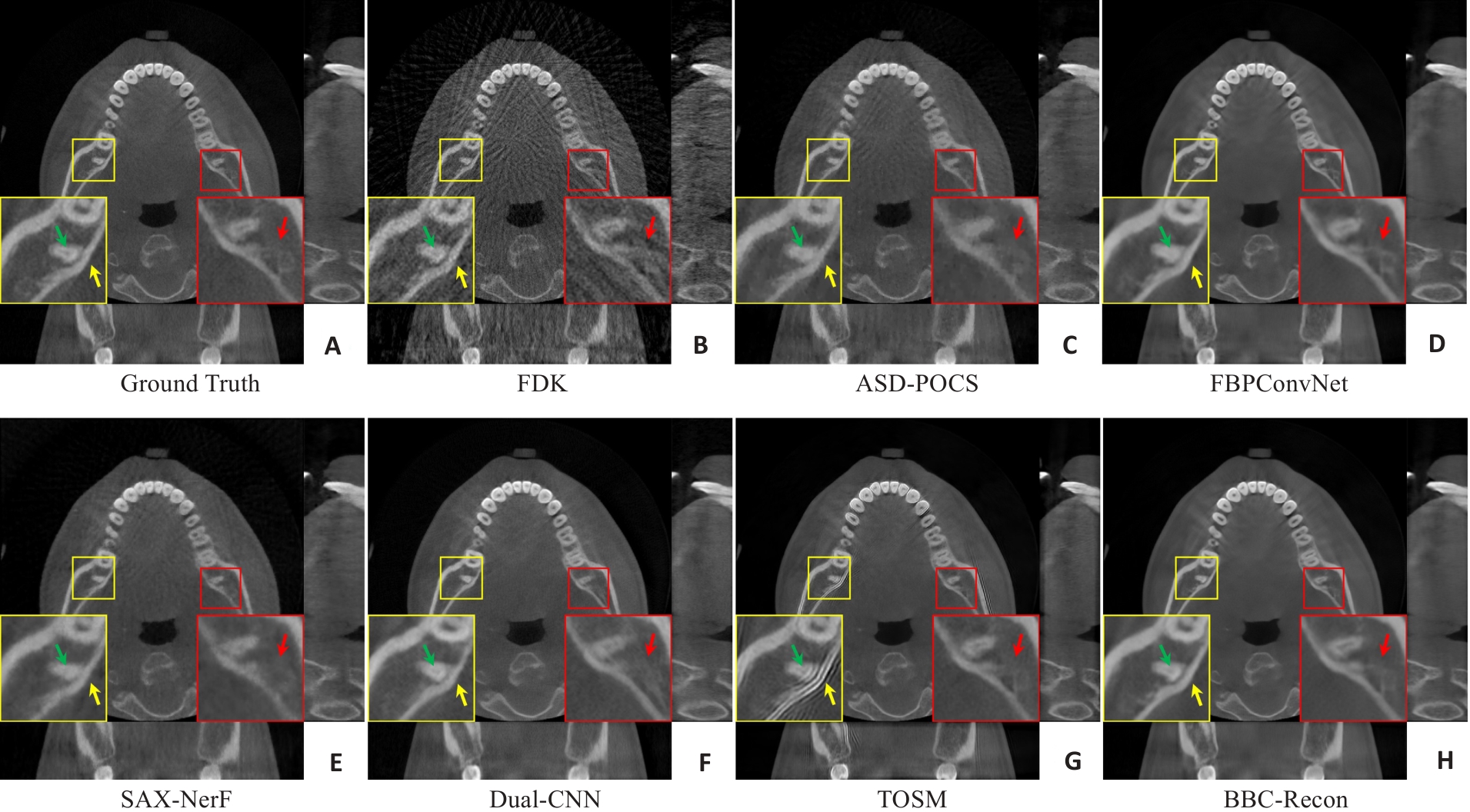
Fig.11 Comparison of 3D imaging results with 75-views of the oral cavity. A: Ground Truth; B: FDK reconstruction; C: ASD-POCS; D: FBPConvNet; E: SAX-NerF; F: Dual-CNN; G: TOSM; H: BBC-Recon (the proposed method).The display window is [-1000,2400]HU.
| Method | Axial | Coronal | Sagittal | ||||||
|---|---|---|---|---|---|---|---|---|---|
| PSNR | SSIM | RMSE | PSNR | SSIM | RMSE | PSNR | SSIM | RMSE | |
| FDK | 26.7048 | 0.9148 | 163.4736 | 26.9566 | 0.7542 | 161.6532 | 26.8140 | 0.7807 | 162.7921 |
| ASD-POCS | 35.4601 | 0.9878 | 59.6062 | 35.9525 | 0.9308 | 58.0802 | 36.2440 | 0.9441 | 57.3296 |
| FBPConvNet | 36.5348 | 0.9905 | 53.5184 | 37.1709 | 0.9348 | 51.8352 | 37.3895 | 0.9545 | 51.2476 |
| SAX-NeRF | 32.3823 | 0.9746 | 85.9164 | 33.1681 | 0.8587 | 82.9146 | 32.9332 | 0.8483 | 84.7201 |
| Dual-CNN | 33.2252 | 0.9792 | 85.9531 | 34.1240 | 0.8796 | 82.3269 | 34.2932 | 0.9009 | 81.6029 |
| TOSM | 0.9567 | ||||||||
| BBC-Recon | 37.7310 | 0.9927 | 47.1424 | 38.3814 | 45.7517 | 38.6036 | 0.9616 | 45.2281 | |
Tab.6 Comparison of 3D imaging quantitative metrics with 150-views for the oral cavity
| Method | Axial | Coronal | Sagittal | ||||||
|---|---|---|---|---|---|---|---|---|---|
| PSNR | SSIM | RMSE | PSNR | SSIM | RMSE | PSNR | SSIM | RMSE | |
| FDK | 26.7048 | 0.9148 | 163.4736 | 26.9566 | 0.7542 | 161.6532 | 26.8140 | 0.7807 | 162.7921 |
| ASD-POCS | 35.4601 | 0.9878 | 59.6062 | 35.9525 | 0.9308 | 58.0802 | 36.2440 | 0.9441 | 57.3296 |
| FBPConvNet | 36.5348 | 0.9905 | 53.5184 | 37.1709 | 0.9348 | 51.8352 | 37.3895 | 0.9545 | 51.2476 |
| SAX-NeRF | 32.3823 | 0.9746 | 85.9164 | 33.1681 | 0.8587 | 82.9146 | 32.9332 | 0.8483 | 84.7201 |
| Dual-CNN | 33.2252 | 0.9792 | 85.9531 | 34.1240 | 0.8796 | 82.3269 | 34.2932 | 0.9009 | 81.6029 |
| TOSM | 0.9567 | ||||||||
| BBC-Recon | 37.7310 | 0.9927 | 47.1424 | 38.3814 | 45.7517 | 38.6036 | 0.9616 | 45.2281 | |
| Method | Test time(s/slice) | |
|---|---|---|
| 150-views | 75-views | |
| ASD-POCS | 124.6289±34.9651 | 40.6434±11.4530 |
| FBPConvNet | 0.0504±0.0067 | 0.0487±0.0071 |
| SAX-NeRF | 5.6568±0.0799 | 2.8322±0.0505 |
| Dual-CNN | 0.0476±0.0028 | 0.0492±0.0028 |
| TOSM | 1062.5625±37.4625 | 1089.9375±31.0622 |
| BBC-Recon | 0.1406±0.0066 | 0.1437±0.0132 |
Tab.7 Comparative analysis of testing durations for different methods
| Method | Test time(s/slice) | |
|---|---|---|
| 150-views | 75-views | |
| ASD-POCS | 124.6289±34.9651 | 40.6434±11.4530 |
| FBPConvNet | 0.0504±0.0067 | 0.0487±0.0071 |
| SAX-NeRF | 5.6568±0.0799 | 2.8322±0.0505 |
| Dual-CNN | 0.0476±0.0028 | 0.0492±0.0028 |
| TOSM | 1062.5625±37.4625 | 1089.9375±31.0622 |
| BBC-Recon | 0.1406±0.0066 | 0.1437±0.0132 |
| Method | Axial | Coronal | Sagittal | ||||||
|---|---|---|---|---|---|---|---|---|---|
| PSNR | SSIM | RMSE | PSNR | SSIM | RMSE | PSNR | SSIM | RMSE | |
| E1 | 35.1979 | 0.9874 | 51.6862 | 35.4067 | 0.9747 | 53.0465 | 34.9260 | 0.9845 | 53.8445 |
| E2 | |||||||||
| E3 | 35.4127 | 0.9880 | 50.4040 | 35.6225 | 0.9761 | 51.9818 | 35.1885 | 0.9854 | 52.2280 |
Tab.8 Comparison of results from ablation experiments
| Method | Axial | Coronal | Sagittal | ||||||
|---|---|---|---|---|---|---|---|---|---|
| PSNR | SSIM | RMSE | PSNR | SSIM | RMSE | PSNR | SSIM | RMSE | |
| E1 | 35.1979 | 0.9874 | 51.6862 | 35.4067 | 0.9747 | 53.0465 | 34.9260 | 0.9845 | 53.8445 |
| E2 | |||||||||
| E3 | 35.4127 | 0.9880 | 50.4040 | 35.6225 | 0.9761 | 51.9818 | 35.1885 | 0.9854 | 52.2280 |
| 1 | Scarfe WC, Farman AG, Sukovic P. Clinical applications of cone-beam computed tomography in dental practice[J]. J Can Dent Assoc, 2006, 72(1): 75-80. |
| 2 | Brenner DJ, Hall EJ. Computed tomography: an increasing source of radiation exposure[J]. N Engl J Med, 2007, 357(22): 2277-84. |
| 3 | Berrington de González A, Mahesh M, Kim KP, et al. Projected cancer risks from computed tomographic scans performed in the United States in 2007[J]. Arch Intern Med, 2009, 169(22): 2071-7. |
| 4 | Feldkamp LA, Davis LC, Kress JW. Practical cone-beam algorithm[J]. J Opt Soc Am A, 1984, 1(6): 612. |
| 5 | Andersen AH, Kak AC. Simultaneous algebraic reconstruction technique (SART): a superior implementation of the art algorithm[J]. Ultrason Imaging, 1984, 6(1): 81-94. |
| 6 | Sidky EY, Pan XC. Image reconstruction in circular cone-beam computed tomography by constrained, total-variation minimization[J]. Phys Med Biol, 2008, 53(17): 4777-807. |
| 7 | Li S, Cao Q, Chen Y, et al. Dictionary learning based sinogram inpainting for CT sparse reconstruction[J]. Optik, 2014, 125(12): 2862-7. |
| 8 | Bertram M, Rose G, Schafer D, et al. Directional interpolation of sparsely sampled cone-beam CT sinogram data[C]//2004 2nd IEEE International Symposium on Biomedical Imaging: Nano to Macro. Arlington, VA, USA. IEEE, 2004: 928-31. |
| 9 | Zhang H, Sonke JJ. Directional sinogram interpolation for sparse angular acquisition in cone-beam computed tomography[J]. J Xray Sci Technol, 2013, 21(4): 481-96. |
| 10 | Muhlich M, Aach T. Analysis of multiple orientations[J]. IEEE Trans Image Process, 2009, 18(7): 1424-37. |
| 11 | Li ZB, Yu LF, Trzasko JD, et al. Adaptive nonlocal means filtering based on local noise level for CT denoising[J]. Med Phys, 2014, 41(1): 011908. |
| 12 | Litjens G, Kooi T, Bejnordi BE, et al. A survey on deep learning in medical image analysis[J]. Med Image Anal, 2017, 42: 60-88. |
| 13 | Razzak MI, Naz S, Zaib A. Deep learning for medical image processing: overview, challenges and future[EB/OL]. 2017: arXiv: 1704.06825. |
| 14 | JinKyong Hwan, McCann MT, Froustey E, et al. Deep convolutional neural network for inverse problems in imaging[J]. IEEE Trans Image Process, 2017, 26(9): 4509-22. |
| 15 | Ronneberger O, Fischer P, Brox T. U-net: Convolutional networks for biomedical image segmentation[C]//Medical image computing and computer-assisted intervention-MICCAI 2015: 18th international conference, Munich, Germany, October 5-9, 2015, proceedings, part III 18. Springer International Publishing, 2015: 234-41. |
| 16 | Chen H, Zhang Y, Kalra MK, et al. Low-dose CT with a residual encoder-decoder convolutional neural network[J]. IEEE Trans Med Imaging, 2017, 36(12): 2524-35. |
| 17 | Zhong WH, Li TL, Hou SK, et al. Unsupervised disentanglement strategy for mitigating artifact in photoacoustic tomography under extremely sparse view[J]. Photoacoustics, 2024, 38: 100613. |
| 18 | Wu WW, Hu DL, Niu C, et al. DRONE: dual-domain residual-based optimization NEtwork for sparse-view CT reconstruction[J]. IEEE Trans Med Imaging, 2021, 40(11): 3002-14. |
| 19 | Wu WW, Guo XD, Chen Y, et al. Deep embedding-attention-refinement for sparse-view CT reconstruction[J]. IEEE Trans Instrum Meas, 2023, 72: 4501111. |
| 20 | Lahiri A, Maliakal G, Klasky ML, et al. Sparse-view cone beam CT reconstruction using data-consistent supervised and adversarial learning from scarce training data[J]. IEEE Trans Comput Imag, 2023, 9: 13-28. |
| 21 | Chao LY, Wang ZW, Zhang HB, et al. Sparse-view cone beam CT reconstruction using dual CNNs in projection domain and image domain[J]. Neurocomputing, 2022, 493: 536-47. |
| 22 | Kim S, Kim B, Lee J, et al. Sparsier2Sparse: self-supervised convolutional neural network-based streak artifacts reduction in sparse-view CT images[J]. Med Phys, 2023, 50(12): 7731-47. |
| 23 | Pan JY, Zhang HY, Wu WF, et al. Multi-domain integrative Swin transformer network for sparse-view tomographic reconstruction[J]. Patterns, 2022, 3(6): 100498. |
| 24 | Genzel M, Gühring I, MacDonald J, et al. Near-exact recovery for tomographic inverse problems via deep learning[J]. Inter Confer Machine Learn, 2022,2022: 7368-81. |
| 25 | Mildenhall B, Srinivasan PP, Tancik M, et al. NeRF: representing scenes as neural radiance fields for view synthesis[C]//European Conference on Computer Vision. Cham: Springer, 2020: 405-421. |
| 26 | Chung H, Ryu D, McCann MT, et al. Solving 3D inverse problems using pre-trained 2D diffusion models[C]//2023 IEEE/CVF Conference on Computer Vision and Pattern Recognition (CVPR). Vancouver, BC, Canada. IEEE, 2023: 22542-51. |
| 27 | Li ZR, Wang YY, Zhang JJ, et al. Two-and-a-half order score-based model for solving 3D ill-posed inverse problems[J]. Comput Biol Med, 2024, 168: 107819. |
| 28 | Liu ZT, Fang Y, Li CJ, et al. Geometry-aware attenuation learning for sparse-view CBCT reconstruction[EB/OL]. 2023: arXiv: 2303. 14739. |
| 29 | Cai YH, Wang JH, Yuille A, et al. Structure-aware sparse-view X-ray 3D reconstruction[C]//2024 IEEE/CVF Conference on Computer Vision and Pattern Recognition (CVPR). Seattle, WA, USA. IEEE, 2024: 11174-83. |
| 30 | Liu ZT, Zhao HX, Qin WH, et al. 3D vessel reconstruction from sparse-view dynamic DSA images via vessel probability guided attenuation learning[EB/OL]. 2024: arXiv: 2405.10705. |
| 31 | Teed Z, Deng J. RAFT: recurrent all-pairs field transforms for optical flow[C]//European Conference on Computer Vision. Cham: Springer, 2020: 402-419. |
| 32 | Kong LT, Jiang BY, Luo DH, et al. IFRNet: intermediate feature refine network for efficient frame interpolation[C]//2022 IEEE/CVF Conference on Computer Vision and Pattern Recognition (CVPR). New Orleans, LA, USA. IEEE, 2022: 1959-68. |
| 33 | Li Z, Zhu ZL, Han LH, et al. AMT: all-pairs multi-field transforms for efficient frame interpolation[C]//2023 IEEE/CVF Conference on Computer Vision and Pattern Recognition (CVPR). Vancouver, BC, Canada. IEEE, 2023: 9801-10. |
| 34 | Charbonnier P, Blanc-Feraud L, Aubert G, et al. Two deterministic half-quadratic regularization algorithms for computed imaging[C]//Proceedings of 1st International Conference on Image Processing. Austin, TX, USA. IEEE, 1994: 168-72. |
| 35 | Moen TR, Chen BY, Holmes DR 3rd, et al. Low-dose CT image and projection dataset[J]. Med Phys, 2021, 48(2): 902-11. |
| 36 | Huang YY, Liu WJ, Yao CQ, et al. A multimodal dental dataset facilitating machine learning research and clinic services[J]. Sci Data, 2024, 11(1): 1291. |
| No related articles found! |
| Viewed | ||||||
|
Full text |
|
|||||
|
Abstract |
|
|||||