Journal of Southern Medical University ›› 2024, Vol. 44 ›› Issue (10): 1985-1994.doi: 10.12122/j.issn.1673-4254.2024.10.17
Siyi CHENG1,2,3,4( ), Zerui CHEN1,2,3,4, Changjiang YU1,2,3,4, Tucheng SUN1,2,3,4, Shuoji ZHU1,2,3,4(), Nanbo LIU1,2,3,4(), Ping ZHU1,2,3,4()
Received:2024-04-25
Online:2024-10-20
Published:2024-10-31
Contact:
Shuoji ZHU, Nanbo LIU, Ping ZHU
E-mail:debarah17@foxmail.com;zhushuoji@gmail.com;liu.nanbo@163.com;tanganqier@163.com
Supported by:Siyi CHENG, Zerui CHEN, Changjiang YU, Tucheng SUN, Shuoji ZHU, Nanbo LIU, Ping ZHU. Intrinsic steady-state pattern of mouse cardiac electrophysiology: analysis using a characterized quantitative electrocardiogram strategy[J]. Journal of Southern Medical University, 2024, 44(10): 1985-1994.
Add to citation manager EndNote|Ris|BibTeX
URL: https://www.j-smu.com/EN/10.12122/j.issn.1673-4254.2024.10.17
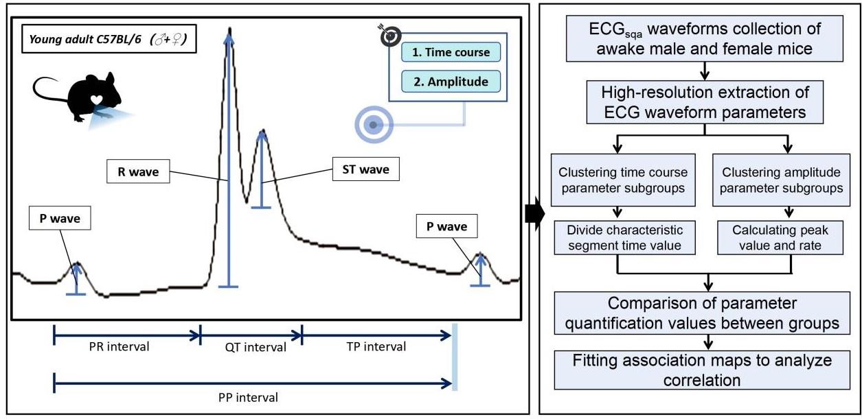
Fig.1 Characteristic parameters and mathematical application pathway of ECGsqa parameters within one single cardiac cycle.
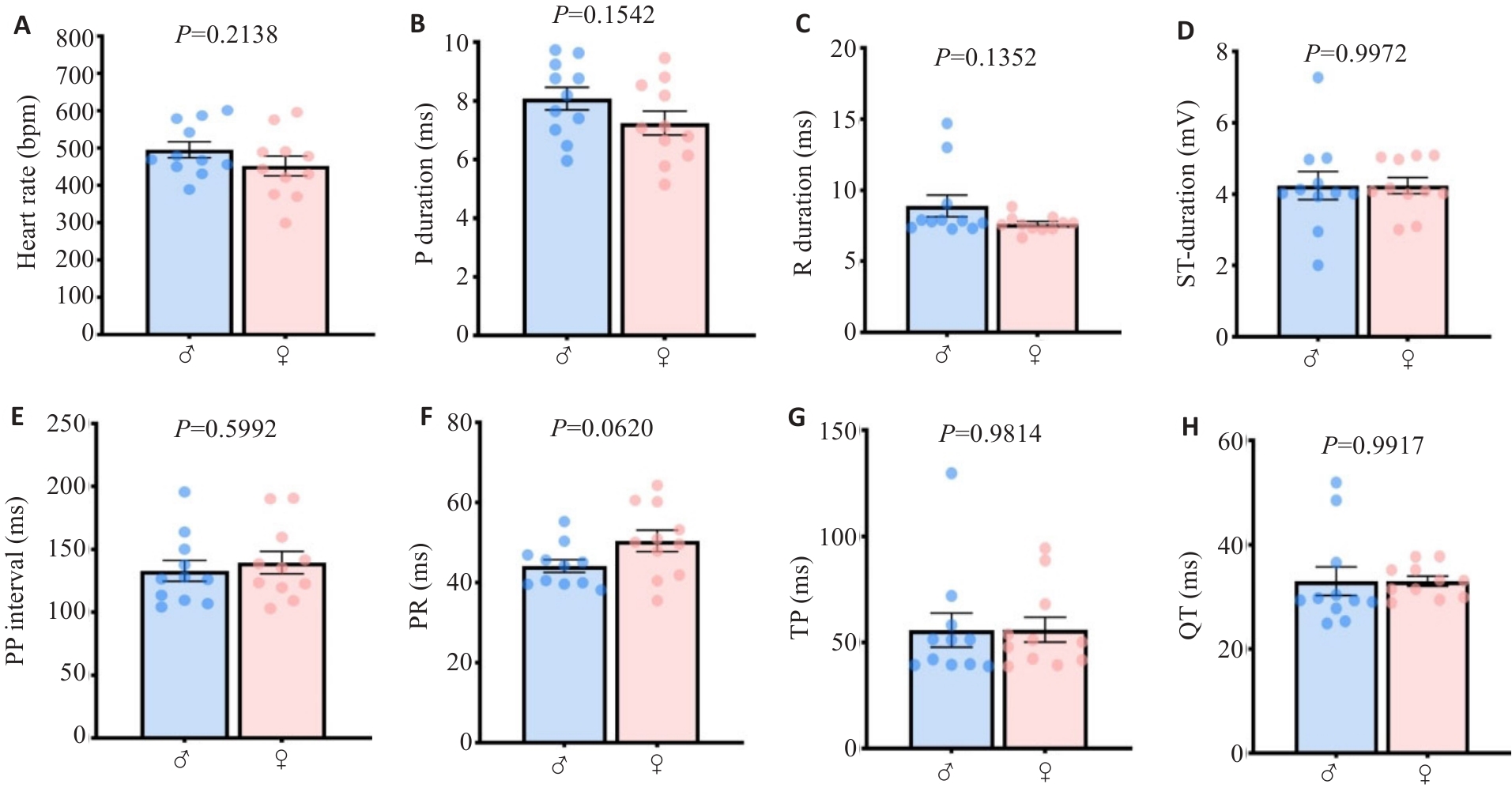
Fig. 2 The main quantified ECGsqa time course parameters in mice are conserved between genders. A: Heart rate. B: P wave rising segment duration. C: R wave rising segment duration. D: ST rising segment duration. E: PP interval. F: PR interval. G: P interval. H: QT interval.
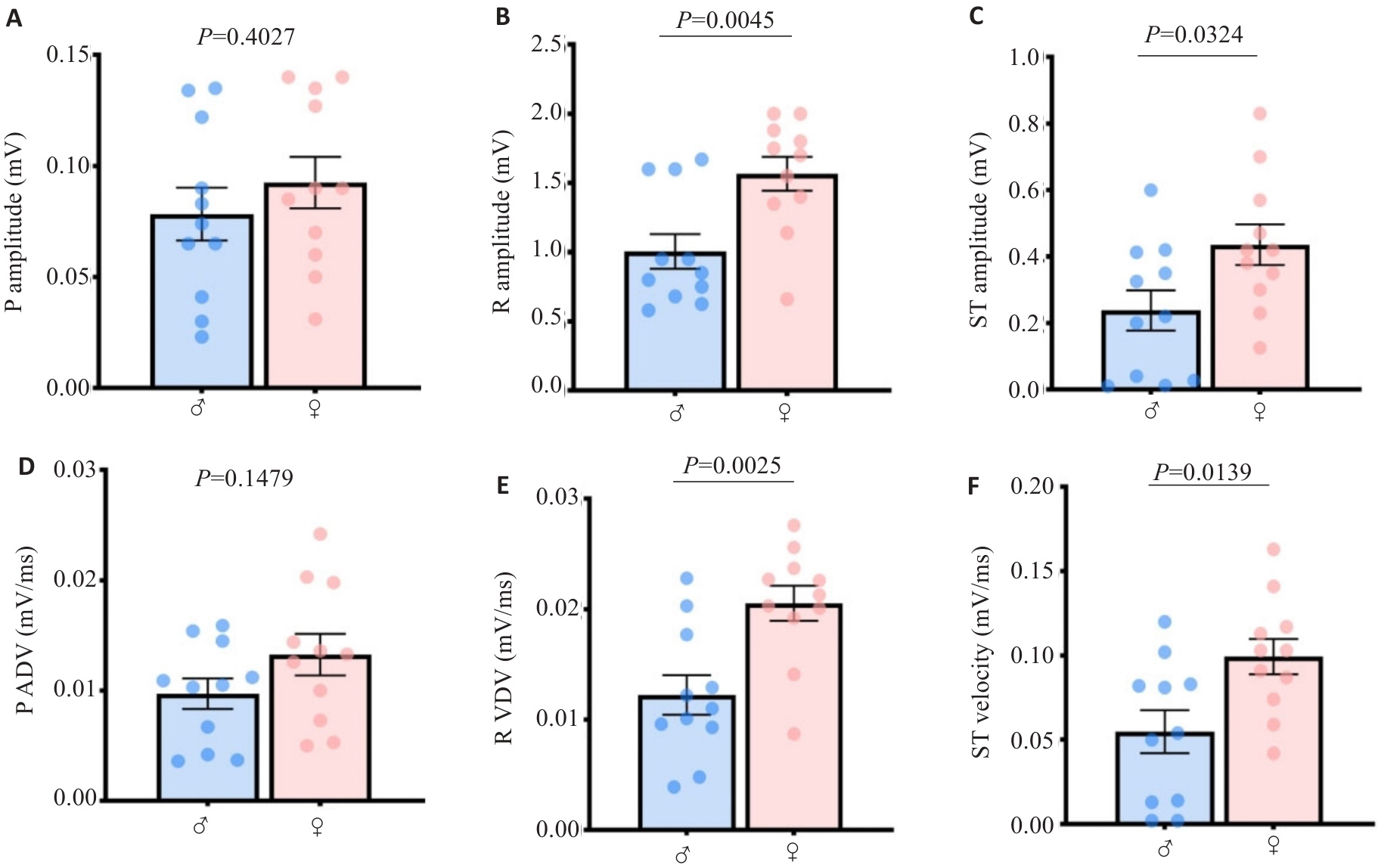
Fig.3 Quantitative values of atrioventricular ECGsqa amplitude and velocity in mice harbor mathematical characteristics of gender differences. A: P wave amplitude. B: R wave amplitude. C: ST segment amplitude. D: P wave velocity. E: R wave velocity. F: ST segment velocity.
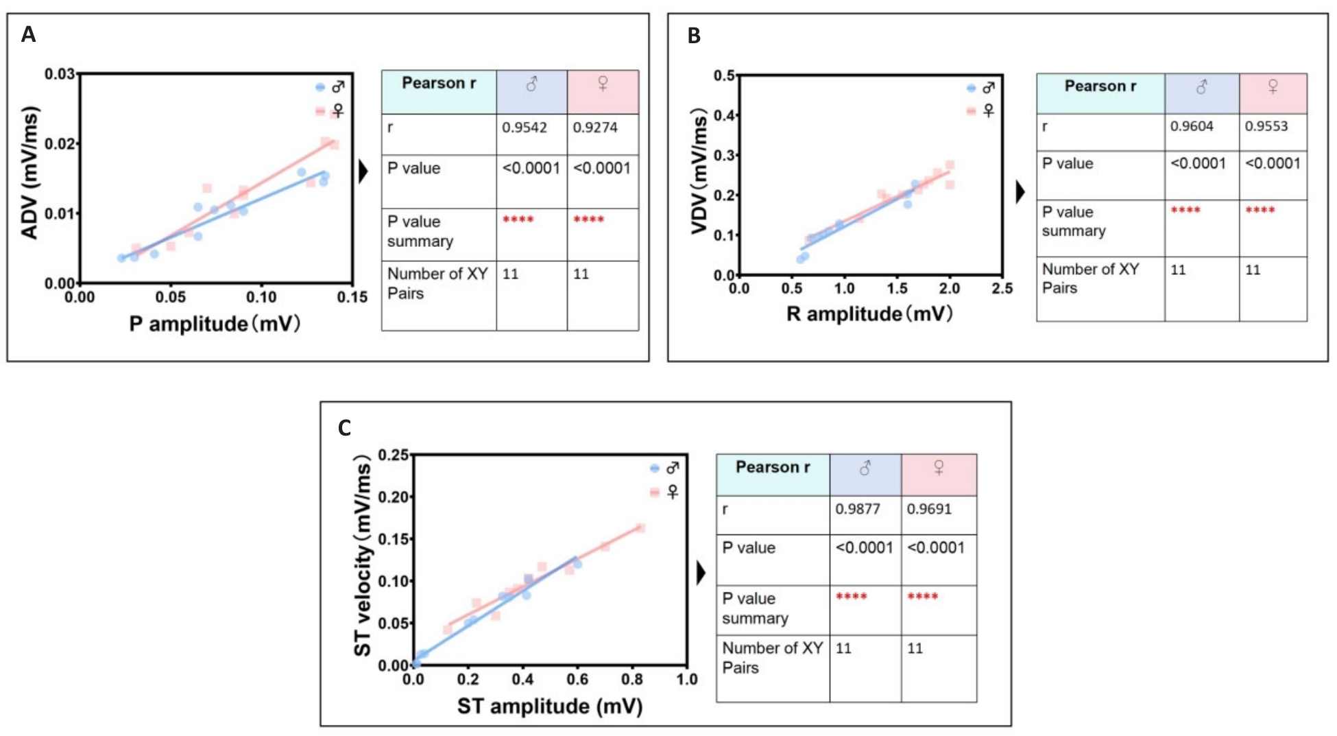
Fig.4 Identification group of the most significant linear correlation parameters in ECGsqa of both male and female mice. A: P wave amplitude is positively correlated with atrial depolarization rate (ADV). B: R wave amplitude is positively correlated with ventricular depolarization rate (VDV). C: ST segment amplitude is positively correlated with ventricular ST segment velocity.
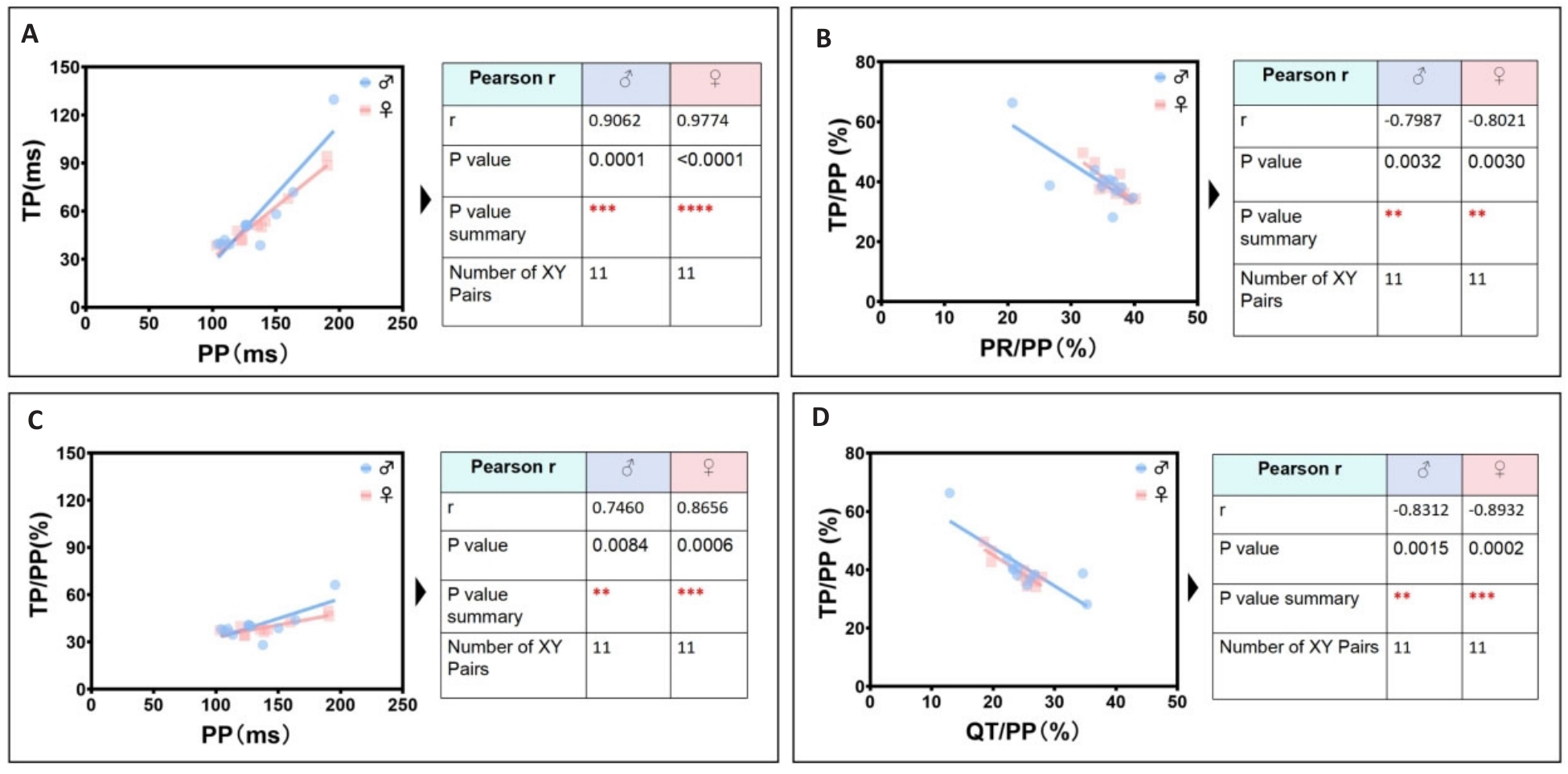
Fig.5 Identification group of secondary significant linear correlation parameters in ECGsqa of both male and female mice. A: PP interval is positively correlated with TP interval. B: PR/PP ratio is negatively correlated with TP/PP ratio. C: PP interval is positively correlated with TP/PP ratio. D: QT/PP ratio is negatively correlated with TP/ PP ratio.
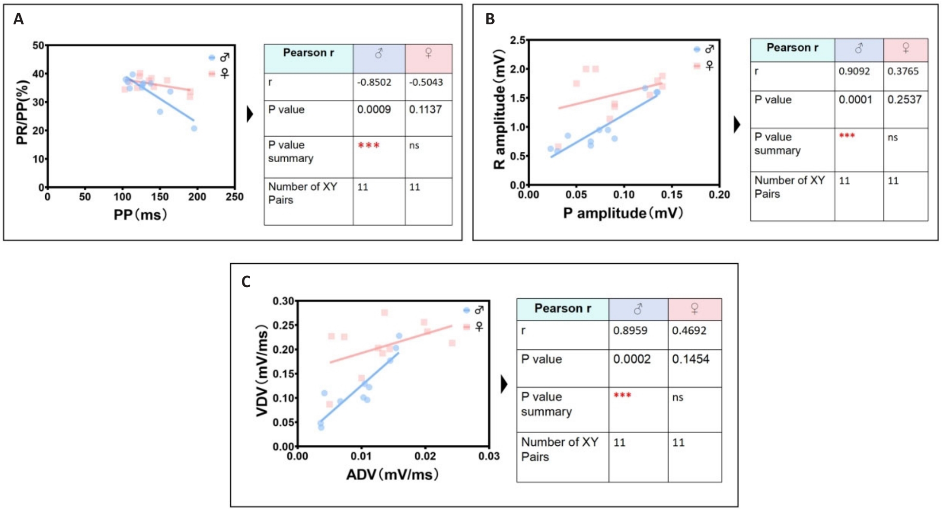
Fig.6 The most significant unique identification group of ECG linear parameters related to male mice. A: Male PP interval is negatively correlated with male PR/PP ratio. B: Male P wave amplitude is positively correlated with male R wave amplitude. C: Male ADV is positively correlated with male VDV.
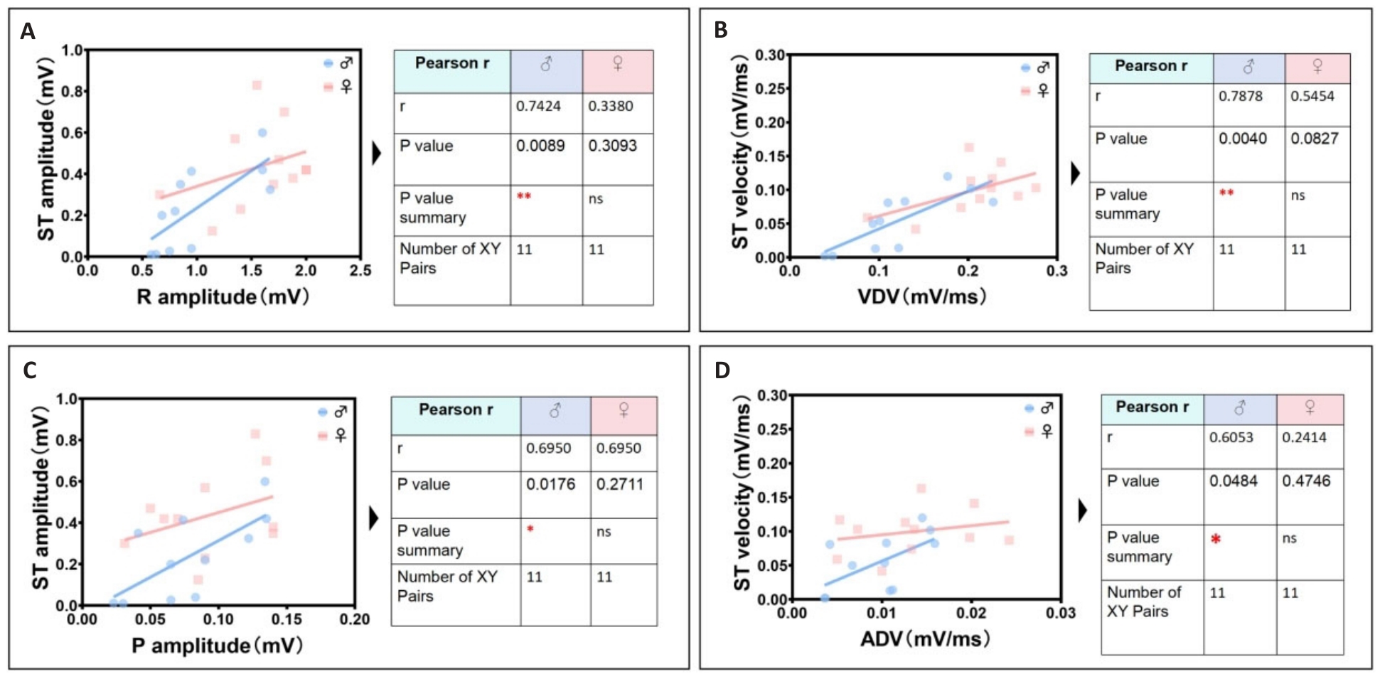
Fig.7 Identification group of unique cardiac electrical linearity-related parameters in male mice. A: Male R wave amplitude is positively correlated with male ST segment amplitude. B: Male VDV is positively correlated with male ST segment velocity. C: Male P wave amplitude is positively correlated with male ST segment amplitude. D: Male ADV is positively correlated with male ST segment velocity.
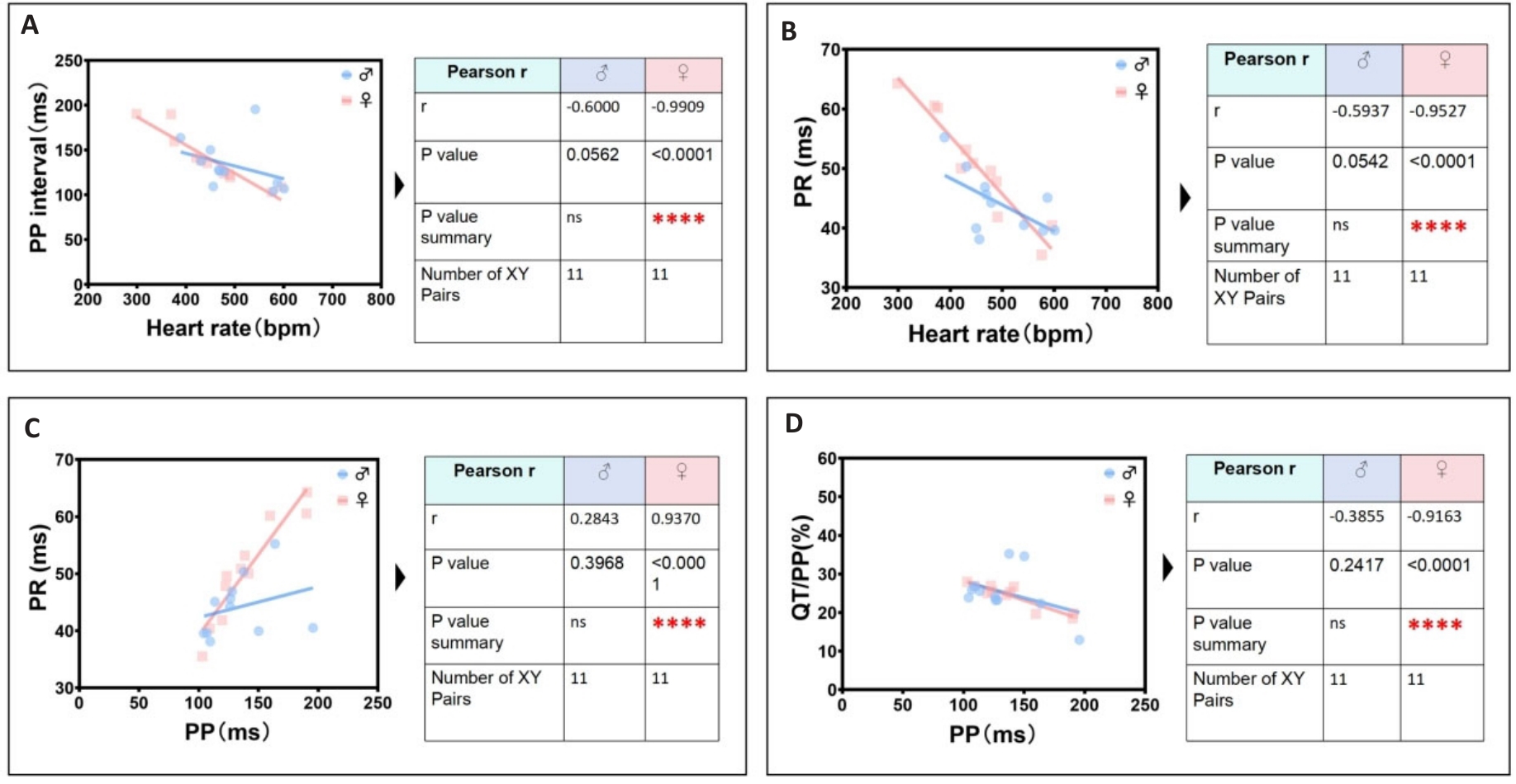
Fig.8 The most significant unique identification group of ECG linearity-related parameters in female mice. A: Female heart rate is negatively correlated with female PP interval. B: Female heart rate is negatively correlated with female PR interval. C: Female PP interval is positively correlated with female PR interval. D: Female PP interval is negatively correlated with female QT/PP ratio.
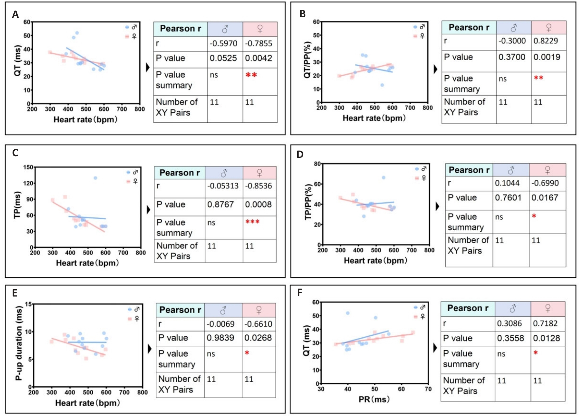
Fig.9 Secondary significant unique identification group of ECG linearity-related parameters in female mice. A: Female heart rate is negatively correlated with female QT interval. B: Female heart rate is positively correlated with female QT/PP ratio. C: Female heart rate is negatively correlated with female TP ratio. D: Female heart rate is negatively correlated with female TP/PP ratio. E: Heart rate is negatively correlated the peak time of rising P wave in female. F: Female PR interval is positively correlated with female QT interval.
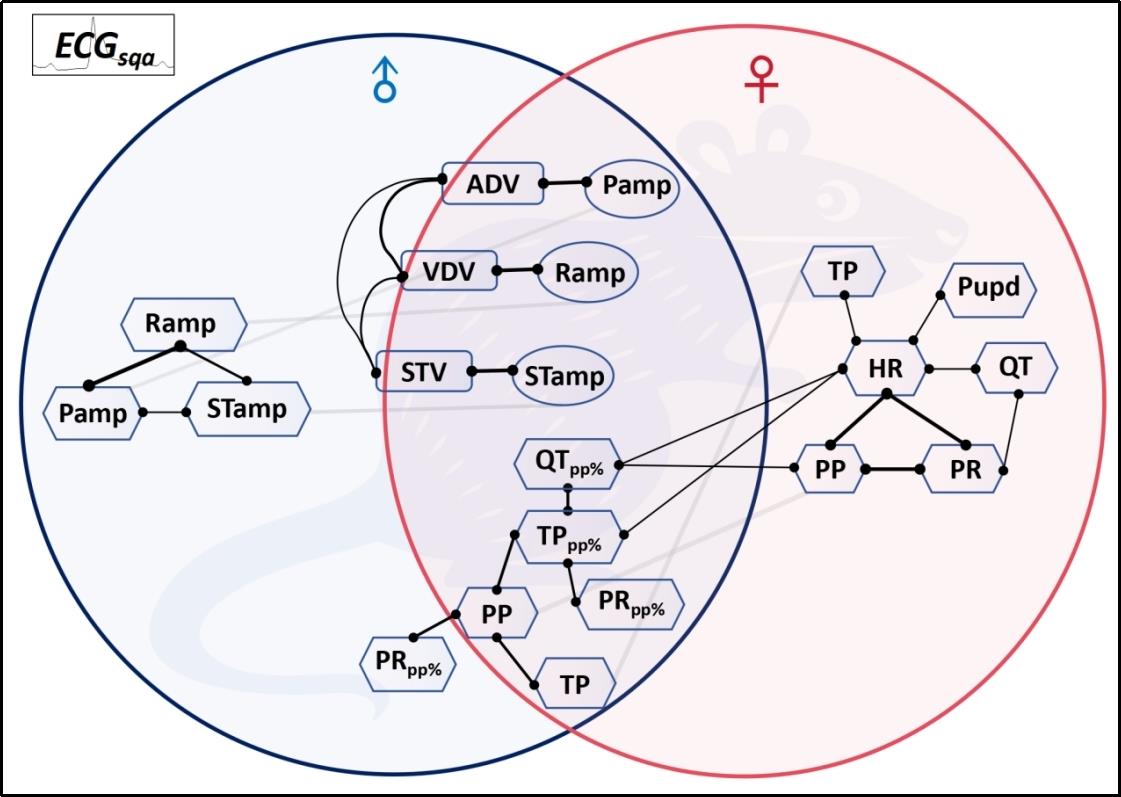
Fig.10 Quantitative ECG correlation parameter network characterizes the intrinsic physiological homeostasis pattern of the mouse cardiac electrical conduction system.
| 1 | Martin SS, Aday AW, Almarzooq ZI, et al. 2024 heart disease and stroke statistics: a report of US and global data from the American heart association[J]. Circulation, 2024, 149(8): e347-60. |
| 2 | Goette A, Auricchio A, Boriani G, et al. EHRA White Paper: knowledge gaps in arrhythmia management-status 2019[J]. Europace, 2019, 21(7): 993-4. |
| 3 | Thomas RJ. Cardiac rehabilitation-challenges, advances, and the road ahead[J]. N Engl J Med, 2024, 390(9): 830-41. |
| 4 | Kowey PR, Naccarelli GV. Antiarrhythmic drug therapy: where do we go from here?[J]. Circulation, 2024, 149(11): 801-3. |
| 5 | Nogami A, Kurita T, Abe H, et al. JCS/JHRS 2019 guideline on non-pharmacotherapy of cardiac arrhythmias[J]. J Arrhythm, 2021, 37(4): 709-870. |
| 6 | Kingma J, Simard C, Drolet B. Overview of cardiac arrhythmias and treatment strategies[J]. Pharmaceuticals, 2023, 16(6): 844. |
| 7 | Fye WB. A history of the origin, evolution, and impact of electrocardiography [J]. Am J Cardiol, 1994, 73(13):937-49. |
| 8 | Jahmunah V, Oh SL, Wei JKE, et al. Computer-aided diagnosis of congestive heart failure using ECG signals‑A review[J]. Phys Med, 2019, 62: 95-104. |
| 9 | Oestereicher MA, Wotton JM, Ayabe S, et al. Comprehensive ECG reference intervals in C57BL/6N substrains provide a generalizable guide for cardiac electrophysiology studies in mice[J]. Mamm Genome, 2023, 34(2): 180-99. |
| 10 | Obergassel J, O'Reilly M, Sommerfeld LC, et al. Effects of genetic background, sex, and age on murine atrial electrophysiology[J]. Europace, 2021, 23(6): 958-69. |
| 11 | Haq KT, Cooper BL, Berk F, et al. The effect of sex and age on ex vivo cardiac electrophysiology: insight from a guinea pig model[J]. Am J Physiol Heart Circ Physiol, 2023, 324(1): H141-54. |
| 12 | Ahmadi P, Afzalian A, Jalali A, et al. Age and gender differences of basic electrocardiographic values and abnormalities in the general adult population; Tehran Cohort Study[J]. BMC Cardiovasc Disord, 2023, 23(1): 303. |
| 13 | Xie M, Zhu SJ, Liu G, et al. A novel quantitative electrocardiography strategy reveals the electroinhibitory effect of tamoxifen on the mouse heart[J]. J Cardiovasc Transl Res, 2023, 16(5): 1232-48. |
| 14 | Jia BZ, Qi YT, Wong-Campos JD, et al. A bioelectrical phase transition patterns the first vertebrate heartbeats[J]. Nature, 2023, 622(7981): 149-55. |
| 15 | Levin M. Bioelectric signaling: Reprogrammable circuits underlying embryogenesis, regeneration, and cancer[J]. Cell, 2021, 184(8): 1971-89. |
| 16 | Gajendragadkar PR, von Ende A, Ibrahim M, et al. Assessment of the causal relevance of ECG parameters for risk of atrial fibrillation: a Mendelian randomisation study[J]. PLoS Med, 2021, 18(5): e1003572. |
| 17 | Amuzescu B, Airini R, Epureanu FB, et al. Evolution of mathematical models of cardiomyocyte electrophysiology[J]. Math Biosci, 2021, 334: 108567. |
| 18 | Lawson BAJ, Drovandi CC, Cusimano N, et al. Unlocking data sets by calibrating populations of models to data density: a study in atrial electrophysiology[J]. Sci Adv, 2018, 4(1): e1701676. |
| 19 | Morotti S, Liu C, Hegyi B, et al. Quantitative cross-species translators of cardiac myocyte electrophysiology: model training, experimental validation, and applications[J]. Sci Adv, 2021, 7(47): eabg0927. |
| 20 | Mazhar F, Bartolucci C, Regazzoni F, et al. A detailed mathematical model of the human atrial cardiomyocyte: integration of electrophysiology and cardiomechanics[J]. J Physiol, 2024, 602(18): 4543-83. |
| 21 | Ntalla I, Weng LC, Cartwright JH, et al. Multi-ancestry GWAS of the electrocardiographic PR interval identifies 202 loci underlying cardiac conduction[J]. Nat Commun, 2020, 11(1): 2542. |
| 22 | van Duijvenboden S, Ramírez J, Young WJ, et al. Genomic and pleiotropic analyses of resting QT interval identifies novel loci and overlap with atrial electrical disorders[J]. Hum Mol Genet, 2021, 30(24): 2513-23. |
| 23 | Liu G, Iden JB, Kovithavongs K, et al. In vivo temporal and spatial distribution of depolarization and repolarization and the illusive murine T wave[J]. J Physiol, 2004, 555(Pt 1): 267-79. |
| 24 | Rodrigues JC, McIntyre B, Dastidar AG, et al. The effect of obesity on electrocardiographic detection of hypertensive left ventricular hypertrophy: recalibration against cardiac magnetic resonance[J]. J Hum Hypertens, 2016, 30(3): 197-203. |
| 25 | de Coster M, Demolder A, de Meyer V, et al. Diagnostic accuracy of R-wave detection by insertable cardiac monitors[J]. Pacing Clin Electrophysiol, 2020, 43(5): 511-7. |
| 26 | Ramírez J, van Duijvenboden S, Young WJ, et al. Common genetic variants modulate the electrocardiographic tpeak-to-tend interval[J]. Am J Hum Genet, 2020, 106(6): 764-78. |
| 27 | Yogasundaram H, Zheng YG, Ly E, et al. Relationship between baseline electrocardiographic measurements and outcomes in patients with high-risk heart failure: insights from the VerICiguaT Global Study in Subjects with Heart Failure with Reduced Ejection Fraction (VICTORIA) trial[J]. Eur J Heart Fail, 2023, 25(10): 1822-30. |
| 28 | Mayourian J, la Cava WG, Vaid A, et al. Pediatric ECG-based deep learning to predict left ventricular dysfunction and remodeling[J]. Circulation, 2024, 149(12): 917-31. |
| 29 | Ardissino M, Patel KHK, Rayes B, et al. Multiple anthropometric measures and proarrhythmic 12-lead ECG indices: a Mendelian randomization study[J]. PLoS Med, 2023, 20(8): e1004275. |
| 30 | Chen LY, Ribeiro ALP, Platonov PG, et al. P wave parameters and indices: a critical appraisal of clinical utility, challenges, and future research-a consensus document endorsed by the international so-ciety of electrocardiology and the international society for holter and noninvasive electrocardiology[J]. Circ Arrhythm Electrophysiol, 2022, 15(4): e010435. |
| 31 | Young WJ, Lahrouchi N, Isaacs A, et al. Genetic analyses of the electrocardiographic QT interval and its components identify additional loci and pathways[J]. Nat Commun, 2022, 13(1): 5144. |
| 32 | Broman MT, Nadadur RD, Perez-Cervantes C, et al. A genomic link from heart failure to atrial fibrillation risk: FOG2 modulates a TBX5/GATA4-dependent atrial gene regulatory network[J]. Circulation, 2024, 149(15): 1205-30. |
| 33 | Frimodt-M ller EK, Soliman EZ, Kizer JR, et al. Lifestyle habits associated with cardiac conduction disease[J]. Eur Heart J, 2023, 44(12): 1058-66. |
| 34 | Gottlieb LA, Larsen K, Halade GV, et al. Prolonged QT intervals in mice with cardiomyocyte-specific deficiency of the molecular clock[J]. Acta Physiol, 2021, 233(1): e13707. |
| 35 | Calò L, Crescenzi C, Martino A, et al. The diagnostic value of the 12-LeadECGin arrhythmogenic LeftVentricularCardiomyopathy: novel ECG signs[J]. JACC Clin Electrophysiol, 2023, 9(12): 2615-27. |
| 36 | Nam JM, Lim JE, Ha TW, et al. Cardiac-specific inactivation of Prdm16 effects cardiac conduction abnormalities and cardiomyopathy-associated phenotypes[J]. Am J Physiol Heart Circ Physiol, 2020, 318(4): H764-77. |
| 37 | Karakayali M, Artac I, Omar T, et al. Assessment of the efficacy of the electrocardiographic P-wave peak time in predicting atrial high rate episode in patients with cardiac implantable electronic devices[J]. J Electrocardiol, 2023, 80: 40-4. |
| 38 | Hennis K, Rötzer RD, Rilling J, et al. In vivo and ex vivo electrophysiological study of the mouse heart to characterize the cardiac conduction system, including atrial and ventricular vulnerability[J]. Nat Protoc, 2022, 17(5): 1189-222. |
| 39 | Litviňuková M, Talavera-López C, Maatz H, et al. Cells of the adult human heart[J]. Nature, 2020, 588(7838): 466-72. |
| 40 | Li Q, Lin ZW, Liu R, et al. Multimodal charting of molecular and functional cell states via in situ electro-sequencing[J]. Cell, 2023, 186(9): 2002-17. e21. |
| 41 | Jagannatha GNP, Antara IMPS, Kosasih AM, et al. P-wave peak time and P-wave dispersion in surface electrocardiography as initial predictors of new-onset atrial fibrillation in early-onset hypertension[J]. Hypertens Res, 2024, 47(1): 137-48. |
| 42 | Feeny AK, Rickard J, Trulock KM, et al. Machine learning of 12-lead QRS waveforms to identify cardiac resynchronization therapy patients with differential outcomes[J]. Circ Arrhythm Electrophysiol, 2020, 13(7): e008210. |
| 43 | Hnatkova K, Andršová I, Novotný T, et al. QRS micro-fragmentation as a mortality predictor[J]. Eur Heart J, 2022, 43(40): 4177-91. |
| 44 | Chen N, Wang L, Jiao JC, et al. RV1+RV3 index to differentiate idiopathic ventricular arrhythmias arising from right ventricular outflow tract and aortic sinus of Valsalva: a multicenter study[J]. J Am Heart Assoc, 2024, 13(7): e033779. |
| 45 | Wallet J, Kimura Y, Blom NA, et al. The R″ wave in V1 and the negative terminal QRS vector in aVF combine to a novel 12-lead ECG algorithm to identify slow conducting anatomical isthmus 3 in patients with tetralogy of Fallot[J]. Europace, 2023, 25(6): euad139. |
| [1] | Na YE, Chenwen WU, Jialin JIANG. A lung sound classification model with a spatial and channel reconstruction convolutional module [J]. Journal of Southern Medical University, 2024, 44(9): 1720-1728. |
| [2] | . Value of ultrasound shear wave elasticity imaging in diagnosis of Hashimoto’s thyroiditis [J]. Journal of Southern Medical University, 2017, 37(05): 683-. |
| [3] | . Assessment of plantar fasciitis using shear wave elastography [J]. Journal of Southern Medical University, 2014, 34(02): 206-. |
| Viewed | ||||||
|
Full text |
|
|||||
|
Abstract |
|
|||||