
南方医科大学学报 ›› 2026, Vol. 46 ›› Issue (6): 1216-1227.doi: 10.12122/j.issn.1673-4254.2026.06.02
• • 上一篇
王伟丽( ), 高怡琳, 洪馨, 陈义珍, 程梦, 张磊, 戴荣(), 王亿平()
收稿日期:2025-12-02
出版日期:2026-06-20
发布日期:2026-06-24
通讯作者:
戴荣,王亿平
E-mail:wang37937379@163.com;azydairong@163.com;wypwyp54@aliyun.com
作者简介:王伟丽,博士,医师,E-mail: wang37937379@163.com
基金资助:
Weili WANG(), Yilin GAO, Xin HONG, Yizhen CHEN, Meng CHENG, Lei ZHANG, Rong DAI(), Yiping WANG()
Received:2025-12-02
Online:2026-06-20
Published:2026-06-24
Contact:
Rong DAI, Yiping WANG
E-mail:wang37937379@163.com;azydairong@163.com;wypwyp54@aliyun.com
Supported by:摘要:
目的 探究清肾颗粒抑制肾纤维化的作用机制,明确其是否通过调控 Akt3 介导的巨噬细胞极化发挥效应。 方法 体内构建单侧输尿管梗阻(UUO)小鼠肾纤维化模型,分为假手术组、模型组、清肾颗粒治疗组[4.0 g·kg-1·d-1)],6只/组,灌胃2周;通过 AAV9 介导肾脏Akt3 敲低,设置 Sham+NC、Sham+shAkt3、Model+NC、Model+shAkt3组;体外以TGF-β1 诱导 HK-2细胞纤维化,用清肾颗粒含药血清干预,同时构建HK-2细胞与THP-1来源巨噬细胞的共培养体系。采用 HE、Masson 染色观察肾脏病理变化;ELISA 检测血清肌酐(Scr)、尿素氮(BUN);Western blotting、RT-qPCR检测Akt3、α-SMA、E-cad、IL-6、TNF-α蛋白及 mRNA 表达;免疫组化、免疫荧光验证蛋白定位;流式细胞术检测巨噬细胞极化表型;CCK-8、EdU 实验评估细胞活力与增殖能力。 结果 体内实验表明,与假手术组相比,模型组小鼠肾功能显著受损,Scr、BUN水平升高(P<0.05),伴肾小管损伤、肾间质胶原纤维沉积,肾组织IL-6、TNF-α的mRNA及α-SMA蛋白表达上调, E-cad蛋白表达下调(P<0.05);与模型组相比,清肾颗粒治疗组上述指标均改善(P<0.05)。AAV9介导肾脏特异性Akt3敲低实验中,与Sham+NC组相比,Model+NC组小鼠肾纤维化加重,肾组织M1型巨噬细胞占比升高(P<0.05),M2型无显著变化(P>0.05);与Model+NC组相比,Model+ shAkt3敲低组肾纤维化减轻,M1型巨噬细胞极化受抑制(P<0.05),M2型仍无明显差异(P>0.05);清肾颗粒显著下调肾组织 Akt3 蛋白表达,降低 M1 型巨噬细胞比例(P<0.05)。体外实验中,与对照组相比,模型组细胞活力、增殖能力下降,Akt3、α-SMA蛋白上调,E-cad蛋白下调,清肾颗粒含药血清组可逆转上述变化(P<0.05);流式细胞术检测显示,TGF-β1诱导的HK-2细胞可促进M0巨噬细胞向M1型极化,清肾颗粒含药血清干预后M1型极化被抑制(P<0.05)。 结论 清肾颗粒通过抑制Akt3介导的M1型巨噬细胞极化,发挥减轻UUO小鼠肾纤维化的作用。
王伟丽, 高怡琳, 洪馨, 陈义珍, 程梦, 张磊, 戴荣, 王亿平. 清肾颗粒通过调控Akt3介导的M1型巨噬细胞极化改善单侧输尿管梗阻小鼠肾纤维化[J]. 南方医科大学学报, 2026, 46(6): 1216-1227.
Weili WANG, Yilin GAO, Xin HONG, Yizhen CHEN, Meng CHENG, Lei ZHANG, Rong DAI, Yiping WANG. Qingshen Granules alleviate renal fibrosis in mice with unilateral ureteral obstruction by regulating Akt3-mediated M1 macrophage polarization[J]. Journal of Southern Medical University, 2026, 46(6): 1216-1227.
| Gene | Forwardprimer (5'→3') | Reverseprimer (5'→3') |
|---|---|---|
| Mo-β-actin | AGTGTGACGTTGACATCCGT | TGCTAGGAGCCAGAGCAGTA |
| Mo-TNF-α | CTCATGCACCACCATCAAGG | ACCTGACCACTCTCCCTTTG |
| Mo-IL-6 | GGAGCCCACCAAGAACGATA | ACCAGCATCAGTCCCAAGAA |
| Mo-α-SMA | GTCCCAGACATCAGGGAGTAA | TCGGATACTTCAGCGTCAGGA |
| Mo-E-cad | GTCCTGGGCAGAGTGAGATT | CGTCTGTCGCCACTTTGAAT |
表1 引物序列
Tab.1 Primer sequences (5' to 3') for RT-qPCR
| Gene | Forwardprimer (5'→3') | Reverseprimer (5'→3') |
|---|---|---|
| Mo-β-actin | AGTGTGACGTTGACATCCGT | TGCTAGGAGCCAGAGCAGTA |
| Mo-TNF-α | CTCATGCACCACCATCAAGG | ACCTGACCACTCTCCCTTTG |
| Mo-IL-6 | GGAGCCCACCAAGAACGATA | ACCAGCATCAGTCCCAAGAA |
| Mo-α-SMA | GTCCCAGACATCAGGGAGTAA | TCGGATACTTCAGCGTCAGGA |
| Mo-E-cad | GTCCTGGGCAGAGTGAGATT | CGTCTGTCGCCACTTTGAAT |
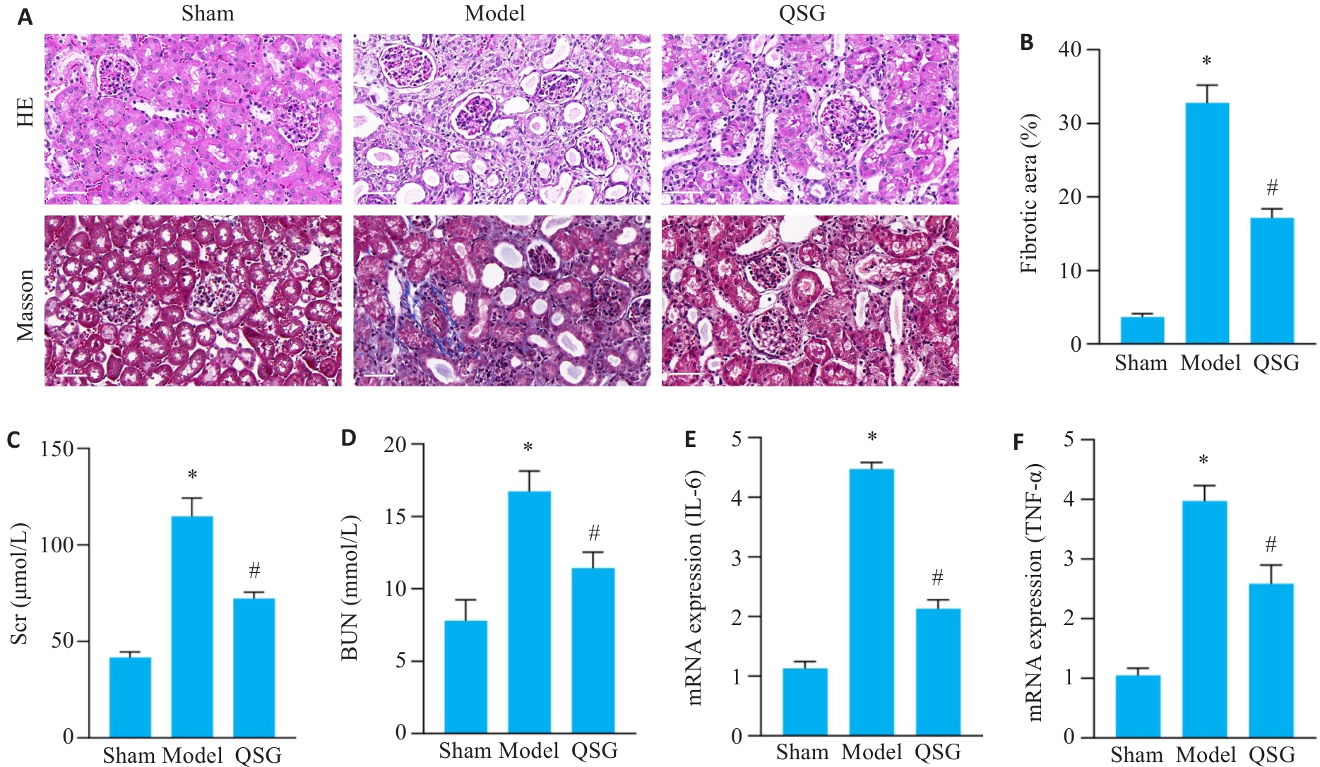
图1 清肾颗粒改善UUO小鼠肾功能,减轻肾纤维化与炎症反应
Fig.1 Qingshen Granules (QSG) improve renal function and alleviate renal fibrosis in UUO mice. A, B: Results of HE staining and Masson staining of the renal tissues of the mice in each group (Scale bar=50 µm, n=6). C, D: Scr and BUN levels of the mice (n=6). E, F: RT-qPCR detection results of IL-6 and TNF-α mRNA in each group (n=6). *P<0.05 vs Sham group; #P<0.05 vs Model group.
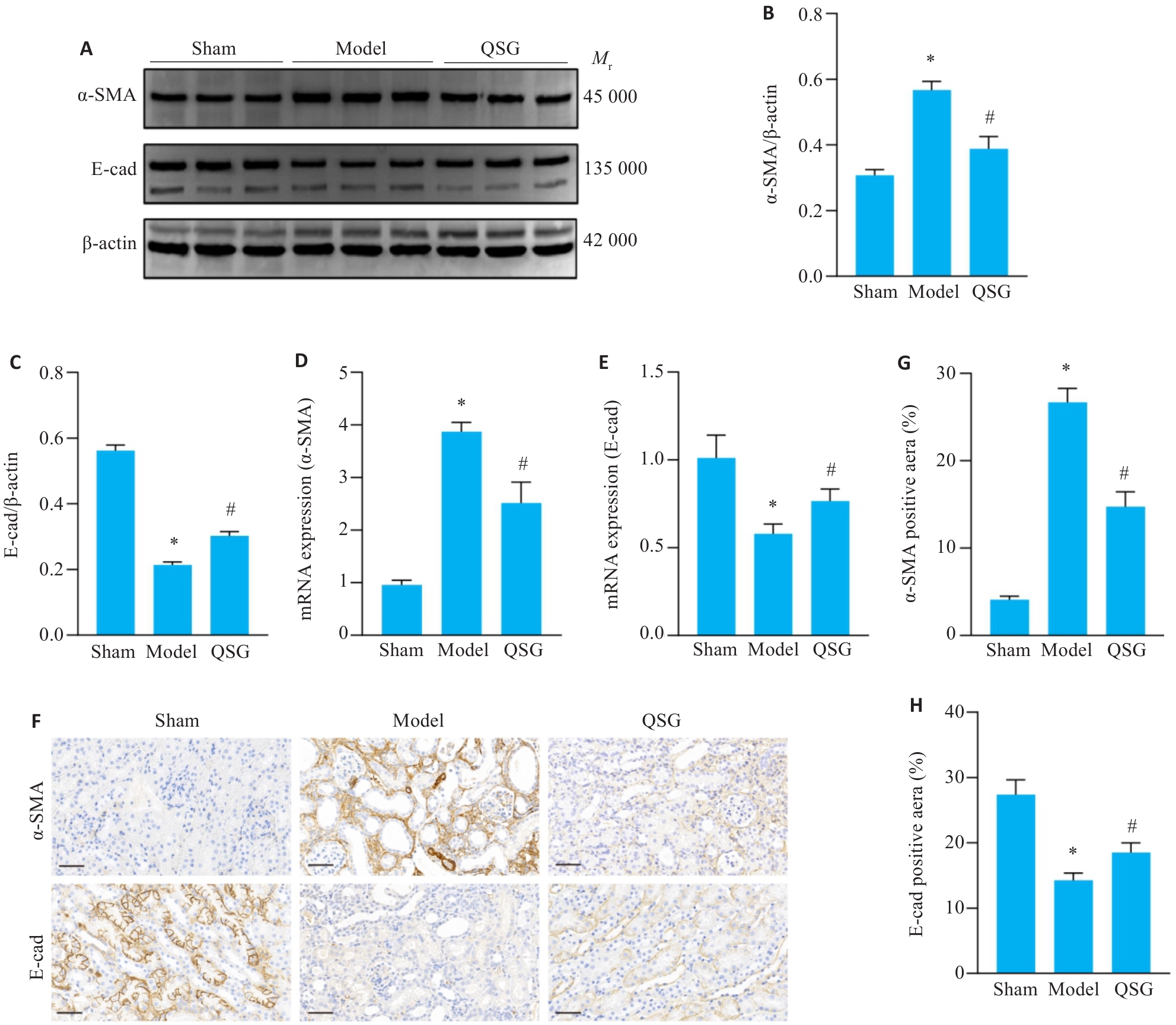
图2 清肾颗粒对UUO小鼠肾组织中α-SMA、E-cad表达的影响
Fig. 2 Effect of QSG on expressions of α-SMA and E-cad in renal tissues of UUO mice. A-C: Western blotting for detecting α-SMA and E-cad expression levels in each group (n=3). D, E: RT-qPCR detection results of α-SMA and E-cad mRNA in each group (n=6). F-H: Immunohistochemical staining results for detecting renal expressions of α-SMA and E-cad (Scale bar=50 µm, n=6). *P<0.05 vs Sham group; #P<0.05 vs Model group.
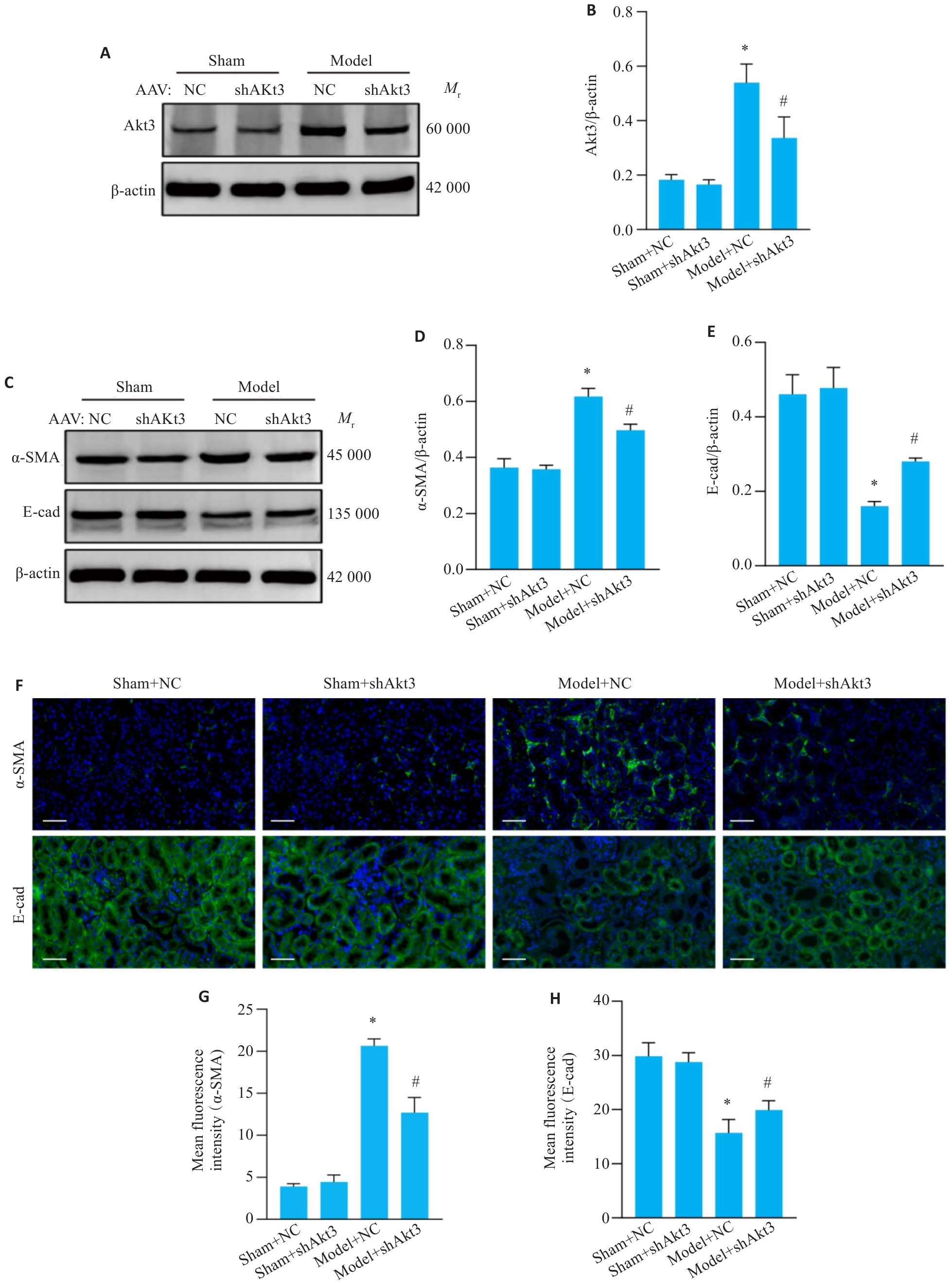
图3 敲低Akt3表达减轻肾纤维化
Fig.3 Knockdown of Akt3 alleviates renal fibrosis in UUO mice. A, B: Western blotting for detecting Akt3 expression level in each group (n=3). C-E: Western blotting for detecting α-SMA and E-cad expression levels in each group (n=3). F-H: Results of immunofluorescence staining of α-SMA and E-cad in renal tissues of the mice in each group (Scale bar = 50 µm, n=6). *P<0.05 vs Sham+NC group; #P<0.05 vs Model+NC group.
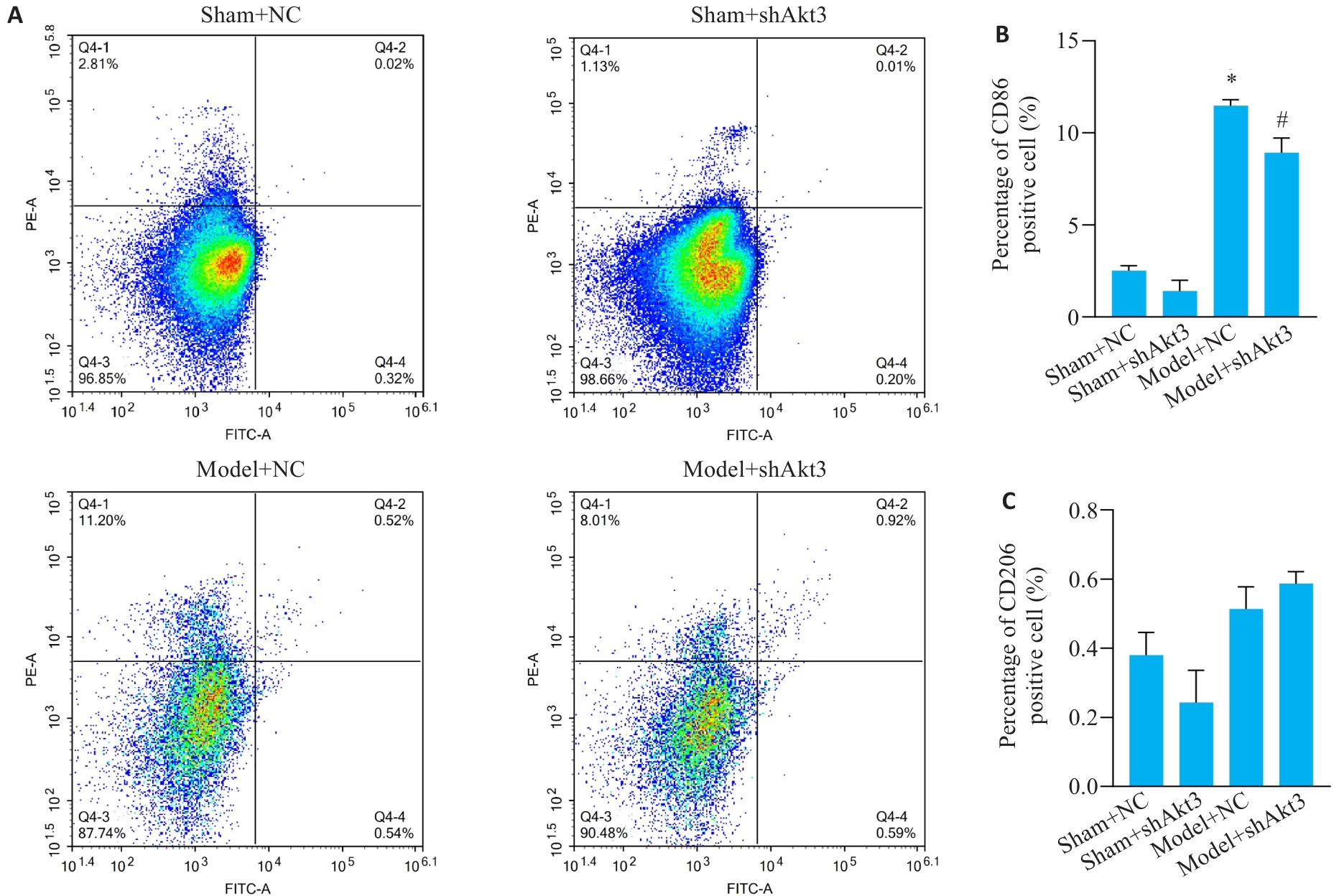
图4 敲低Akt3表达抑制M1巨噬细胞极化
Fig.4 Knockdown of Akt3 expression inhibits M1 macrophage polarization. A-C: Flow cytometric analysis of M1/M2 macrophage expressions in the renal tissues of the mice in each group (n=3). *P<0.05 vs Sham+NC group; #P<0.05 vs Model+NC group.
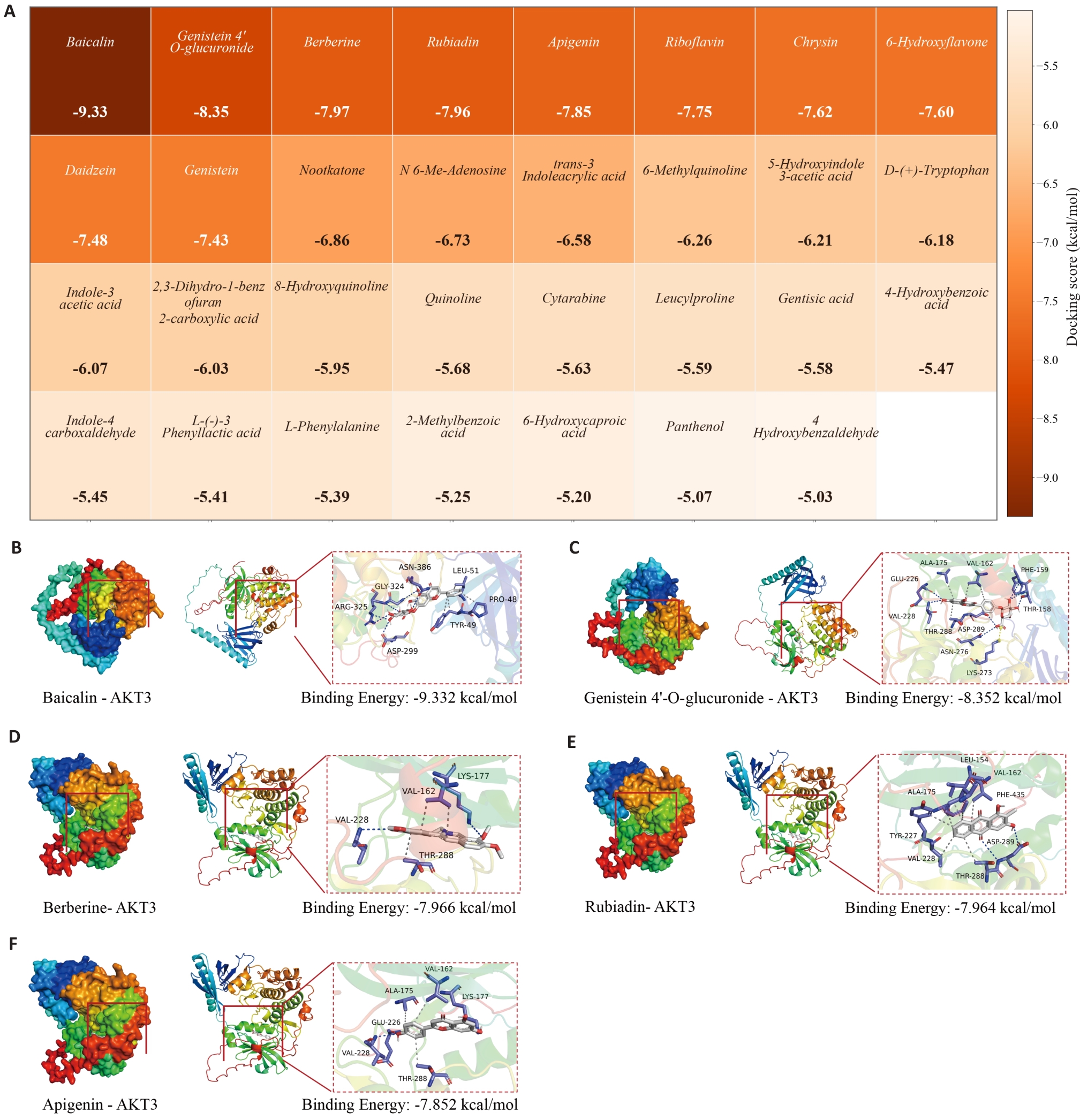
图5 清肾颗粒主要入血成分与Akt3的分子对接结果
Fig.5 Molecular docking results of the major blood-absorbed components of QSG with Akt3. A: Heatmap of molecular docking binding energies for active components of QSG with binding energy <-5.0 kcal/mol against Akt3. B-F: Docking modes and binding site diagrams of Baicalin (B), Genistein 4'-O-glucuronide (C), Berberine (D), Rubiadin (E), and Apigenin (F) with Akt3.
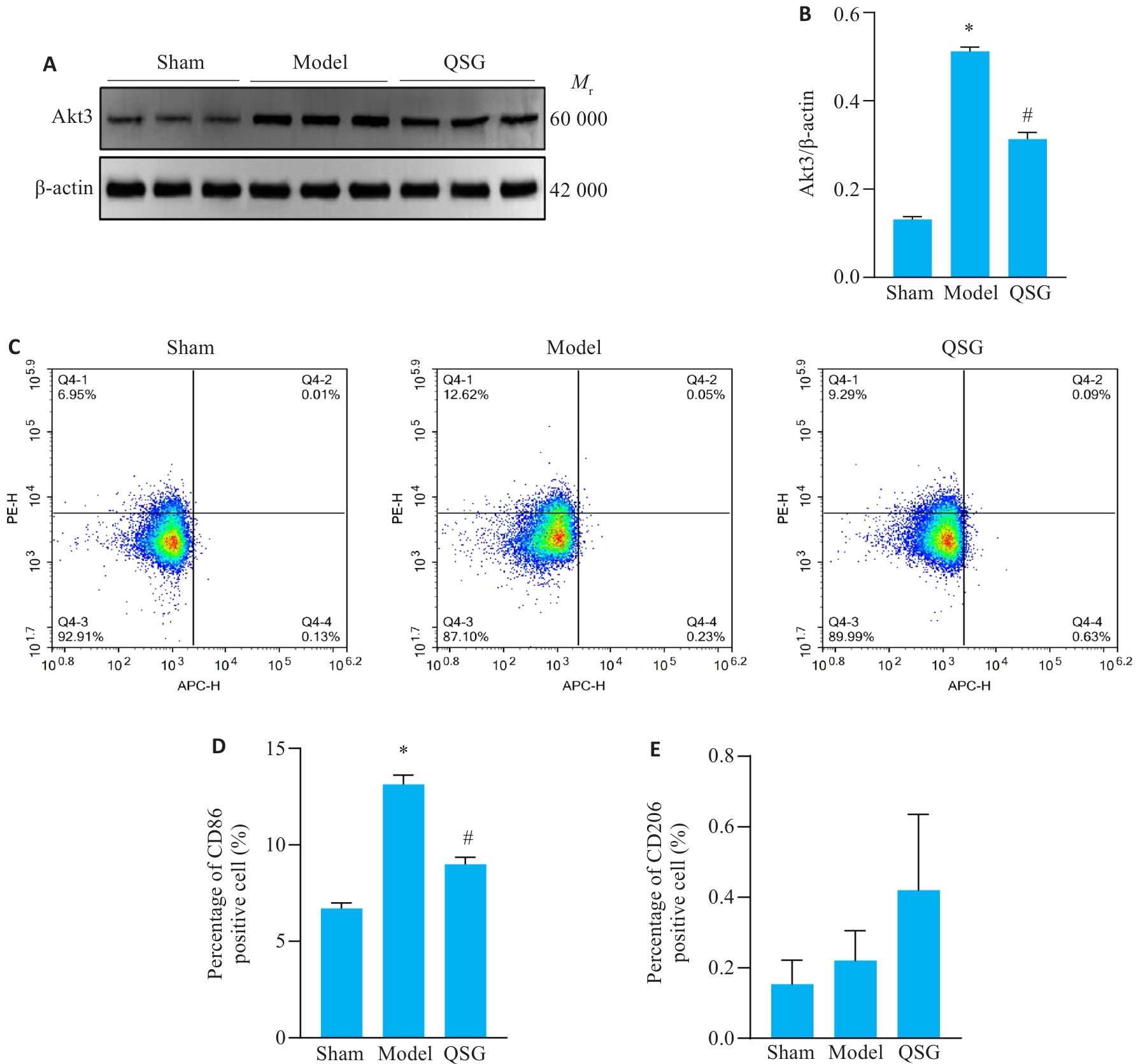
图6 清肾颗粒下调Akt3表达,抑制M1巨噬细胞极化
Fig.6 QSG downregulates Akt3 expression and inhibits M1 macrophage polarization in the renal tissues of UUO mice. A, B: Western blotting for detecting Akt3 expression in each group (n=3). C-E: M1/M2 macrophage expression in the renal tissues of the mice in each group (n=3). *P<0.05 vs Sham group; #P<0.05 vs Model group.
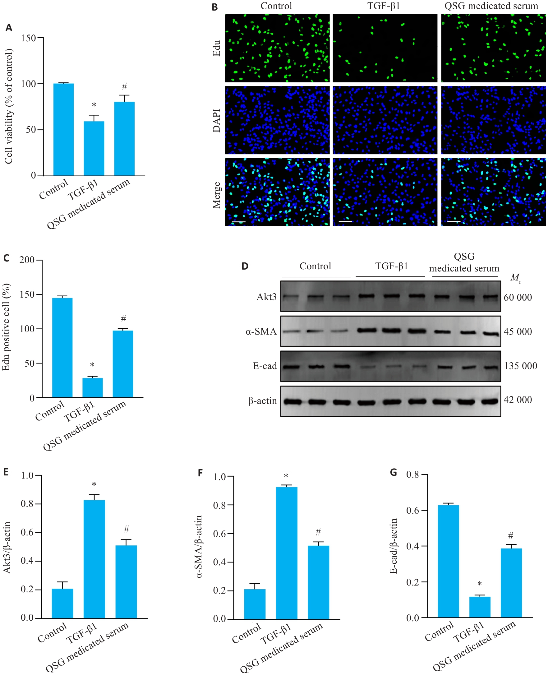
图7 清肾颗粒含药血清下调Akt3表达,抑制 TGF-β1诱导的HK-2细胞纤维化
Fig.7 QSG-medicated serum inhibits Akt3 expression and TGF-β1-induced HK-2 cell fibrosis. A: Cell viability of cells in each group (n=6). B, C: EdU cell proliferation assay (Scale bar=100 μm, n=3). D-G: Western blotting for Akt3, α-SMA, and E-cad expressions in cells from each group (n=3). * P<0.05 vs Control group; #P<0.05 vs TGF-β1 group.
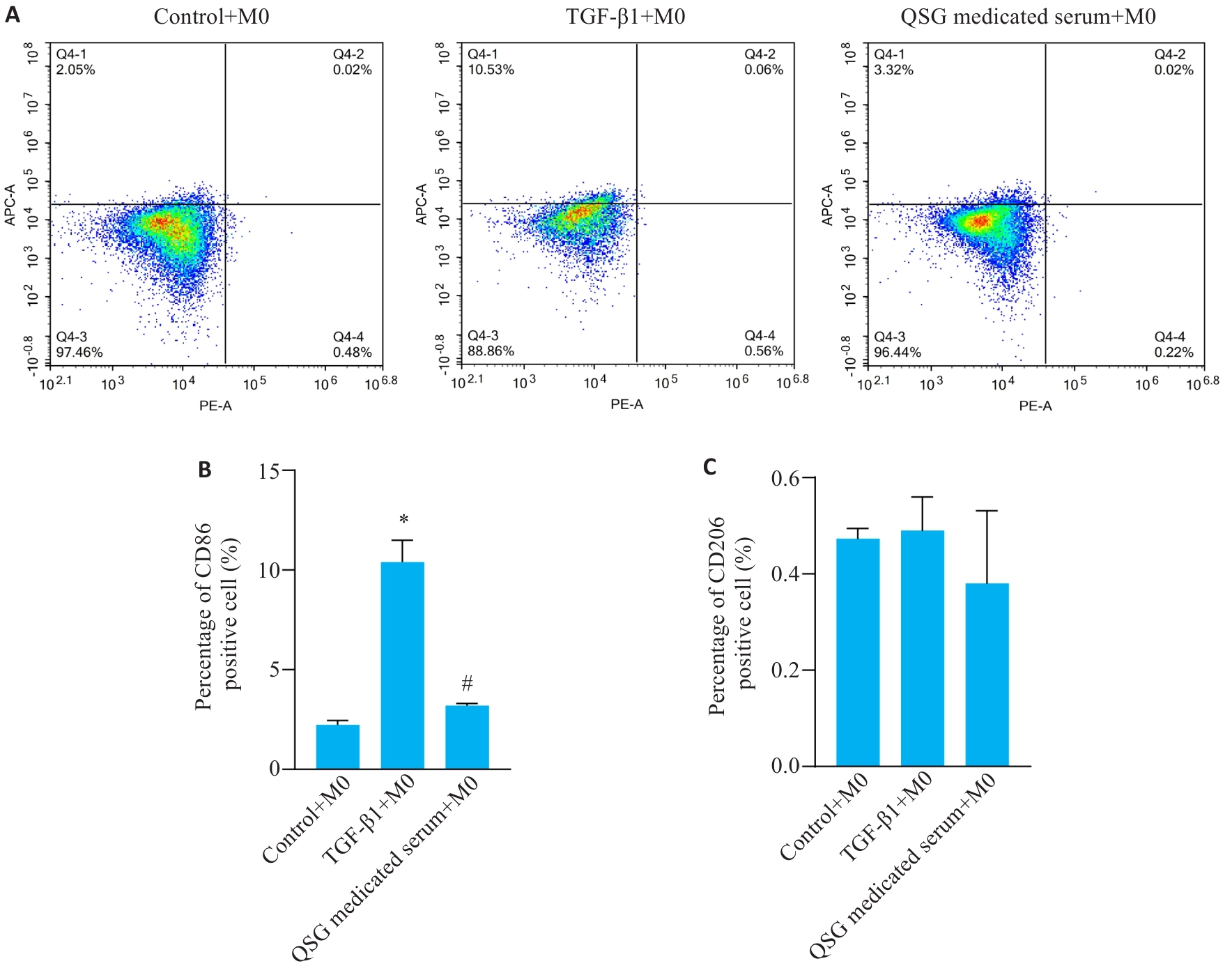
图8 清肾颗粒含药血清抑制 M1巨噬细胞极化
Fig.8 QSG-medicated serum inhibits M1 macrophage polarization. A-C: Flow cytometric analysis of M1/M2 macrophage expression levels in THP-1 cells from each group (n=3). *P<0.05 vs Control group; #P<0.05 vs TGF-β1 group.
| [1] | Stevens PE, Ahmed SB, Carrero JJ, et al. KDIGO 2024 clinical practice guideline for the evaluation and management of chronic kidney disease[J]. Kidney Int, 2024, 105(4): S117-314. doi:10.1016/j.kint.2023.10.018 |
| [2] | Li L, Fu HY, Liu YH. The fibrogenic niche in kidney fibrosis: components and mechanisms[J]. Nat Rev Nephrol, 2022, 18(9): 545-57. doi:10.1038/s41581-022-00590-z |
| [3] | Liu YH. Cellular and molecular mechanisms of renal fibrosis[J]. Nat Rev Nephrol, 2011, 7(12): 684-96. doi:10.1038/nrneph.2011.149 |
| [4] | Zhou D, Liu YH. Understanding the mechanisms of kidney fibrosis[J]. Nat Rev Nephrol, 2016, 12(2): 68-70. doi:10.1038/nrneph.2015.215 |
| [5] | Ruiz-Ortega M, Rayego-Mateos S, Lamas S, et al. Targeting the progression of chronic kidney disease[J]. Nat Rev Nephrol, 2020, 16(5): 269-88. doi:10.1038/s41581-019-0248-y |
| [6] | Grams ME, Coresh J, Matsushita K, et al. Estimated glomerular filtration rate, albuminuria, and adverse outcomes [J]. JAMA, 2023, 330(13): 1266-77. doi:10.1001/jama.2023.17002 |
| [7] | Liyanage T, Toyama T, Hockham C, et al. Prevalence of chronic kidney disease in Asia: a systematic review and analysis[J]. BMJ Glob Health, 2022, 7(1): e007525. doi:10.1136/bmjgh-2021-007525 |
| [8] | Locati M, Curtale G, Mantovani A. Diversity, mechanisms, and significance of macrophage plasticity[J]. Annu Rev Pathol Mech Dis, 2020, 15: 123-47. doi:10.1146/annurev-pathmechdis-012418-012718 |
| [9] | Nakagawa M, Karim MR, Izawa T, et al. Immunophenotypical characterization of M1/M2 macrophages and lymphocytes in cisplatin-induced rat progressive renal fibrosis[J]. Cells, 2021, 10(2): 257. doi:10.3390/cells10020257 |
| [10] | Li X, Lu ZQ, Song CM, et al. Activation of M1 macrophages in sepsis-induced acute kidney injury in response to heparin-binding protein[J]. PLoS One, 2018, 13(5): e0196423. doi:10.1371/journal.pone.0196423 |
| [11] | Pan BX, Liu GH, Jiang ZP, et al. Regulation of renal fibrosis by macrophage polarization[J]. Cell Physiol Biochem, 2015, 35(3): 1062-9. doi:10.1159/000373932 |
| [12] | Li MO, Flavell RA. Contextual regulation of inflammation: a duet by transforming growth factor-beta and interleukin-10[J]. Immunity, 2008, 28(4): 468-76. doi:10.1016/j.immuni.2008.03.003 |
| [13] | Masure S, Haefner B, Wesselink JJ, et al. Molecular cloning, expression and characterization of the human serine/threonine kinase Akt-3[J]. Eur J Biochem, 1999, 265(1): 353-60. doi:10.1046/j.1432-1327.1999.00774.x |
| [14] | Sandirasegarane L, Kester M. Enhanced stimulation of Akt-3/protein kinase B-γ in human aortic smooth muscle cells[J]. Biochem Biophys Res Commun, 2001, 283(1): 158-63. doi:10.1006/bbrc.2001.4739 |
| [15] | Xie SS, Yang H. SP1 activates AKT3 to facilitate the development of diabetic nephropathy[J]. J Endocrinol Investig, 2025, 48(5): 1269-81. doi:10.1007/s40618-025-02530-7 |
| [16] | Li PY, Hao XH, Liu JX, et al. miR-29a-3p regulates autophagy by targeting Akt3-mediated mTOR in SiO2-induced lung fibrosis[J]. Int J Mol Sci, 2023, 24(14): 11440. doi:10.3390/ijms241411440 |
| [17] | 包娅琼, 茅燕萍, 王亿平, 等. 清肾颗粒辅助治疗慢性肾脏病湿热证效果观察[J]. 山东医药, 2024, 64(8): 65-8. doi:10.3969/j.issn.1002-266X.2024.08.016 |
| [18] | 呼 琴, 金 华. 清肾颗粒通过调控miR-23b及Nrf2通路改善慢性肾脏病湿热证患者的肾功能: 基于网络药理学和临床试验[J]. 南方医科大学学报, 2025(9): 1867-79. |
| [19] | 孔 敏, 王亿平, 金 华, 等. 清肾颗粒治疗慢性肾脏病合并脂代谢异常湿热证疗效观察[J]. 安徽中医药大学学报, 2022, 41(2): 21-6. doi:10.3969/j.issn.2095-7246.2022.02.006 |
| [20] | 戴 荣, 曹泽平, 刘传娇, 等. 清肾颗粒通过调控外泌体、miR-330-3p以及CREBBP表达抑制小鼠肾纤维化[J]. 南方医科大学学报, 2024, 44(8): 1431-40. doi:10.12122/j.issn.1673-4254.2024.08.01 |
| [21] | 金 华, 张叶青, 呼 琴, 等. 清肾颗粒对大鼠NRK-52E细胞转分化模型miR-23b和PINK1/Parkin通路的影响[J]. 中国药理学通报, 2024, 40(1): 162-70. |
| [22] | 张 磊, 金 华, 王 东, 等. 清肾颗粒含药血清通过抑制氧化应激介导的NF-κB信号通路减轻高糖诱导的肾小管上皮细胞转分化[J]. 中国病理生理杂志, 2022, 38(7): 1234-45. doi:10.3969/j.issn.1000-4718.2022.07.011 |
| [23] | Martínez-Klimova E, Aparicio-Trejo OE, Tapia E, et al. Unilateral ureteral obstruction as a model to investigate fibrosis-attenuating treatments[J]. Biomolecules, 2019, 9(4): 141. doi:10.3390/biom9040141 |
| [24] | 王 宏, 刘琳娜, 江 茜, 等. 大黄-丹参药对延缓单侧输尿管梗阻大鼠肾纤维化的研究[J]. 中国临床药理学杂志, 2023, 39(3): 368-72. |
| [25] | Manning BD, Cantley LC. AKT/PKB signaling: navigating downstream[J]. Cell, 2007, 129(7): 1261-74. doi:10.1016/j.cell.2007.06.009 |
| [26] | Takahashi H, Rokudai S, Kawabata-Iwakawa R, et al. AKT3 is a novel regulator of cancer-associated fibroblasts in head and neck squamous cell carcinoma[J]. Cancers, 2021, 13(6): 1233. doi:10.3390/cancers13061233 |
| [27] | Ding L, Biswas S, Morton RE, et al. Akt3 deficiency in macrophages promotes foam cell formation and atherosclerosis in mice[J]. Cell Metab, 2012, 15(6): 861-72. doi:10.1016/j.cmet.2012.04.020 |
| [28] | Arner EN, Alzhanova D, Westcott JM, et al. AXL-TBK1 driven AKT3 activation promotes metastasis[J]. Sci Signal, 2024, 17(867): eado6057. doi:10.1126/scisignal.ado6057 |
| [29] | Tian Y, Li J, Cheung TC, et al. Akt3 promotes cancer stemness in triple-negative breast cancer through YB1-Snail/Slug signaling axis[J]. Genes Dis, 2023, 10(2): 301-6. doi:10.1016/j.gendis.2022.08.006 |
| [30] | Zhang TT, Ding H, Wang Y, et al. Akt3-mTOR regulates hippocampal neurogenesis in adult mouse[J]. J Neurochem, 2021, 159(3): 498-511. doi:10.1111/jnc.15441 |
| [31] | An N, Xu MZ, Chen RM, et al. HBP21 alleviates sepsis-induced acute kidney injury by targeting PI3K/AKT-mediated M1 macrophage polarization[J]. Mediat Inflamm, 2025, 2025: 9021628. doi:10.1155/mi/9021628 |
| [32] | Vergadi E, Ieronymaki E, Lyroni K, et al. Akt signaling pathway in macrophage activation and M1/M2 polarization[J]. J Immunol, 2017, 198(3): 1006-14. doi:10.4049/jimmunol.1601515 |
| [33] | Fang C, Zhong R, Lu S, et al. TREM2 promotes macrophage polarization from M1 to M2 and suppresses osteoarthritis through the NF-κB/CXCL3 axis[J]. Int J Biol Sci, 2024, 20(6): 1992-2007. doi:10.7150/ijbs.91519 |
| [34] | Li AH, Niu YX, Xu WH, et al. Sophora flos extract ameliorates inflammatory bowel disease via inhibiting the PI3K/AKT/mTOR signaling pathway activation and modulating M1/M2 polarization in macrophages[J]. Phytomedicine, 2025, 146: 157095. doi:10.1016/j.phymed.2025.157095 |
| [1] | 陈义珍, 王伟丽, 程梦, 张威, 高怡琳, 洪馨, 张磊, 戴荣, 王亿平. 清肾颗粒通过调控糖酵解重编程及组蛋白 H3K18 乳酸化减轻小鼠肾纤维化[J]. 南方医科大学学报, 2026, 46(3): 582-591. |
| [2] | 李楠, 张亮, 郭俏凤, 周越, 刘长江. 有氧运动通过调控miR-221-3p介导的脂肪组织巨噬细胞极化改善小鼠胰岛素抵抗[J]. 南方医科大学学报, 2026, 46(1): 74-82. |
| [3] | 呼琴, 金华. 清肾颗粒通过调控miR-23b及Nrf2通路改善慢性肾脏病湿热证患者的肾功能:基于网络药理学和临床试验[J]. 南方医科大学学报, 2025, 45(9): 1867-1879. |
| [4] | 张兆君, 吴琼, 谢苗苗, 叶洳吟, 耿晨晨, 石纪雯, 杨清玲, 王文锐, 石玉荣. 层状双氢氧化物负载si-NEAT1通过miR-133b/PD-L1轴调控乳腺癌紫杉醇耐药及巨噬细胞极化[J]. 南方医科大学学报, 2025, 45(8): 1718-1731. |
| [5] | 牛民主, 殷丽霞, 乔通, 尹林, 张可妮, 胡建国, 宋传旺, 耿志军, 李静. 旱莲苷A通过调控JAK2/STAT3通路抑制M1型巨噬细胞极化改善葡聚糖硫酸钠诱导的小鼠结肠炎[J]. 南方医科大学学报, 2025, 45(6): 1297-1306. |
| [6] | 涂舒谕, 陈祥宇, 李程辉, 黄丹萍, 张莉. 补阳还五汤通过调控外泌体miR-590-5p介导的巨噬细胞极化延缓大鼠血管衰老[J]. 南方医科大学学报, 2025, 45(6): 1251-1259. |
| [7] | 俞佳雯, 周薏, 钱春美, 穆蓝, 阙任烨. 铁过载诱导的小鼠肝纤维化过程影响巨噬细胞M2极化[J]. 南方医科大学学报, 2025, 45(4): 684-691. |
| [8] | 张芡, 刘博文, 雷丽, 王晔, 张馨月, 毛樟坤, 唐鹏, 张金梅, 杨佳宜, 彭彦茜, 刘泽. 丝氨酸蛋白酶抑制剂E1过表达通过诱导M2型巨噬细胞极化促进三阴性乳腺癌细胞增殖与紫杉醇耐药[J]. 南方医科大学学报, 2025, 45(12): 2551-2560. |
| [9] | 卢晓宇, 刘智慧, 刘烨, 庞天霄, 卞蓉, 郭玲, 何学红. 参芪泄浊饮通过调控Rap1/MAPK/FoxO3a信号通路改善氧化应激及炎症反应延缓大鼠肾纤维化[J]. 南方医科大学学报, 2025, 45(12): 2585-2597. |
| [10] | 刘新新, 徐迎芮, 盛红娜, 刘昊. 人源脐带间充质干细胞移植通过Chi3l1抑制M1型巨噬细胞极化减轻1型糖尿病小鼠的炎症反应[J]. 南方医科大学学报, 2025, 45(12): 2738-2746. |
| [11] | 戴荣, 曹泽平, 刘传娇, 葛永, 程梦, 王伟丽, 陈义珍, 张磊, 王亿平. 清肾颗粒通过调控外泌体、miR-330-3p以及CREBBP表达抑制小鼠肾纤维化[J]. 南方医科大学学报, 2024, 44(8): 1431-1440. |
| [12] | 李敬怡, 杨思圆, 韩 振, 江天乐, 朱 耀, 周子航, 周静萍. Akt2抑制剂促进大鼠根尖周炎症微环境中巨噬细胞的极化:基于降低miR-155-5p的表达[J]. 南方医科大学学报, 2023, 43(4): 568-576. |
| [13] | 张梦莹, 李 志, 裴纬亚, 李雪琴, 杨 辉, 朱小龙, 吕 坤. M2型巨噬细胞来源的外泌体lncRNA NR_028113.1通过激活JAK2/STAT3通路促进巨噬细胞的极化[J]. 南方医科大学学报, 2023, 43(3): 393-399. |
| [14] | 刘 敏, 金 华, 呼 琴, 陈 诺, 张叶青, 王亿平. 清肾颗粒含药血清通过miR-23b-5p靶向Nrf2通路减轻NRK-52E细胞转分化[J]. 南方医科大学学报, 2023, 43(12): 2078-2085. |
| [15] | 沈炳香, 王法财, 周 艺, 李 婷, 何春远, 赵为陈. 人参皂苷Rh2调控盘状结构域受体1对糖尿病大鼠肾纤维化和细胞凋亡的影响[J]. 南方医科大学学报, 2021, 41(7): 1107-1113. |
| 阅读次数 | ||||||
|
全文 |
|
|||||
|
摘要 |
|
|||||
