南方医科大学学报 ›› 2025, Vol. 45 ›› Issue (6): 1289-1296.doi: 10.12122/j.issn.1673-4254.2025.06.18
苏维1( ), 赖厚桦2, 唐昕1, 周群1, 唐亚纯1, 符浩1, 陈选才1(
 )
)
收稿日期:2024-09-13
出版日期:2025-06-20
发布日期:2025-06-27
通讯作者:
陈选才
E-mail:suwei022069@163.com;Chenxuancai2015@163.com
作者简介:苏 维,硕士,住院医师,E-mail: suwei022069@163.com
基金资助:
Wei SU1( ), Houhua LAI2, Xin TANG1, Qun ZHOU1, Yachun TANG1, Hao FU1, Xuancai CHEN1(
 )
)
Received:2024-09-13
Online:2025-06-20
Published:2025-06-27
Contact:
Xuancai CHEN
E-mail:suwei022069@163.com;Chenxuancai2015@163.com
摘要:
目的 探究Apelin(APLN)对膀胱癌细胞增殖、迁移和血管生成的影响,并分析可能的作用机制。 方法 使用GEO数据筛选出膀胱癌组织及细胞中的差异表达基因。收集60例膀胱癌组织及癌旁组织样本,分析患者的临床及病理参数。体外培养膀胱癌J82细胞或人脐静脉内皮细胞HUVEC,转染相关质粒。并将其分为对照组、si-NC组、si-APLN组、oe-NC组、oe-APLN组、oe-APLN+si-NC组及oe-APLN+si-FGFR1组。qRT-PCR及Western blotting实验检测APLN在膀胱癌组织及细胞中的表达情况,平板克隆实验检测J82细胞增殖情况,Transwell小室实验检测J82细胞迁移情况,血管生成实验检测HUVEC细胞血管生成情况,Western blotting实验检测FGF2表达以及FGFR1磷酸化水平。 结果 APLN mRNA及蛋白表达在膀胱癌组织中的表达较正常组更高(P<0.05)。此外,与SV-HUC-1相比,膀胱癌细胞(T24和J82)中APLN mRNA及蛋白表达上调(P<0.05)。临床病理特征分析结果显示,APLN表达与膀胱癌的侵袭、远端转移及TNM分期密切相关(P<0.05)。si-APLN组J82细胞增殖和迁移率较si-APIN组下降,HUVEC细胞总血管生成长度减少(P<0.05)。与oe-NC组相比,oe-APLN组J82细胞增殖和迁移率上升,FGF2表达以及FGFR1磷酸化水平上升,HUVEC细胞总血管生成长度增加(P<0.05)。与oe-APLN+si-NC组相比,oe-APLN+si-FGF2组J82细胞增殖和迁移率下降,FGF2表达以及FGFR1磷酸化水平下降,HUVEC细胞总血管生成长度减少(P<0.05)。与oe-APLN+si-NC组相比,oe-APLN+si-FGFR1组J82细胞增殖和迁移率下降,FGFR1表达以及FGFR1磷酸化水平下降,HUVEC细胞总血管生成长度减少(P<0.05)。 结论 APLN可能通过促进FGF2/FGFR1通路活化,促进J82细胞增殖、迁移及HUVEC血管生成。
苏维, 赖厚桦, 唐昕, 周群, 唐亚纯, 符浩, 陈选才. Apelin通过激活 FGF2/FGFR1通路促进膀胱癌增殖、迁移及血管生成[J]. 南方医科大学学报, 2025, 45(6): 1289-1296.
Wei SU, Houhua LAI, Xin TANG, Qun ZHOU, Yachun TANG, Hao FU, Xuancai CHEN. Apelin promotes proliferation, migration, and angiogenesis in bladder cancer by activating the FGF2/FGFR1 pathway[J]. Journal of Southern Medical University, 2025, 45(6): 1289-1296.
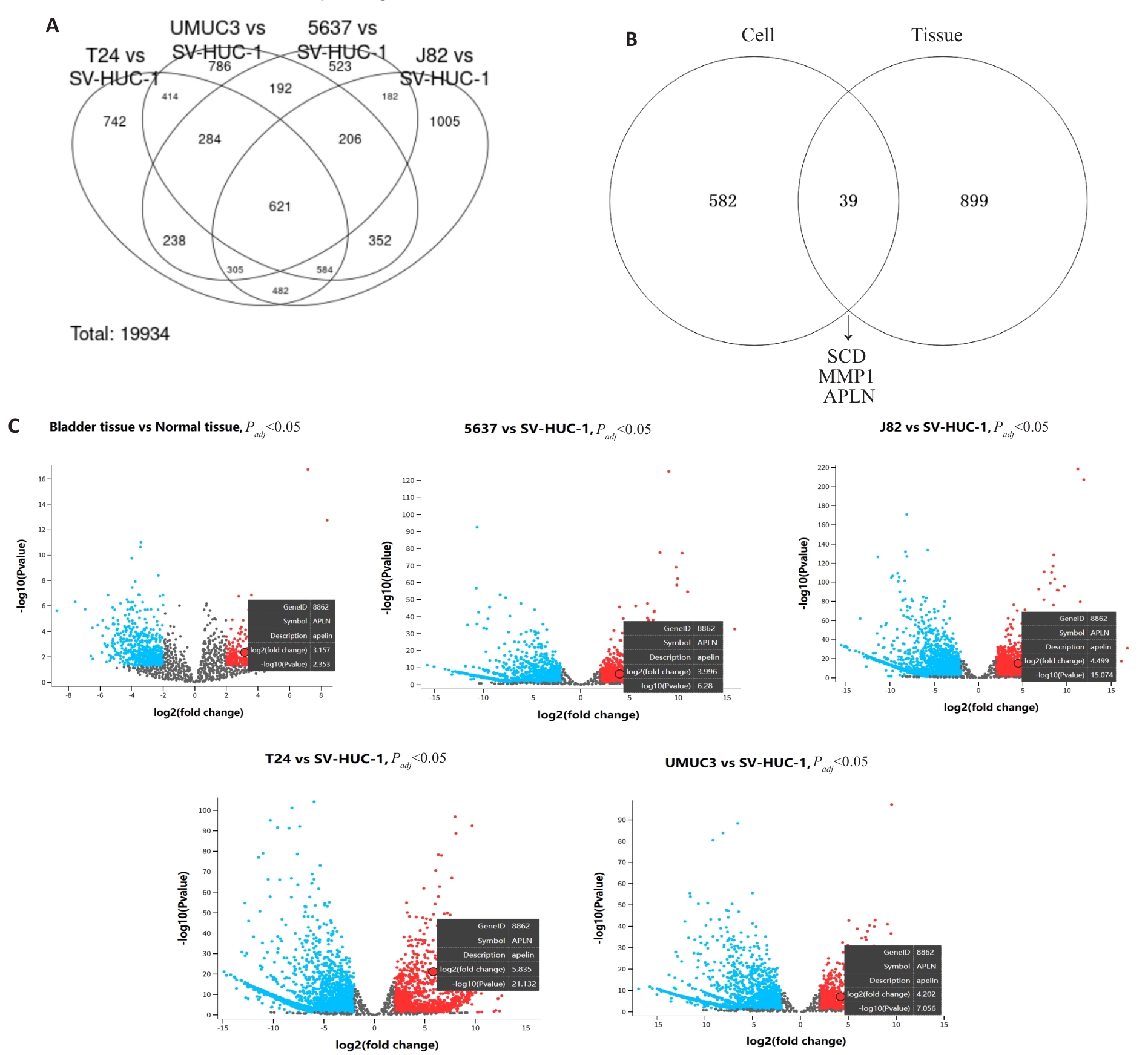
图1 GEO数据库筛选差异表达基因
Fig.1 Identification of differentially expressed genes (DEGs) in bladder cancer tissues and cells from GEO datasets. A: Venn diagram showing common DEGs between SV-HUC-1 and 4 bladder cancer cell lines (5637, UMUC3, T24 and J82) in the GSE231383 dataset. B: Venn diagram identifying common DEGs across two GEO datasets. C: Volcano plot of DEGs from two independent GEO datasets, showing upregulated APLN expression in bladder cancer tissues/cells (screening criteria: |log FC|≥2, P<0.05).
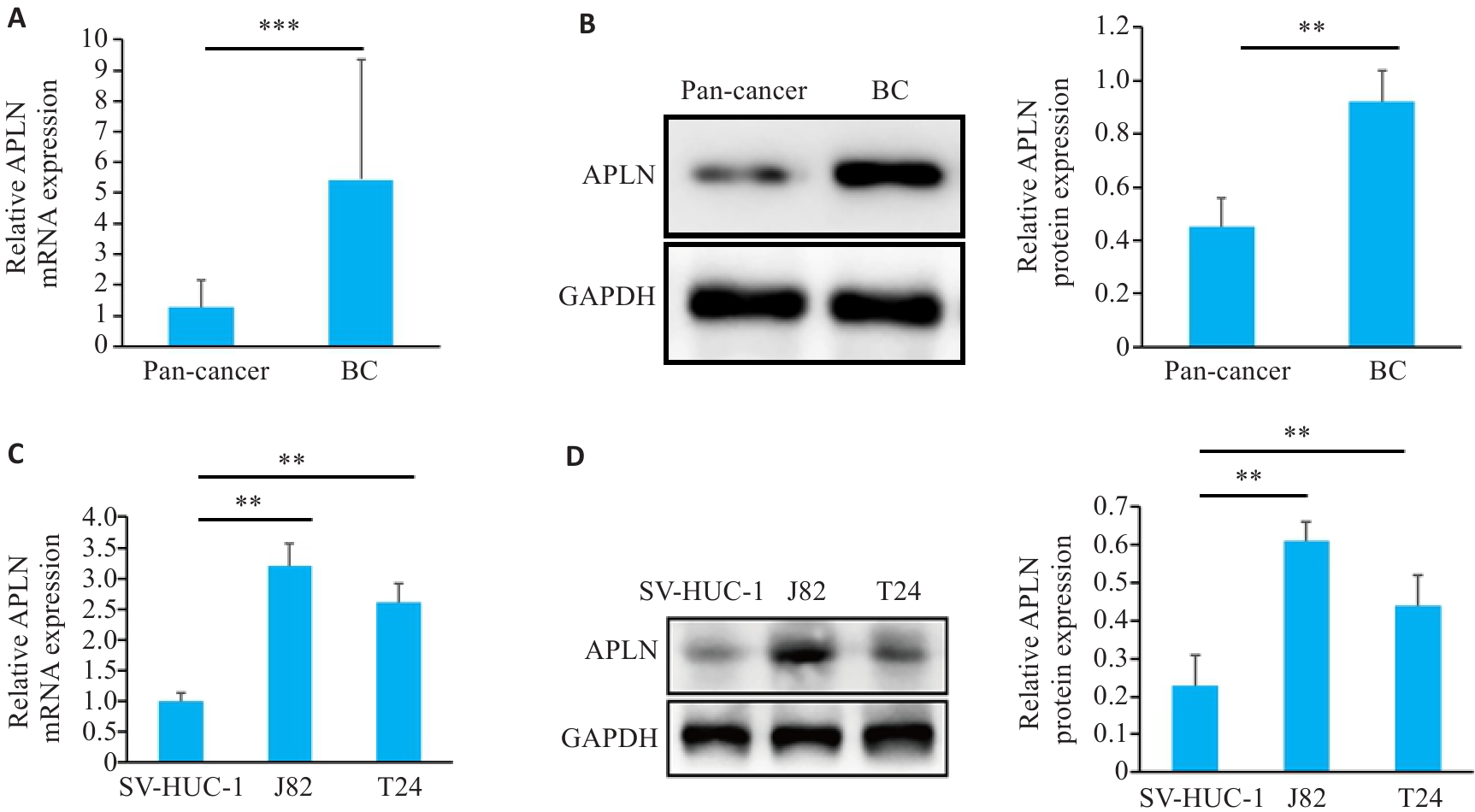
图2 APLN在膀胱癌组织和细胞中的表达情况
Fig.2 Apelin (APLN) expression in bladder cancer tissues and cells. A: qRT-PCR for detecting APLN mRNA expression in bladder cancer tissues and normal tissues (n=60). B: Western blotting for detecting APLN protein expression in bladder cancer tissues and normal tissues (n=5). C, D: qRT-PCR and Western blotting for detecting APLN expression in bladder cancer cells and SV-HUC-1 cells. Each experiment was repeated 3 times. **P<0.01, ***P<0.001.
| Characteristics | Case | APLN Expression | P | |
|---|---|---|---|---|
| High (n=30) | Low (n=30) | |||
| Age(year) | 0.796 | |||
| ≥60 | 32 | 15 | 13 | |
| <60 | 28 | 15 | 17 | |
| Gender | 0.158 | |||
| Female | 18 | 6 | 12 | |
| Male | 42 | 24 | 18 | |
| Tumor size (cm) | 0.438 | |||
| <3 | 28 | 12 | 16 | |
| ≥3 | 32 | 18 | 14 | |
| Invasion | 0.019 | |||
| T0-T2 | 30 | 10 | 20 | |
| T3-T4 | 30 | 20 | 10 | |
| Distant metastasis | 0.037 | |||
| Yes | 33 | 21 | 22 | |
| No | 27 | 9 | 8 | |
| TNM stage | 0.035 | |||
| 0-II | 35 | 13 | 22 | |
| III-IV | 25 | 17 | 8 | |
| Histological grade | 0.438 | |||
| High/intermediate | 28 | 16 | 12 | |
| Low | 32 | 14 | 18 | |
表1 APLN mRNA表达与膀胱癌患者临床病理特征的关系
Tab.1 Correlation of APLN mRNA expression with clinico-pathological features of patients with bladder cancer (n)
| Characteristics | Case | APLN Expression | P | |
|---|---|---|---|---|
| High (n=30) | Low (n=30) | |||
| Age(year) | 0.796 | |||
| ≥60 | 32 | 15 | 13 | |
| <60 | 28 | 15 | 17 | |
| Gender | 0.158 | |||
| Female | 18 | 6 | 12 | |
| Male | 42 | 24 | 18 | |
| Tumor size (cm) | 0.438 | |||
| <3 | 28 | 12 | 16 | |
| ≥3 | 32 | 18 | 14 | |
| Invasion | 0.019 | |||
| T0-T2 | 30 | 10 | 20 | |
| T3-T4 | 30 | 20 | 10 | |
| Distant metastasis | 0.037 | |||
| Yes | 33 | 21 | 22 | |
| No | 27 | 9 | 8 | |
| TNM stage | 0.035 | |||
| 0-II | 35 | 13 | 22 | |
| III-IV | 25 | 17 | 8 | |
| Histological grade | 0.438 | |||
| High/intermediate | 28 | 16 | 12 | |
| Low | 32 | 14 | 18 | |
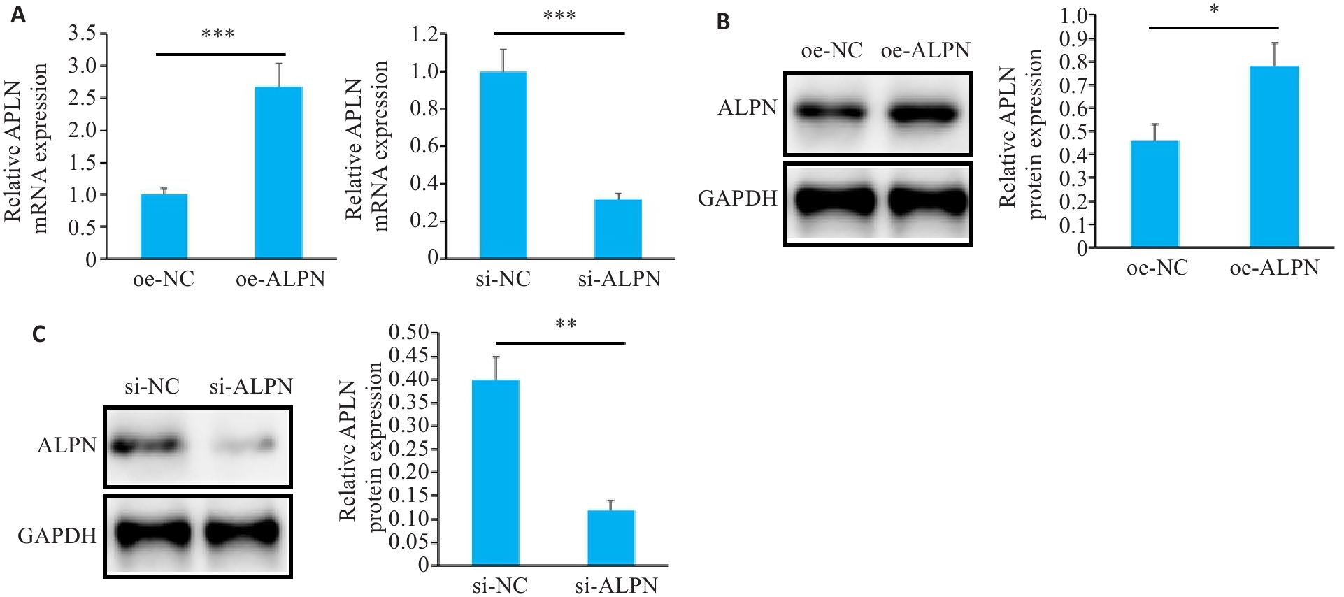
图3 转染后J82细胞中APLN表达情况
Fig.3 APLN expression in J82 cells after transfection. A: qRT-PCR for detecting APLN mRNA expression in each group of J82 cells. B, C: Western blotting for detecting APLN protein expression in each group of J82 cells. Each experiment was repeated 3 times. *P<0.05, **P<0.01, ***P<0.001.
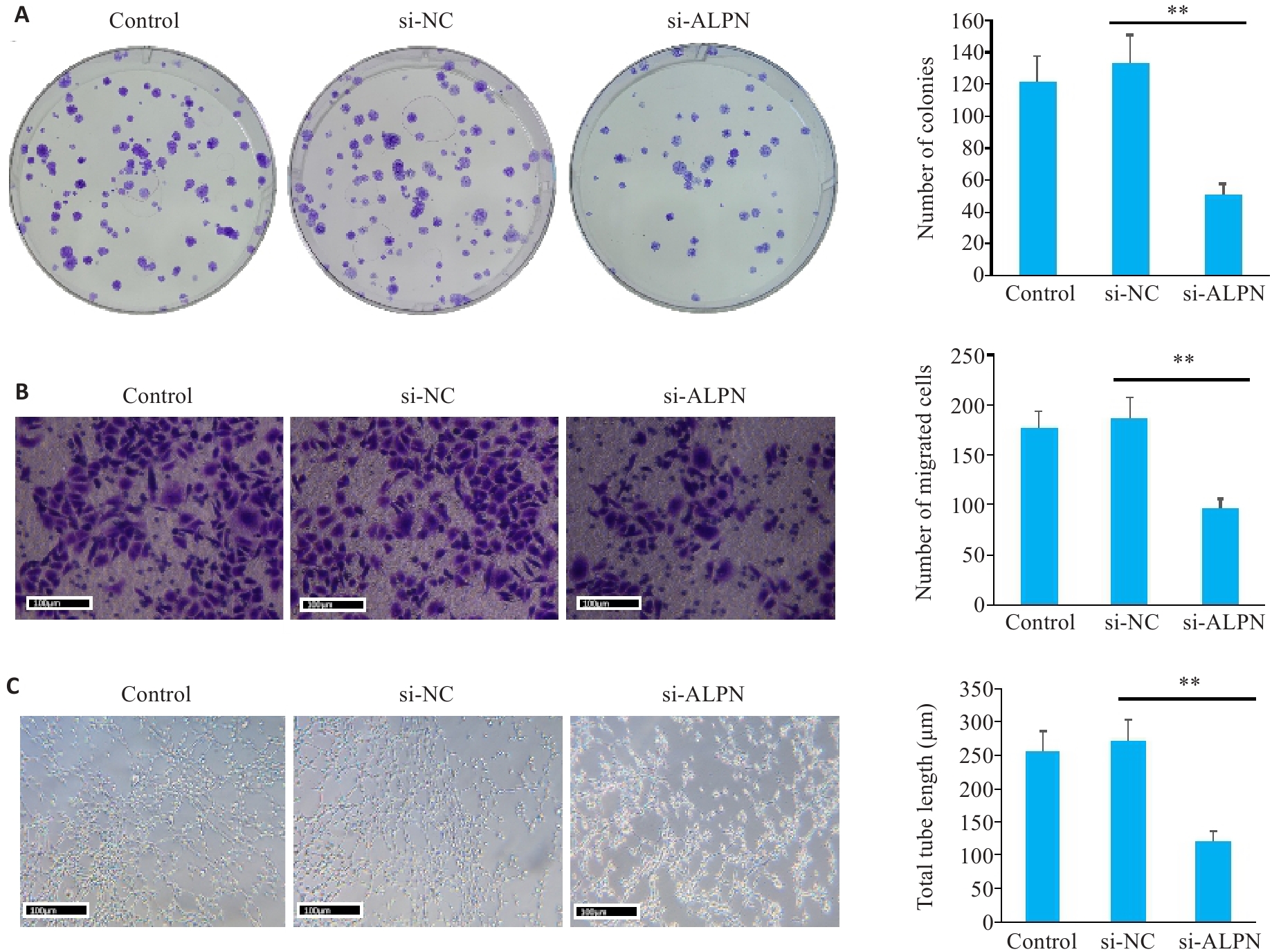
图4 转染后J82细胞增殖、迁移和血管生成情况
Fig.4 Proliferation, migration and angiogenesis of J82 cells after transfection. A: Plate cloning assayfor assessing J82 cell proliferation in each group. B: Transwell assay for evaluating cell migration in each group. C: Angiogenesis assay for assessing HUVEC tube formation in each group. Each experiment was repeated 3 times. Scale bar=100 μm. **P<0.01.
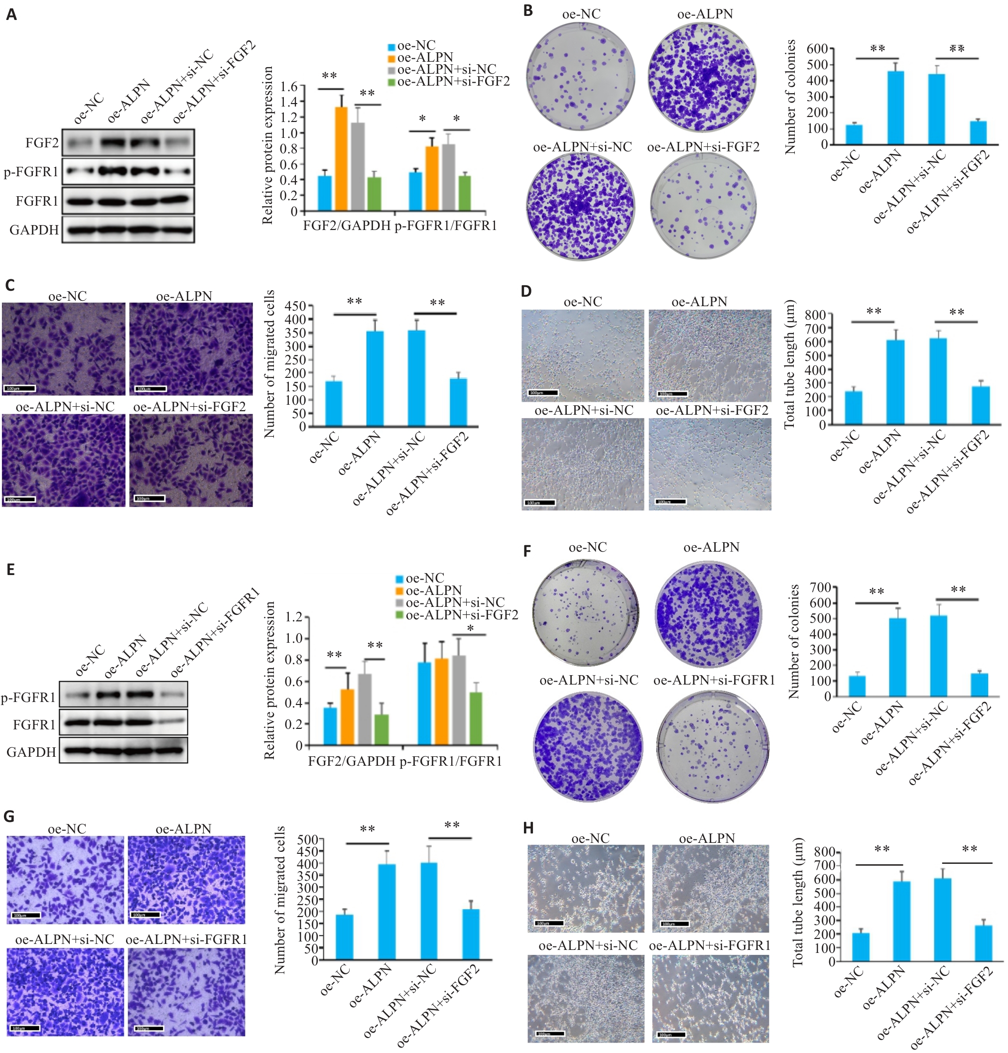
图5 转染后J82细胞增殖、迁移和血管生成情况
Fig.5 Proliferation, migration, angiogenesis and FGF2/FGFR1 signaling in J82 cells after transfection. A, E: Western blotting for detecting FGF2 expression and FGFR1 phosphorylation in different groups. B, F: Plate cloning assay for assessing J82 cell proliferation. C, G: Transwell assay for evaluating cell migration. D, H: Angiogenesis assay for assessing HUVEC tube formation. Each experiment was repeated 3 times. Scale bar=100 μm. *P<0.05, **P<0.01.
| 1 | Lee YL, Chien TW, Wang JC. Using Sankey diagrams to explore the trend of article citations in the field of bladder cancer: Research achievements in China higher than those in the United States[J]. Medicine: Baltimore, 2022, 101(34): e30217. doi:10.1097/md.0000000000030217 |
| 2 | Witjes JA, Bruins HM, Cathomas R, et al. European association of urology guidelines on muscle-invasive and metastatic bladder cancer: summary of the 2020 guidelines[J]. Eur Urol, 2021, 79(1): 82-104. doi:10.1016/j.eururo.2020.03.055 |
| 3 | Tsai YS, Wu TY, Ou CH, et al. Dynamic changes of quality of life in muscle-invasive bladder cancer survivors[J]. BMC Urol, 2022, 22(1): 126. doi:10.1186/s12894-022-01084-7 |
| 4 | Ciummo SL, Sorrentino C, Fieni C, et al. Interleukin-30 subverts prostate cancer-endothelium crosstalk by fostering angiogenesis and activating immunoregulatory and oncogenic signaling pathways[J]. J Exp Clin Cancer Res, 2023, 42(1): 336. doi:10.1186/s13046-023-02902-y |
| 5 | Xie J, Zhang H, Wang K, et al. M6A-mediated-upregulation of lncRNA BLACAT3 promotes bladder cancer angiogenesis and hematogenous metastasis through YBX3 nuclear shuttling and enhancing NCF2 transcription[J]. Oncogene, 2023, 42(40): 2956-70. doi:10.1038/s41388-023-02814-3 |
| 6 | Ruan R, Li L, Li X, et al. Unleashing the potential of combining FGFR inhibitor and immune checkpoint blockade for FGF/FGFR signaling in tumor microenvironment[J]. Mol Cancer, 2023, 22(1): 60. doi:10.1186/s12943-023-01761-7 |
| 7 | Liu G, Chen T, Ding Z, et al. Inhibition of FGF-FGFR and VEGF-VEGFR signalling in cancer treatment[J]. Cell Prolif, 2021, 54(4): e13009. doi:10.1111/cpr.13009 |
| 8 | Hosni S, Kilian V, Klümper N, et al. Adipocyte precursor-derived NRG1 promotes resistance to FGFR inhibition in urothelial carcinoma[J]. Cancer Res, 2024, 84(5): 725-40. doi:10.1158/0008-5472.can-23-1398 |
| 9 | Chen JC, Shih HC, Lin CY, et al. microRNA-631 resensitizes doxorubicin-resistant chondrosarcoma cells by targeting apelin[J]. Int J Mol Sci, 2023, 24(1): 839. doi:10.3390/ijms24010839 |
| 10 | Frisch A, Kälin S, Monk R, et al. Apelin controls angiogenesis-dependent glioblastoma growth[J]. Int J Mol Sci, 2020, 21(11): E4179. doi:10.3390/ijms21114179 |
| 11 | Lv DG, Luo XL, Chen Z, et al. Apelin/APJ signaling activates autophagy to promote human lung adenocarcinoma cell migration[J]. Life Sci, 2021, 281: 119763. doi:10.1016/j.lfs.2021.119763 |
| 12 | Trang NTN, Lai CY, Tsai HC, et al. Apelin promotes osteosarcoma metastasis by upregulating PLOD2 expression via the Hippo signaling pathway and hsa_circ_0000004/miR-1303 axis[J]. Int J Biol Sci, 2023, 19(2): 412-25. doi:10.7150/ijbs.77688 |
| 13 | Hennigs JK, Cao AQ, Li CG, et al. PPARγ-p53-mediated vasculoregenerative program to reverse pulmonary hypertension[J]. Circ Res, 2021, 128(3): 401-18. doi:10.1161/circresaha.119.316339 |
| 14 | Cardoso Dos Santos LM, Azar P, Brun C, et al. Apelin is expressed in intimal smooth muscle cells and promotes their phenotypic transition[J]. Sci Rep, 2023, 13(1): 18736. doi:10.1038/s41598-023-45470-z |
| 15 | Shen J, Feng J, Wu Z, et al. Apelin prevents and alleviates crystalline silica-induced pulmonary fibrosis via inhibiting transforming growth factor beta 1-triggered fibroblast activation[J]. Int J Biol Sci, 2023, 19(13): 4004-19. doi:10.7150/ijbs.81436 |
| 16 | Jin S, Wang Y, Ma L, et al. Feedback interaction between apelin and endoplasmic reticulum stress in the rat myocardium[J]. J Cardiovasc Pharmacol, 2023, 81(1): 21-34. doi:10.1097/fjc.0000000000001369 |
| 17 | Wang YH, Wang G, Liu XJ, et al. Inhibition of APLN suppresses cell proliferation and migration and promotes cell apoptosis in esophageal cancer cells in vitro, through activating PI3K/mTOR signaling pathway[J]. Eur J Histochem, 2022, 66(3):308-15. doi:10.4081/ejh.2022.3336 |
| 18 | Chen Y, Lin X, Zheng J, et al. APLN: a potential novel biomarker for cervical cancer[J]. Sci Prog, 2021, 104(2): 368504211011341. doi:10.1177/00368504211011341 |
| 19 | Sugimoto K, Yokokawa T, Misaka T, et al. High-fat diet attenuates the improvement of hypoxia-induced pulmonary hypertension in mice during reoxygenation[J]. BMC Cardiovasc Disord, 2021, 21(1): 331. doi:10.21203/rs.3.rs-139169/v1 |
| 20 | Zhou Y, Xu R, Luo J, et al. Dysregulation of miR-204-5p/APLN axis affects malignant progression and cell stemness of esophageal cancer[J]. Mutat Res, 2022, 825: 111791. doi:10.1016/j.mrfmmm.2022.111791 |
| 21 | Wang Q, Wang BY, Zhang WJ, et al. APLN promotes the proliferation, migration, and glycolysis of cervical cancer through the PI3K/AKT/mTOR pathway[J]. Arch Biochem Biophys, 2024, 755: 109983. doi:10.1016/j.abb.2024.109983 |
| 22 | Larionova I, Kazakova E, Gerashchenko T, et al. New angiogenic regulators produced by TAMs: perspective for targeting tumor angiogenesis[J]. Cancers: Basel, 2021, 13(13): 3253. doi:10.3390/cancers13133253 |
| 23 | Anna G, Alessio N, Davide C, et al. Role of TGFβ1 and WNT6 in FGF2 and BMP4-driven endothelial differentiation of murine embryonic stem cells[J]. Angiogenesis, 2021, 25(1): 1-16. doi:10.1007/s10456-021-09815-4 |
| 24 | Huang YC, Chen WC, Yu CL, et al. FGF2 drives osteosarcoma metastasis through activating FGFR1-4 receptor pathway-mediated ICAM-1 expression[J]. Biochem Pharmacol, 2023, 218: 115853. doi:10.1016/j.bcp.2023.115853 |
| 25 | Wang Y, Sun Q, Ye Y, et al. FGF-2 signaling in nasopharyngeal carcinoma modulates pericyte-macrophage crosstalk and metastasis[J]. JCI Insight, 2022, 7(10): e157874. doi:10.1172/jci.insight.157874 |
| 26 | Zheng C, Shi CJ, Du LJ, et al. Expression of fibroblast growth factor receptor like 1 protein in oral squamous cell carcinoma and its influence on tumor cell proliferation and migration[J]. Hua Xi Kou Qiang Yi Xue Za Zhi, 2020, 38(5): 558-65. doi:10.7518/hxkq.2020.05.015 |
| 27 | Cheng C, Wang J, Xu P, et al. Gremlin1 is a therapeutically targetable FGFR1 ligand that regulates lineage plasticity and castration resistance in prostate cancer[J]. Nat Cancer, 2022, 3(5): 565-80. doi:10.1038/s43018-022-00380-3 |
| 28 | Giordano M, Decio A, Battistini C, et al. L1CAM promotes ovarian cancer stemness and tumor initiation via FGFR1/SRC/STAT3 signaling[J]. J Exp Clin Cancer Res, 2021, 40(1): 319. doi:10.1186/s13046-021-02117-z |
| 29 | Biswas PK, Kwak Y, Kim A, et al. TTYH3 modulates bladder cancer proliferation and metastasis via FGFR1/H-ras/A-raf/MEK/ERK pathway[J]. Int J Mol Sci, 2022, 23(18): 10496. doi:10.3390/ijms231810496 |
| 30 | Nian C, Gan X, Liu Q, et al. Synthesis and anti-gastric cancer activity by targeting FGFR1 pathway of novel asymmetric bis-Chalcone compounds[J]. Curr Med Chem, 2024, 31(39): 6521-41. doi:10.2174/0109298673298420240530093525 |
| [1] | 张宏博, 闫梦宇, 张建东, 孙培旺, 汪蕊, 郭园园. 吡非尼酮抑制调节性T细胞延缓小鼠膀胱癌进展[J]. 南方医科大学学报, 2025, 45(7): 1513-1518. |
| [2] | 李煜桐, 宋杏钰, 孙蕊旭, 董璇, 刘宏伟. PYCR1的泛癌分析及其对膀胱癌化疗和免疫治疗应答的潜在预测价值[J]. 南方医科大学学报, 2025, 45(4): 880-892. |
| [3] | 陈守峰, 张舒超, 樊伟林, 孙 巍, 刘贝贝, 刘建民, 郭园园. 吡非尼酮联合PD-L1抑制剂抑制小鼠异位膀胱肿瘤的生长[J]. 南方医科大学学报, 2024, 44(2): 210-216. |
| [4] | 李立强, 郭园园, 王成勇, 常睿, 孙巍, 高五岳, 王超, 刘贝贝. miR-204-5p通过靶向负性调控RAB22A促进膀胱癌细胞的恶性生物学行为[J]. 南方医科大学学报, 2024, 44(11): 2235-2242. |
| [5] | 罗 瑞, 田龙海, 杨永曜. 高良姜素通过下调lncRNA H19的表达抑制ox-LDL诱导的人源正常主动脉内皮细胞血管生成活性[J]. 南方医科大学学报, 2024, 44(1): 52-59. |
| [6] | 颜秋霞, 曾 鹏, 黄树强, 谭翠钰, 周秀琴, 乔 静, 赵晓英, 冯 玲, 朱振杰, 张国志, 胡 鸿, 陈彩蓉. RBMX通过下调PKM2抑制膀胱癌细胞的增殖、迁移、侵袭和糖酵解[J]. 南方医科大学学报, 2024, 44(1): 9-16. |
| [7] | 张晓林, 吴浩松, 汪 盛. SLC12A8通过JAK/STAT途径促进膀胱癌细胞的增殖、侵袭与迁移并触发上皮间充质转化[J]. 南方医科大学学报, 2023, 43(9): 1613-1621. |
| [8] | 蔡涛浓, 卢江丽, 林志君, 罗明睿, 梁海滔, 秦自科, 叶云林. 单中心非肌层浸润性膀胱癌患者行卡介苗灌注治疗的疗效分析[J]. 南方医科大学学报, 2023, 43(3): 488-494. |
| [9] | 吴小凤, 詹日明, 程大钊, 陈 黎, 王天雨, 唐旭东. 非小细胞肺癌细胞外泌体源性FZD10促进体外血管生成[J]. 南方医科大学学报, 2022, 42(9): 1351-1358. |
| [10] | 刘俊平, 石宇彤, 吴敏敏, 徐梦岐, 张凤梅, 何志强, 唐 敏. JAG1影响血管生成并促进三阴性乳腺癌细胞的迁移、侵袭和粘附[J]. 南方医科大学学报, 2022, 42(7): 1100-1108. |
| [11] | 付小聪, 余光创, 郭艳芳. 组蛋白赖氨酸去甲基化酶家族在膀胱癌中的表达模式及其潜在作用:基于多组学分析[J]. 南方医科大学学报, 2022, 42(12): 1822-1831. |
| [12] | 姚 越, 张 冲, 熊言骏, 韩 兵, 高幸福, 汪 盛. miR-let-7c-5p通过靶向HMGA2抑制膀胱癌细胞侵袭和迁移[J]. 南方医科大学学报, 2021, 41(7): 1022-1029. |
| [13] | 植 曦, 周俊豪, 田 湖, 周冉冉, 黄振辉, 刘存东. SHOX2体外增强膀胱癌细胞的迁移、侵袭和干细胞特性[J]. 南方医科大学学报, 2021, 41(7): 995-1001. |
| [14] | 曹 圆, 许 凯, 陈玢屾, 王奕铭, 李炳坤, 李朝明, 徐 鹏. DNMT3b 基因在人膀胱癌组织中的表达及与临床预后的相关性[J]. 南方医科大学学报, 2020, 40(09): 1295-1300. |
| [15] | 余 淦, 周 辉, 许 凯, 孟丽荣, 郎 斌. 靶向TUG1的mir-29c-3p通过调节CAPN7的表达影响膀胱癌细 胞的迁移和侵袭[J]. 南方医科大学学报, 2020, 40(09): 1325-1331. |
| 阅读次数 | ||||||
|
全文 |
|
|||||
|
摘要 |
|
|||||