南方医科大学学报 ›› 2024, Vol. 44 ›› Issue (8): 1599-1604.doi: 10.12122/j.issn.1673-4254.2024.08.19
温小慧1,2( ), 黄诗雅1,2, 刘学红1,2, 李坤寅3, 关永格3
收稿日期:2024-01-24
出版日期:2024-08-20
发布日期:2024-09-06
通讯作者:
关永格
E-mail:18816781135@163.com
作者简介:温小慧,博士,E-mail: 18816781135@163.com
基金资助:
Xiaohui WEN1,2( ), Shiya HUANG1,2, Xuehong LIU1,2, Kunyin LI3, Yongge GUAN3
Received:2024-01-24
Online:2024-08-20
Published:2024-09-06
Contact:
Yongge GUAN
E-mail:18816781135@163.com
摘要:
目的 基于Notch1信号观察子宫腺肌病(AM)在位内膜及Ishikawa细胞系糖酵解相关因子和蛋白的表达,并探讨子宫腺肌病的发生机制。 方法 收集AM(AM组,n=8)及子宫肌瘤(CON组,n=8)在位子宫内膜组织,通过临床特征评估两组子宫内膜的可比性, qRT-PCR和Western blot检测其Notch1信号及糖酵解相关因子和蛋白的表达,通过葡萄糖和乳酸试验检测其葡萄糖和乳酸的含量;建立过表达Notch1的Ishikawa细胞慢病毒稳转株, CCK-8检测其细胞存活率, Transwell迁移和侵袭实验检测其迁移和侵袭能力,细胞外酸化速率检测其糖酵解储备。 结果 与CON组子宫内膜组织比较,AM组糖类抗原125(CA125)水平明显高于CON组(P<0.01),AM患者在位子宫内膜组织Notch1、HK2和PDHA的mRNA相对表达量增加(P<0.01或P<0.05),Notch1、GLUT1、HK2、PKM和PDHA蛋白相对表达量均显著升高(P<0.01或P<0.05),葡萄糖含量显著下降、乳酸水平显著升高(P<0.01或P<0.05)。与对照病毒空载体(ov-NC)比较,过表达Notch1的Ishikawa细胞慢病毒稳转株(ov-Notch1)的细胞存活率显著升高(P<0.05),细胞迁移和侵袭细胞数量显著增加(P<0.05),酵解容量和酵解储备显著升高(P<0.01或P<0.05)。 结论 Notch1信号可能通过调控细胞的增殖、迁移、侵袭和糖酵解过程,进而参与AM的发生发展。
温小慧, 黄诗雅, 刘学红, 李坤寅, 关永格. Notch1信号通过调控细胞增殖、迁移、侵袭和糖酵解参与子宫腺肌病的发生及发展[J]. 南方医科大学学报, 2024, 44(8): 1599-1604.
Xiaohui WEN, Shiya HUANG, Xuehong LIU, Kunyin LI, Yongge GUAN. Role of Notch 1 signaling and glycolysis in the pathogenic mechanism of adenomyosis[J]. Journal of Southern Medical University, 2024, 44(8): 1599-1604.
| Primer | Sequence (5' to 3') |
|---|---|
| Notch1 | TGGACCAGATTGGGGAGTTC |
| GCACACTCGTCTGTGTTGAC | |
| GLUT1 | TGGCATCAACGCTGTCTTCT |
| AGCCAATGGTGGCATACACA | |
| HK2 | GTGAATCGGAGAGGTCCCAC |
| GCTAACTTCGGCCACAGGAT | |
| PKM | ATGTCGAAGCCCCATAGTGAA |
| TGGGTGGTGAATCAATGTCCA | |
| PDHA | ATGGAATGGGAACGTCTGTTG |
| CCTCTCGGACGCACAGGATA | |
| GAPDH | CAGGAGGCATTGCTGATGAT |
| GAAGGCTGGGGCTCATTT |
表1 引物序列
Tab.1 Primer sequences for RT-qPCR
| Primer | Sequence (5' to 3') |
|---|---|
| Notch1 | TGGACCAGATTGGGGAGTTC |
| GCACACTCGTCTGTGTTGAC | |
| GLUT1 | TGGCATCAACGCTGTCTTCT |
| AGCCAATGGTGGCATACACA | |
| HK2 | GTGAATCGGAGAGGTCCCAC |
| GCTAACTTCGGCCACAGGAT | |
| PKM | ATGTCGAAGCCCCATAGTGAA |
| TGGGTGGTGAATCAATGTCCA | |
| PDHA | ATGGAATGGGAACGTCTGTTG |
| CCTCTCGGACGCACAGGATA | |
| GAPDH | CAGGAGGCATTGCTGATGAT |
| GAAGGCTGGGGCTCATTT |
| Group | Age (year) | Uterine volume (cm3) | Number of pregnancies | Number of abortions | CA125 (U/mL) |
|---|---|---|---|---|---|
| CON | 46.63±2.78 | 280.38 (128.75-1009.46) | 3.00 (2.00-3.00) | 0.00 (0.00-1.75) | 14.44 (12.16-23.82) |
| AM | 44.88±2.95 | 636.08 (396.14-1020.82) | 3.50 (2.25-5.00) | 1.50 (0.25-3.50) | 45.01 (28.51-73.14)** |
表2 子宫内膜来源患者的基线特征
Tab.2 Baseline characteristics of the patients with adenomyosis and uterine fibroids [n=8, Mean±SD or M (P25-P75)]
| Group | Age (year) | Uterine volume (cm3) | Number of pregnancies | Number of abortions | CA125 (U/mL) |
|---|---|---|---|---|---|
| CON | 46.63±2.78 | 280.38 (128.75-1009.46) | 3.00 (2.00-3.00) | 0.00 (0.00-1.75) | 14.44 (12.16-23.82) |
| AM | 44.88±2.95 | 636.08 (396.14-1020.82) | 3.50 (2.25-5.00) | 1.50 (0.25-3.50) | 45.01 (28.51-73.14)** |
| Group | Notch1 | GLUT1 | HK2 | PKM | PDHA |
|---|---|---|---|---|---|
| CON | 1.31±0.78 | 0.71 (0.65-1.13) | 1.01 (0.50-5.26) | 1.01 (0.29-3.63) | 1.78±1.68 |
| AM | 5.12±3.46* | 3.04 (1.21-3.85) | 3.31 (1.58-12.38)* | 3.10 (1.40-8.02) | 6.92±5.05* |
表3 在位子宫内膜组织Notch1信号及糖酵解相关因子mRNA的表达
Tab.3 Relative mRNA expressions of Notch1 signaling pathway and glycolysis-related factors in the endometrial tissue [n=8, Mean±SD or M (P25-P75)]
| Group | Notch1 | GLUT1 | HK2 | PKM | PDHA |
|---|---|---|---|---|---|
| CON | 1.31±0.78 | 0.71 (0.65-1.13) | 1.01 (0.50-5.26) | 1.01 (0.29-3.63) | 1.78±1.68 |
| AM | 5.12±3.46* | 3.04 (1.21-3.85) | 3.31 (1.58-12.38)* | 3.10 (1.40-8.02) | 6.92±5.05* |

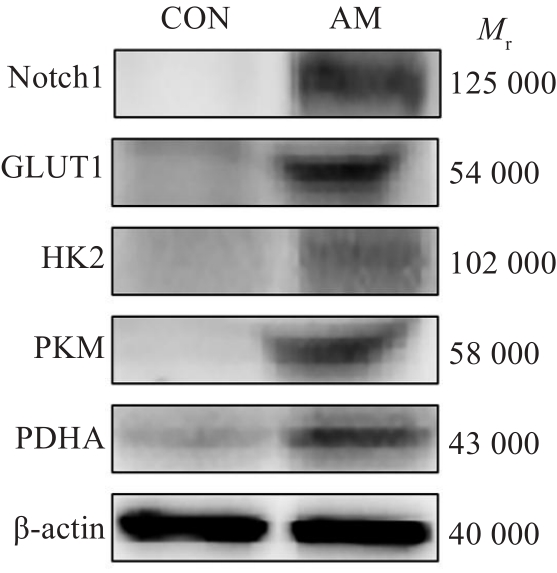
图1 在位子宫内膜组织Notch1信号及糖酵解相关蛋白的表达
Fig.1 Expression of Notch1 signal and glycolysis-related proteins in the endometrial tissues detected by Western blotting.
| Group | Notch1 | GLUT1 | HK2 | PKM | PDHA |
|---|---|---|---|---|---|
| CON | 0.21±0.11 | 0.15±0.09 | 0.85±0.07 | 0.16±0.05 | 0.21±003 |
| AM | 0.60±0.28** | 0.36±0.22* | 0.24±0.13* | 0.31±0.14* | 0.40±0.14* |
表4 在位子宫内膜组织Notch1信号及糖酵解相关蛋白的表达
Tab.4 Protein expressions of Notch1 signaling pathway and glycolysis-related factors in the endometrial tissue (n=7, Mean±SD)
| Group | Notch1 | GLUT1 | HK2 | PKM | PDHA |
|---|---|---|---|---|---|
| CON | 0.21±0.11 | 0.15±0.09 | 0.85±0.07 | 0.16±0.05 | 0.21±003 |
| AM | 0.60±0.28** | 0.36±0.22* | 0.24±0.13* | 0.31±0.14* | 0.40±0.14* |
| Group | Glucose | Lactate |
|---|---|---|
| CON | 88.8±24.82 | 50.16±4.09 |
| AM | 50.51±23.50* | 74.34±8.00** |
表5 在位子宫内膜组织葡萄糖、乳酸含量
Tab.5 Glucose and lactic acid levels in the endometrial tissue (n=5, Mean±SD)
| Group | Glucose | Lactate |
|---|---|---|
| CON | 88.8±24.82 | 50.16±4.09 |
| AM | 50.51±23.50* | 74.34±8.00** |

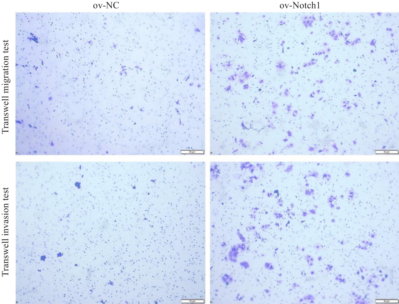
图2 过表达Notch1信号对Ishikawa细胞系迁移、侵袭的影响
Fig.2 Effects of overexpression of Notch1 signaling on migration and invasion of Ishikawa cells (Original magnification: ×200).
| Group | Migration cells | Invasion cells |
|---|---|---|
| ov-NC | 4.78±1.68 | 9.89±4.00 |
| ov-Notch1 | 64.56±17.67* | 140.89±48.49* |
表6 过表达Notch1信号对Ishikawa细胞系迁移、侵袭细胞数的影响
Tab.6 Effects of overexpression of Notch1 signaling on migration and invasion of Ishikawa cells (n=3, Mean±SD)
| Group | Migration cells | Invasion cells |
|---|---|---|
| ov-NC | 4.78±1.68 | 9.89±4.00 |
| ov-Notch1 | 64.56±17.67* | 140.89±48.49* |
| Group | Fermentation capacity | Fermentation reserve |
|---|---|---|
| ov-NC | 10.43±1.27 | 9.61±1.88 |
| ov-Notch1 | 17.13±1.68** | 16.20±1.62* |
表7 过表达Notch1信号对Ishikawa细胞系糖酵解能力的影响
Tab.7 Effect of overexpression of Notch1 signaling on glycolysis capacity of Ishikawa cells (n=3, Mean±SD)
| Group | Fermentation capacity | Fermentation reserve |
|---|---|---|
| ov-NC | 10.43±1.27 | 9.61±1.88 |
| ov-Notch1 | 17.13±1.68** | 16.20±1.62* |
| 1 | Guo SW. The pathogenesis of adenomyosis vis-à-vis endometriosis[J]. J Clin Med, 2020, 9(2): 485. |
| 2 | Harada T, Khine YM, Kaponis A, et al. The impact of adenomyosis on women's fertility[J]. Obstet Gynecol Surv, 2016, 71(9): 557-68. |
| 3 | Vannuccini S, Tosti C, Carmona F, et al. Pathogenesis of adenomyosis: an update on molecular mechanisms[J]. Reprod Biomed Online, 2017, 35(5): 592-601. |
| 4 | Mehasseb MK, Taylor AH, Pringle JH, et al. Enhanced invasion of stromal cells from adenomyosis in a three-dimensional coculture model is augmented by the presence of myocytes from affected uteri[J]. Fertil Steril, 2010, 94(7): 2547-51. |
| 5 | Struble J, Reid S, Bedaiwy MA. Adenomyosis: a clinical review of a challenging gynecologic condition[J]. J Minim Invasive Gynecol, 2016, 23(2): 164-85. |
| 6 | Wang J, Deng XH, Yang Y, et al. Expression of GRIM-19 in adenomyosis and its possible role in pathogenesis[J]. Fertil Steril, 2016, 105(4): 1093-101. |
| 7 | Yoo JY, Ku BJ, Kim TH, et al. β-catenin activates TGF-β-induced epithelial-mesenchymal transition in adenomyosis[J]. Exp Mol Med, 2020, 52(10): 1754-65. |
| 8 | Gu NH, Li GJ, Yang BX, et al. Hypo-expression of tuberin promotes adenomyosis via the mTOR1-autophagy axis[J]. Front Cell Dev Biol, 2021, 9: 710407. |
| 9 | Stratopoulou CA, Donnez J, Dolmans MM. Origin and pathogenic mechanisms of uterine adenomyosis: what is known so far[J]. Reprod Sci, 2021, 28(8): 2087-97. |
| 10 | Zhai JY, Li S, Sen S, et al. Transcriptomic analysis supports collective endometrial cell migration in the pathogenesis of adenomyosis[J]. Reprod Biomed Online, 2022, 45(3): 519-30. |
| 11 | 邓显光, 阮 慧, 李 恋. 有氧糖酵解在乳腺癌中的作用及中医药干预研究进展[J]. 中国实验方剂学杂志, 2024, 30(13): 1-13. |
| 12 | Hirschhaeuser F, Sattler UGA, Mueller-Klieser W. Lactate: a metabolic key player in cancer[J]. Cancer Res, 2011, 71(22): 6921-5. |
| 13 | Jin L, Chun J, Pan C, et al. Phosphorylation-mediated activation of LDHA promotes cancer cell invasion and tumour metastasis[J]. Oncogene, 2017, 36(27): 3797-806. |
| 14 | Yan XL, Zhang XB, Ao R, et al. Effects of shRNA-mediated silencing of PKM2 gene on aerobic glycolysis, cell migration, cell invasion, and apoptosis in colorectal cancer cells[J]. J Cell Biochem, 2017, 118(12): 4792-803. |
| 15 | 余 功, 陈江涛, 胡 桥, 等. 清燥救肺汤对荷Lewis小鼠肺癌细胞糖酵解关键限速酶HK2, PFK2, PKM2的影响[J]. 中国实验方剂学杂志, 2020, 26(4): 54-8. |
| 16 | 蔡 哲, 刘繁荣. 淋巴瘤发病机制中Notch1的作用及研究进展[J]. 临床与实验病理学杂志, 2023, 39(3): 343-6. |
| 17 | Mitsuhashi Y, Horiuchi A, Miyamoto T, et al. Prognostic significance of Notch signalling molecules and their involvement in the invasiveness of endometrial carcinoma cells[J]. Histopathology, 2012, 60(5): 826-37. |
| 18 | Brustugun OT. A NOTCH added to metabolomics[J]. Br J Cancer, 2019, 121(1): 3-4. |
| 19 | Liu YQ, Wang XY, Wan L, et al. TIPE2 inhibits the migration and invasion of endometrial cells by targeting β‑catenin to reverse epithelial-mesenchymal transition[J]. Hum Reprod, 2020, 35(6): 1377-90. |
| 20 | Jin TT, Li MQ, Li T, et al. The inactivation of hippo signaling pathway promotes the development of adenomyosis by regulating EMT, proliferation, and apoptosis of cells[J]. Reprod Sci, 2023, 30(9): 2715-27. |
| 21 | Peterson R, Minchella P, Cui W, et al. RPLP1 is up-regulated in human adenomyosis and endometrial adenocarcinoma epithelial cells and is essential for cell survival and migration in vitro [J]. Int J Mol Sci, 2023, 24(3): 2690. |
| 22 | Pollacco J, Sacco K, Portelli M, et al. Molecular links between endometriosis and cancer[J]. Gynecol Endocrinol, 2012, 28(8): 577-81. |
| 23 | Wang BY, Yang Y, Deng XH, et al. Interaction of M2 macrophages and endometrial cells induces downregulation of GRIM-19 in endometria of adenomyosis[J]. Reprod Biomed Online, 2020, 41(5): 790-800. |
| 24 | Yu O, Schulze-Rath R, Grafton J, et al. Adenomyosis incidence, prevalence and treatment: united States population-based study 2006-2015[J]. Am J Obstet Gynecol, 2020, 223(1): 94. e1-94. e10. |
| 25 | Larsen SB, Lundorf E, Forman A, et al. Adenomyosis and junctional zone changes in patients with endometriosis[J]. Eur J Obstet Gynecol Reprod Biol, 2011, 157(2): 206-11. |
| 26 | Maruyama S, Imanaka S, Nagayasu M, et al. Relationship between adenomyosis and endometriosis; Different phenotypes of a single disease?[J]. Eur J Obstet Gynecol Reprod Biol, 2020, 253: 191-7. |
| 27 | Qi SS, Zhao XB, Li MJ, et al. Aberrant expression of Notch1/numb/snail signaling, an epithelial mesenchymal transition related pathway, in adenomyosis[J]. Reprod Biol Endocrinol, 2015, 13: 96. |
| 28 | Kasvandik S, Samuel K, Peters M, et al. Deep quantitative proteomics reveals extensive metabolic reprogramming and cancer-like changes of ectopic endometriotic stromal cells[J]. J Proteome Res, 2016, 15(2): 572-84. |
| 29 | Zhang MM, Wang SX, Tang L, et al. Downregulated circular RNA hsa_circ_0067301 regulates epithelial-mesenchymal transition in endometriosis via the miR-141/Notch signaling pathway[J]. Biochem Biophys Res Commun, 2019, 514(1): 71-7. |
| 30 | Moriyama H, Moriyama M, Isshi H, et al. Role of Notch signaling in the maintenance of human mesenchymal stem cells under hypoxic conditions[J]. Stem Cells Dev, 2014, 23(18): 2211-24. |
| 31 | 王享利. 缺氧对前列腺癌细胞糖酵解及迁移侵袭能力的影响[J]. 中国现代医学杂志, 2016, 26(23): 32-6. |
| 32 | Kuwabara S, Yamaki M, Yu HQ, et al. Notch signaling regulates the expression of glycolysis-related genes in a context-dependent manner during embryonic development[J]. Biochem Biophys Res Commun, 2018, 503(2): 803-8. |
| [1] | 周海忆, 何斯怡, 韩瑞芳, 关永格, 董丽娟, 宋阳. 艾灸通过调控miR-223-3p/NLRP3焦亡通路修复薄型子宫内膜[J]. 南方医科大学学报, 2025, 45(7): 1380-1388. |
| [2] | 郑孟冬, 刘妍, 刘娇娇, 康巧珍, 王婷. 蛋白4.1R对肝细胞HL-7702增殖、凋亡以及糖酵解的影响[J]. 南方医科大学学报, 2024, 44(7): 1355-1360. |
| [3] | 潘甚豪, 李炎坤, 伍哲维, 毛玉玲, 王春艳. 子宫内膜异位症患者新鲜胚胎移植临床妊娠率预测模型的建立与验证[J]. 南方医科大学学报, 2024, 44(7): 1407-1415. |
| [4] | 曾玉燕, 贾金金, 卢 洁, 曾 诚, 耿红玲, 陈 颐. 子宫腺肌病组织中的雌激素、雌激素受体、miR-21:致病作用和调控作用[J]. 南方医科大学学报, 2024, 44(4): 627-635. |
| [5] | 满 豪, 王建伟, 吴 毛, 邵 阳, 杨俊锋, 李绍烁, 吕锦业, 周 悦. 脊髓康通过激活星形胶质细胞的YAP/PKM2信号轴促进脊髓损伤大鼠神经功能的恢复[J]. 南方医科大学学报, 2024, 44(4): 636-643. |
| [6] | 王梓凝, 杨 明, 李双磊, 迟海涛, 王军惠, 肖苍松. 心肌梗死后心肌纤维化小鼠心肌线粒体功能和能量代谢重塑相关性的转录组学分析[J]. 南方医科大学学报, 2024, 44(4): 666-674. |
| [7] | 王 娟, 杨雯钦, 刘 进, 石金凤, 肖 萍, 李美香. 脂联素通过上调PPARα/HOXA10通路改善多囊卵巢综合征大鼠的子宫内膜容受性[J]. 南方医科大学学报, 2024, 44(2): 298-307. |
| [8] | 王圆圆, 来天娇, 褚丹霞, 白晶, 严淑萍, 秦海霞, 郭瑞霞. 醋酸甲地孕酮联合盐酸二甲双胍作为早期子宫内膜样腺癌和子宫内膜非典型增生患者保留生育功能治疗:一项前瞻性研究[J]. 南方医科大学学报, 2024, 44(11): 2055-2062. |
| [9] | 颜秋霞, 曾 鹏, 黄树强, 谭翠钰, 周秀琴, 乔 静, 赵晓英, 冯 玲, 朱振杰, 张国志, 胡 鸿, 陈彩蓉. RBMX通过下调PKM2抑制膀胱癌细胞的增殖、迁移、侵袭和糖酵解[J]. 南方医科大学学报, 2024, 44(1): 9-16. |
| [10] | 冯 雯, 赖跃兴, 王 静, 徐 萍. 长链非编码RNA ABHD11-AS1促进胃癌细胞糖酵解并加速肿瘤恶性进展[J]. 南方医科大学学报, 2023, 43(9): 1485-1492. |
| [11] | 王秋生, 张 震, 王 炼, 汪 煜, 姚新宇, 王月月, 张小凤, 葛思堂, 左芦根. 高表达DAP5促进胃癌细胞的糖代谢并与不良预后相关[J]. 南方医科大学学报, 2023, 43(7): 1063-1070. |
| [12] | 王会杰, 孙珍贵, 赵文英, 耿 彪. S100A10可促进肺腺癌细胞的增殖和侵袭:基于激活Akt-mTOR信号通路[J]. 南方医科大学学报, 2023, 43(5): 733-740. |
| [13] | 喻靖文, 杨美洁, 蒋 莉, 肖智博, 李 霜, 陈锦云. 子宫腺肌病术前MR T2WI信号特征与HIFU消融疗效密切相关:一项倾向性评分配对队列研究[J]. 南方医科大学学报, 2023, 43(4): 597-603. |
| [14] | 吴超英, 程文俊. 载脂蛋白E通过激活ERK/MMP9信号通路促进子宫内膜癌细胞的迁移[J]. 南方医科大学学报, 2023, 43(2): 232-241. |
| [15] | 朱秀君, 蔡林儿, 肖 静. 聚集性林奇综合征家族一例报告[J]. 南方医科大学学报, 2022, 42(8): 1263-1266. |
| 阅读次数 | ||||||
|
全文 |
|
|||||
|
摘要 |
|
|||||