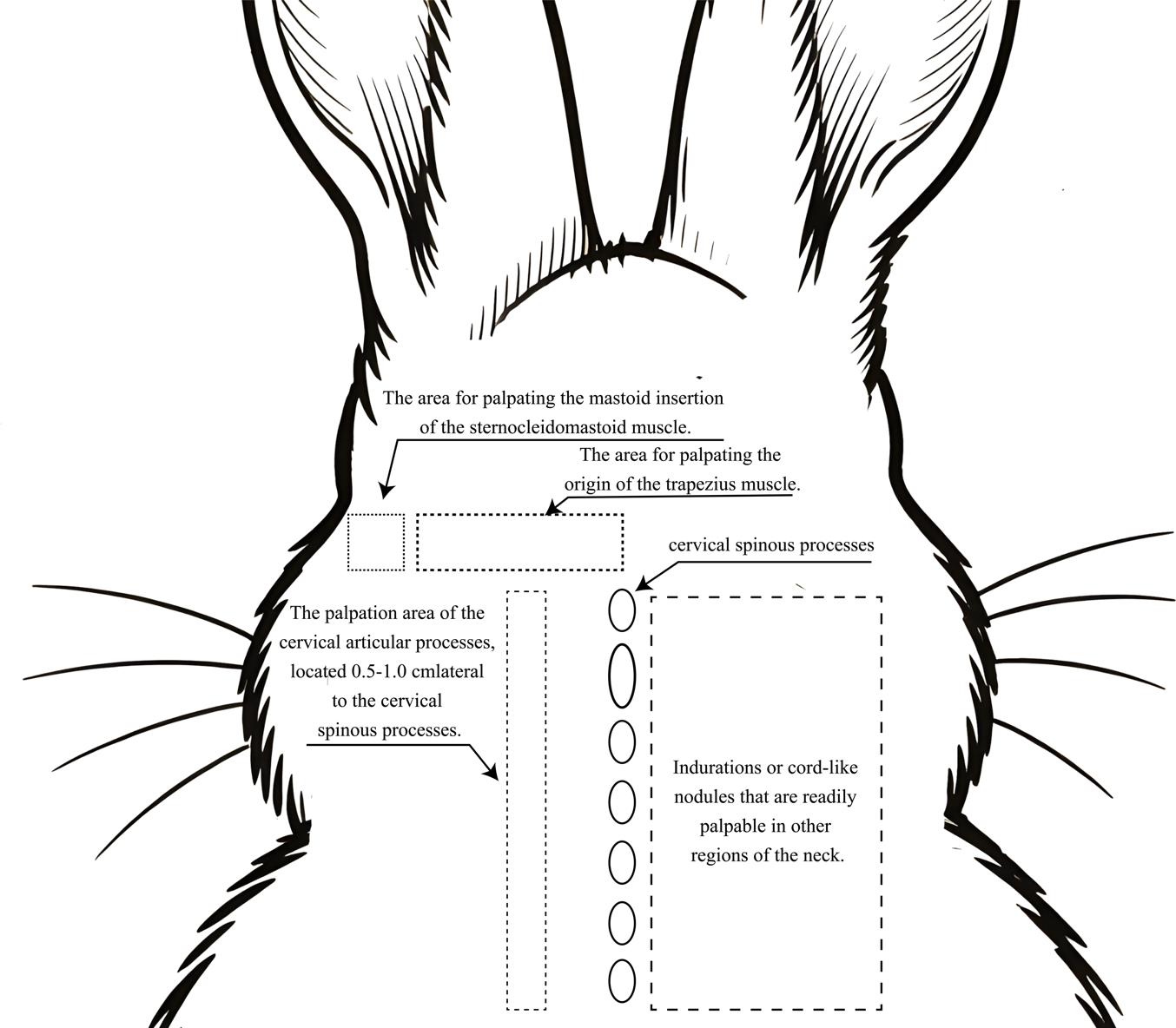
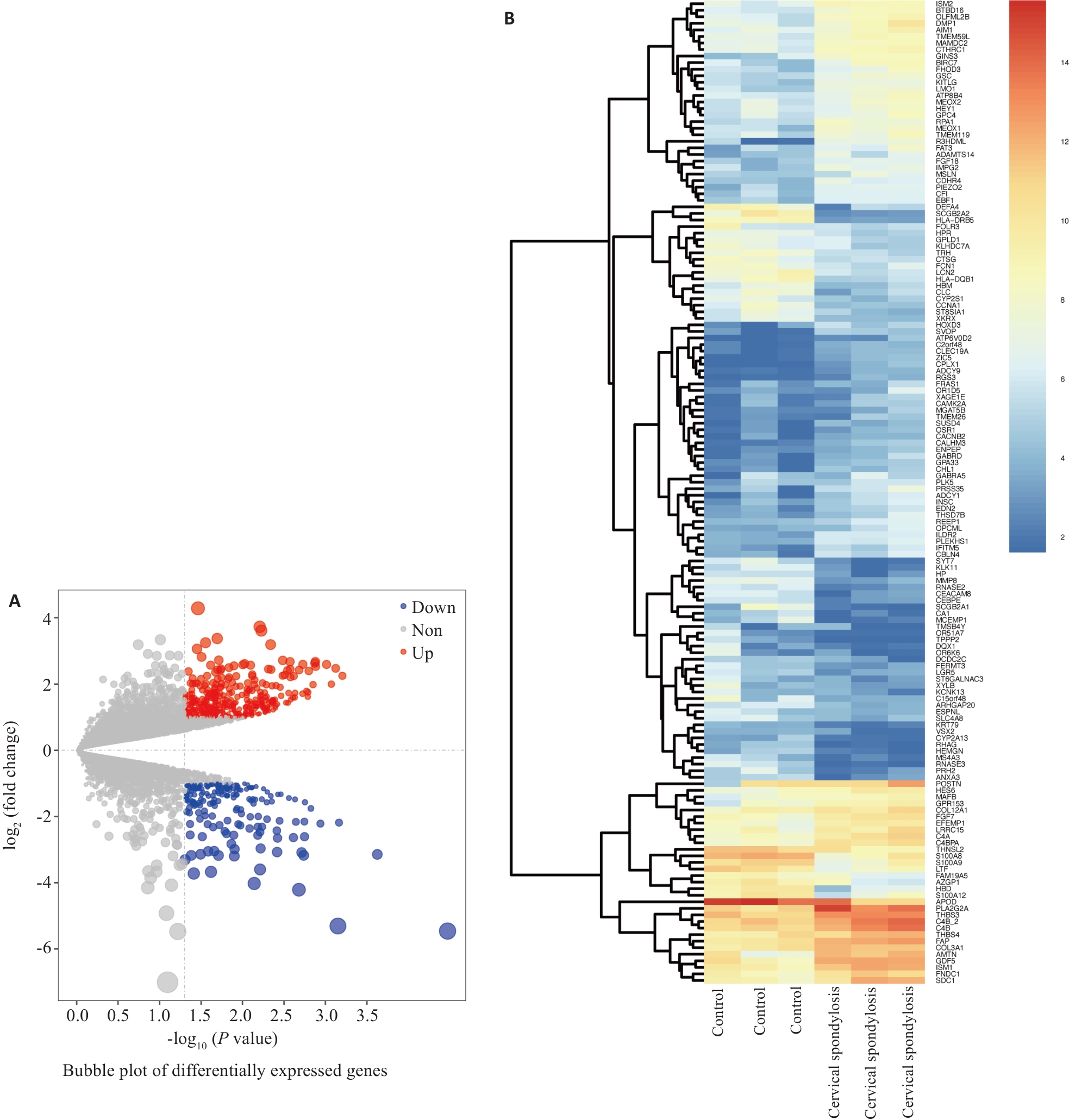
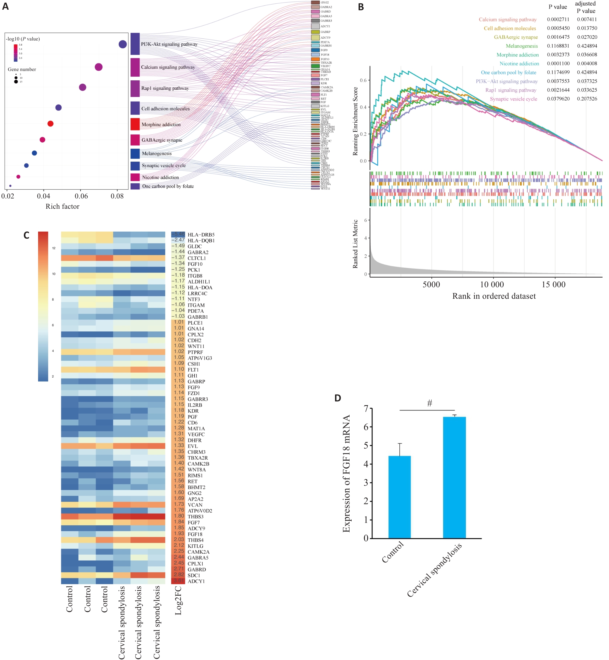
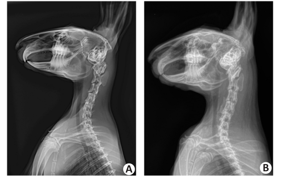
| [1] |
章 薇, 李金香, 娄必丹, 等. 中医康复临床实践指南·项痹(颈椎病)[J]. 康复学报, 2020, 30(5): 337-42.
|
| [2] |
Theodore N. Degenerative cervical spondylosis[J]. N Engl J Med, 2020, 383(2): 159-68. doi:10.1056/nejmra2003558
|
| [3] |
崔学军, 姚 敏. 颈椎病中西医结合诊疗专家共识[J]. 世界中医药, 2023, 18(7): 918-22.
|
| [4] |
Lv YW, Tian W, Chen DF, et al. The prevalence and associated factors of symptomatic cervical Spondylosis in Chinese adults: a community-based cross-sectional study[J]. BMC Musculoskelet Disord, 2018, 19(1): 325. doi:10.1186/s12891-018-2234-0
|
| [5] |
Li SY, Liu TY, Yang Q, et al. Knowledge, attitude, and practice toward cervical spondylosis among the healthy general population[J]. BMC Public Health, 2025, 25(1): 1014. doi:10.1186/s12889-025-22051-5
|
| [6] |
Chen W, Chen H, Zheng DD, et al. Gene-hydrogel micro-environment regulates extracellular matrix metabolism balance in nucleus pulposus[J]. Adv Sci (Weinh), 2019, 7(1): 1902099. doi:10.1002/advs.202070001
|
| [7] |
Tsuchiya K, Okano I, Guven AE, et al. Quantitative assessment of cervical disc degeneration using disc signal intensity index[J]. Spine J, 2025, 25(5): 903-10. doi:10.1016/j.spinee.2024.11.017
|
| [8] |
刘福水, 金玉立, 谢洪武, 等. 针刀干预对颈椎病兔软骨终板基质金属蛋白酶13、聚集蛋白聚糖、Ⅱ型胶原的影响[J]. 中华中医药杂志, 2020, 35(6): 3146-50.
|
| [9] |
胡万春, 刘 通, 张畅畅, 等. 针刀疗法适应证及优势病种分析[J]. 天津中医药大学学报, 2025, 44(4): 342-8.
|
| [10] |
刘福水, 方 婷, 洪 滔, 等. 针刀干预对颈椎病兔颈后伸肌细胞PI3K/Akt信号通路的影响[J]. 中华中医药杂志, 2020, 35(2): 918-22.
|
| [11] |
李沅骋, 李开平, 宋子琪, 等. 针刀疗法对兔退变颈椎间盘组织中p38MAPK蛋白与IL-1表达的影响[J]. 中华中医药学刊, 2022, 40(11): 250-4, 296-298.
|
| [12] |
余家阔, 吴毅文, 戴先进, 等. 颈椎病生物力学发病机制实验研究[J]. 安徽医科大学学报, 1990, 25(1): 47-51.
|
| [13] |
郭长青, 刘福水, 马惠芳, 等. 针刀干预对颈椎病兔颈肌细胞凋亡的影响[J]. 针刺研究, 2014, 39(1): 68-72.
|
| [14] |
游建宇, 刘福水, 陈明人. 基于筋骨理论探讨针刀在颈椎病防治中的应用[J]. 中华中医药杂志, 2022, 37(2): 753-5.
|
| [15] |
张怡瑾, 李 辉, 陈子颖, 等. 基于“筋出槽,骨错缝” 病机探析推拿治疗颈椎病的调衡作用[J]. 中医杂志, 2025, 66(13): 1326.
|
| [16] |
陈 斌, 刘 洪, 张良志, 等. 可视化针刀抑制髓核细胞凋亡从而改善颈椎病兔的椎间盘退变[J]. 针刺研究, 2022, 47(11): 1005-11.
|
| [17] |
李浩林, 陈 平, 王海东, 等. 基于PI3K/AKT/NFATc1通路探讨针刀疏筋解结术抑制类风湿关节炎大鼠骨破坏的作用及机制[J]. 针刺研究, 2025, 50(2): 131-40.
|
| [18] |
贾小飞, 冉 丽, 马小双, 等. 针刀调控软骨细胞自噬维持骨关节炎软骨细胞的稳态[J]. 中国组织工程研究, 2024, 28(34): 5452-7.
|
| [19] |
Zhang GZ, Li L, Yang ZL, et al. TMT-based proteomics analysis of senescent nucleus pulposus from patients with intervertebral disc degeneration[J]. Int J Mol Sci, 2023, 24(17): 13236. doi:10.3390/ijms241713236
|
| [20] |
Peng FS, Sun MT, Jing XZ, et al. Piezo1 promotes intervertebral disc degeneration through the Ca2+/F-actin/Yap signaling axis[J]. Mol Med, 2025, 31(1): 90. doi:10.1186/s10020-025-01147-z
|
| [21] |
胡斌涵, 陈 灿, 黄茂畅, 等. 超声引导针刀松解椎间孔内口对兔软骨终板基质稳态的影响[J]. 中国组织工程研究, 2025, 29(18): 3758-66.
|
| [22] |
邵欣欣, 李鑫茹, 潘丽婕, 等. 基于Piezo1/p38MAPK/ERK5力敏感通路探讨针刀干预膝关节骨关节炎兔膝关节软骨退化的作用机制[J/OL]. 中国针灸, 2025: 1-15. doi/10.13703/j.0255-2930.20241023-k0004.
|
| [23] |
Yang QH, Feng JF, Xu HY, et al. TRIM29 alleviates intervertebral disc degeneration through the PI3K/AKT/mTOR pathway[J]. Sci Rep, 2025, 15(1): 24797. doi:10.1038/s41598-025-10272-y
|
| [24] |
Li HZ, Zhang JL, Yuan DL, et al. Role of signaling pathways in age-related orthopedic diseases: focus on the fibroblast growth factor family[J]. Mil Med Res, 2024, 11(1): 40. doi:10.1186/s40779-024-00544-5
|
| [25] |
Chen TQ, Peng Y, Hu WJ, et al. Irisin enhances chondrogenic differentiation of human mesenchymal stem cells via Rap1/PI3K/AKT axis[J]. Stem Cell Res Ther, 2022, 13(1): 392. doi:10.1186/s13287-022-03092-8
|
| [26] |
Wen ZH, Li SY, Liu Y, et al. An engineered M2 macrophage-derived exosomes-loaded electrospun biomimetic periosteum promotes cell recruitment, immunoregulation, and angiogenesis in bone regeneration[J]. Bioact Mater, 2025, 50: 95-115. doi:10.1016/j.bioactmat.2025.03.027
|
| [27] |
Wei YL, Jin ZF, Zhang H, et al. The transient receptor potential channel, vanilloid 5, induces chondrocyte apoptosis via Ca2+ CaMKII-dependent MAPK and Akt/mTOR pathways in a rat osteoarthritis model[J]. Cell Physiol Biochem, 2018, 51(5): 2309-23. doi:10.1159/000495874
|
| [28] |
Yao XD, Zhang JM, Jing XZ, et al. Fibroblast growth factor 18 exerts anti-osteoarthritic effects through PI3K-AKT signaling and mitochondrial fusion and fission[J]. Pharmacol Res, 2019, 139: 314-24. doi:10.1016/j.phrs.2018.09.026
|
| [29] |
Sun KQ, Sun JY, Yan C, et al. Sympathetic neurotransmitter, VIP, delays intervertebral disc degeneration via FGF18/FGFR2-mediated activation of Akt signaling pathway[J]. Adv Biol (Weinh), 2024, 8(3): e2300250. doi:10.1002/adbi.202300250
|
| [30] |
Ng JQ, Jafarov TH, Little CB, et al. Loss of Grem1-lineage chondrogenic progenitor cells causes osteoarthritis[J]. Nat Commun, 2023, 14(1): 6909. doi:10.1038/s41467-023-42199-1
|
| [31] |
Chen MY, Lu Y, Liu YH, et al. Injectable microgels with hybrid exosomes of chondrocyte-targeted FGF18 gene-editing and self-renewable lubrication for osteoarthritis therapy[J]. Adv Mater, 2024, 36(16): e2312559. doi:10.1002/adma.202312559
|
| [32] |
Kong KY, Li BX, Chang YY, et al. Delivery of FGF18 using mRNA-LNP protects the cartilage against degeneration via alleviating chondrocyte senescence[J]. J Nanobiotechnology, 2025, 23(1): 34. doi:10.1186/s12951-025-03103-9
|
 ), KHALIUNAA Tumurbaatar2, 曹奇光2, 杨煜乾2, 任长安1, 朱金超2, 赵小兰2, 曹锂2, 邓彪2, 王小乐1(