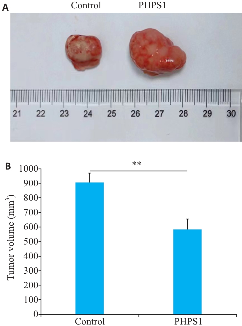
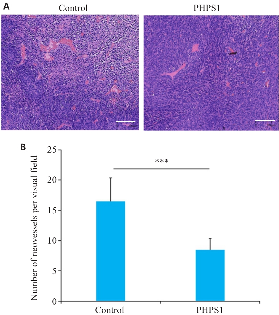
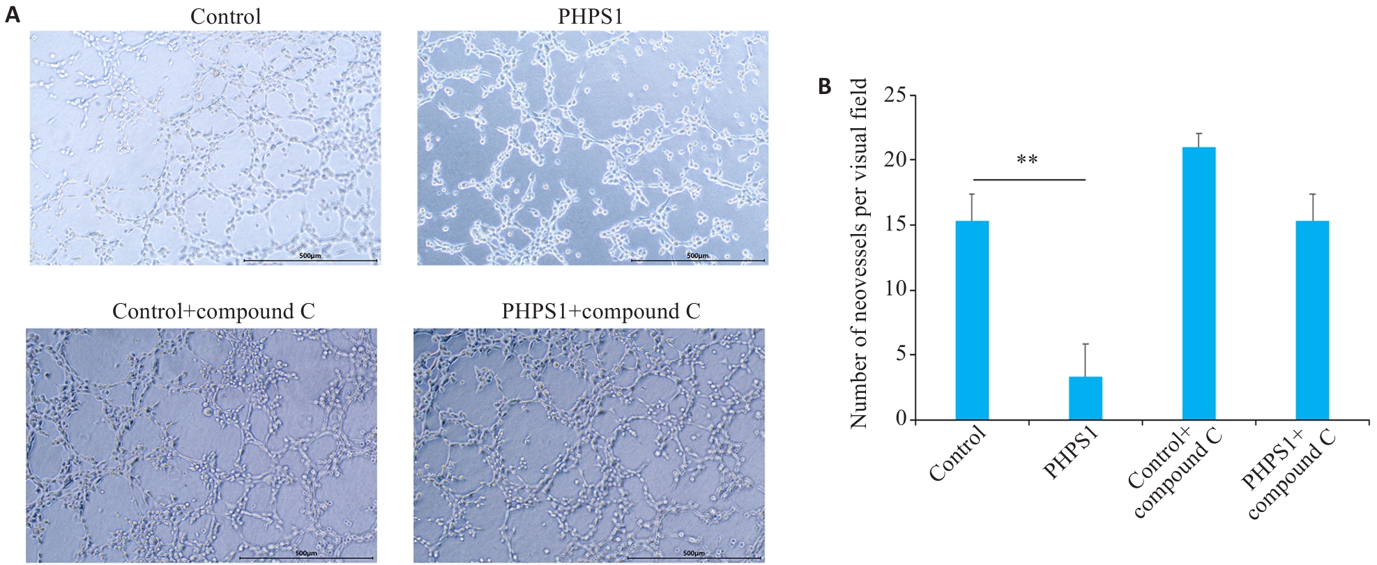
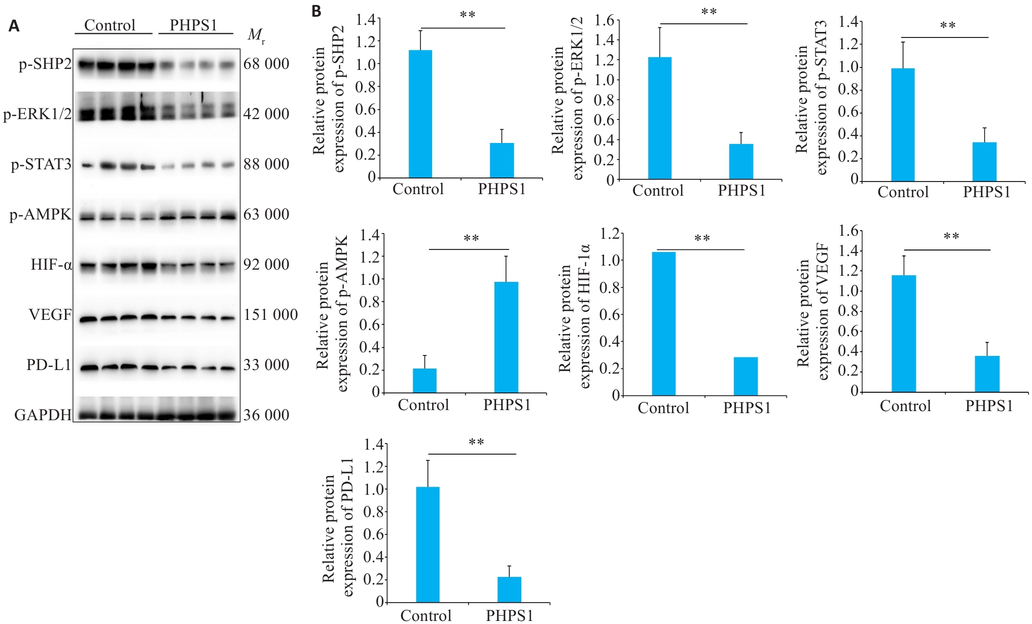
| 1 |
Renu K, Vinayagam S, Veeraraghavan VP, et al. Molecular crosstalk between the immunological mechanism of the tumor microenvironment and epithelial-mesenchymal transition in oral cancer[J]. Vaccines, 2022, 10(9): 1490.
|
| 2 |
Grégoire V, Grau C, Lapeyre M, et al. Target volume selection and delineation (T and N) for primary radiation treatment of oral cavity, oropharyngeal, hypopharyngeal and laryngeal squamous cell carcinoma[J]. Oral Oncol, 2018, 87: 131-7.
|
| 3 |
Porceddu SV, Daniels C, Yom SS, et al. Head and neck cancer international group (HNCIG) consensus guidelines for the delivery of postoperative radiation therapy in complex cutaneous squamous cell carcinoma of the head and neck (cSCCHN)[J]. Int J Radiat Oncol Biol Phys, 2020, 107(4): 641-51.
|
| 4 |
Woo SB. Oral epithelial dysplasia and premalignancy[J]. Head Neck Pathol, 2019, 13(3): 423-39.
|
| 5 |
Bai YC, Boath J, White GR, et al. The balance between differentiation and terminal differentiation maintains oral epithelial homeostasis[J]. Cancers, 2021, 13(20): 5123.
|
| 6 |
Niogret C, Birchmeier W, Guarda G. SHP-2 in lymphocytes' cytokine and inhibitory receptor signaling[J]. Front Immunol, 2019, 10: 2468.
|
| 7 |
Wu XL, Guan SY, Lu YG, et al. Macrophage-derived SHP-2 inhibits the metastasis of colorectal cancer via Tie2-PI3K signals[J]. Oncol Res, 2023, 31(2): 125-39.
|
| 8 |
Feng GS. Shp2-mediated molecular signaling in control of embryonic stem cell self-renewal and differentiation[J]. Cell Res, 2007, 17(1): 37-41.
|
| 9 |
Spaulding HR, Yan Z. AMPK and the adaptation to exercise[J]. Annu Rev Physiol, 2022, 84: 209-27.
|
| 10 |
Ciccarese F, Zulato E, Indraccolo S. LKB1/AMPK pathway and drug response in cancer: a therapeutic perspective[J]. Oxid Med Cell Longev, 2019, 2019: 8730816.
|
| 11 |
Hsu CC, Peng DN, Cai Z, et al. AMPK signaling and its targeting in cancer progression and treatment[J]. Semin Cancer Biol, 2022, 85: 52-68.
|
| 12 |
Karampitsakos T, Galaris A, Barbayianni I, et al. SH2 domain-containing phosphatase-SHP2 attenuates fibrotic responses through negative regulation of mitochondrial metabolism in lung fibroblasts[J]. Diagnostics, 2023, 13(6): 1166.
|
| 13 |
Jiang JH, Hu BJ, Chung CS, et al. SHP2 inhibitor PHPS1 ameliorates acute kidney injury by Erk1/2-STAT3 signaling in a combined murine hemorrhage followed by septic challenge model[J]. Mol Med, 2020, 26(1): 89.
|
| 14 |
Salem IH, Plante S, Gounni AS, et al. A shift in the IL-6/STAT3 signalling pathway imbalance towards the SHP2 pathway in severe asthma results in reduced proliferation process[J]. Cell Signal, 2018, 43: 47-54.
|
| 15 |
Tan SZ, Li DP, Zhu X. Cancer immunotherapy: pros, cons and beyond[J]. Biomed Pharmacother, 2020, 124: 109821.
|
| 16 |
Sobierajska K, Ciszewski WM, Sacewicz-Hofman I, et al. Endothelial cells in the tumor microenvironment[J]. Adv Exp Med Biol, 2020, 1234: 71-86.
|
| 17 |
Xu ZY, Guo CY, Ye QL, et al. Endothelial deletion of SHP2 suppresses tumor angiogenesis and promotes vascular normalization[J]. Nat Commun, 2021, 12(1): 6310.
|
| 18 |
Chen H, Cresswell GM, Libring S, et al. Tumor cell-autonomous SHP2 contributes to immune suppression in metastatic breast cancer[J]. Cancer Res Commun, 2022, 2(10): 1104-18.
|
| 19 |
Zhao H, Ren XJ, Kong RY, et al. Auxilin regulates intestinal stem cell proliferation through EGFR[J]. Stem Cell Reports, 2022, 17(5): 1120-37.
|
| 20 |
Herzig S, Shaw RJ. AMPK: guardian of metabolism and mitochondrial homeostasis[J]. Nat Rev Mol Cell Biol, 2018, 19(2): 121-35.
|
| 21 |
Chun Y, Kim J. AMPK-mTOR signaling and cellular adaptations in hypoxia[J]. Int J Mol Sci, 2021, 22(18): 9765.
|
| 22 |
Cha JH, Yang WH, Xia WY, et al. Metformin promotes antitumor immunity via endoplasmic-reticulum-associated degradation of PD-L1[J]. Mol Cell, 2018, 71(4): 606-20. e7.
|
| 23 |
Cha JH, Chan LC, Li CW, et al. Mechanisms controlling PD-L1 expression in cancer[J]. Mol Cell, 2019, 76(3): 359-70.
|
| 24 |
Chen DS, Mellman I. Elements of cancer immunity and the cancer-immune set point[J]. Nature, 2017, 541(7637): 321-30.
|
| 25 |
Yao H, Lan J, Li CS, et al. Inhibiting PD-L1 palmitoylation enhances T-cell immune responses against tumours[J]. Nat Biomed Eng, 2019, 3(4): 306-17.
|
| 26 |
Hanahan D, Weinberg RA. Hallmarks of cancer: the next generation[J]. Cell, 2011, 144(5): 646-74.
|
| 27 |
Mabeta P, Steenkamp V. The VEGF/VEGFR axis revisited: implications for cancer therapy[J]. Int J Mol Sci, 2022, 23(24): 15585.
|
| 28 |
Wang ZQ, Zhao P, Tian KH, et al. TMEM9 promotes lung adenocarcinoma progression via activating the MEK/ERK/STAT3 pathway to induce VEGF expression[J]. Cell Death Dis, 2024, 15(4): 295.
|
| 29 |
Lee SH, Golinska M, Griffiths JR. HIF-1-independent mechanisms regulating metabolic adaptation in hypoxic cancer cells[J]. Cells, 2021, 10(9): 2371.
|
| 30 |
Ma S, Lu CC, Yang LY, et al. ANXA2 promotes esophageal cancer progression by activating MYC-HIF1A-VEGF axis[J]. J Exp Clin Cancer Res, 2018, 37(1): 183.
|
 ), 刘昕2, 刘健2(