2. 南方医科大学口腔医院//广东省口腔医院,种植中心,广东 广州 510280;
3. 南方医科大学深圳口腔医院,修复科,广东 广州 518001;
4. 南方医科大学口腔医院//广东省口腔医院,正畸科,广东 广州 510280
2. Department of Implant, Stomatological Hospital, Southern Medical University/ Guangdong Provincial Stomatological Hospital, Guangzhou 510280, China;
3. Department of Prosthodontics, Shenzhen stomatological Hospital, Southern Medical University, Shenzhen 518001, China;
4. Department of Orthodontics, Stomatological Hospital, Southern Medical University/ Guangdong Provincial Stomatological Hospital, Guangzhou 510280, China
多生牙是指位于颌骨中由牙胚发育而来且具有牙齿结构的额外牙[1-2],可发生于上下颌骨的任何位置,好发于上颌。多生牙在恒牙列和乳牙列中均可发生,多发于混合牙列期。因其可能占据正常牙齿的空间位置,常造成邻牙移位[3]、扭转、阻萌[4]、牙根吸收[5]或伴发牙源性囊性病损等,所以有必要拔除埋伏多生牙[6-9]。
由于埋伏多生牙邻近正常牙齿及重要解剖结构,手术过程可能损伤邻近组织。术者主要依靠术前锥形束CT[10-11],锥形束CT的应用虽然能在术前判断多生牙的初步位置[12-14],但无法将数据准确传递到口内中[10-11],由于完全骨埋伏多生牙的位置翻瓣后不可见,特别是对于内外侧骨板较厚,与重要邻近结构贴合紧的多生牙,仅凭医生的临床经验,容易造成多生牙位置与手术位置存在一定程度的偏差[15-16],术者为了获取足够的术野,增加翻瓣的面积和去骨的范围,加大了手术创伤[17],这不仅增加了手术时间及难度,甚至会造成邻近重要解剖结构的损伤,增加患者的术后反应。近年来数字化打印技术[18-20]及导航辅助系统[21-23]的应用,为牙槽外科的发展带来了新的契机。为了解决多生牙拔除术中及术后的问题,笔者尝试使用一种数字化定位导板,通过简单的操作、稳定的固位、准确的定位,探讨其在上颌前牙区唇侧埋伏多生牙拔除中的应用,评价其应用效果,旨在促进微创牙槽外科进一步发展。
1 资料和方法 1.1 临床资料选取2019年3月~2019年8月于南方医科大学口腔医院颌面外科病房就诊的多生牙患者30例为研究对象。纳入标准:(1)多生牙位于上颌前牙区,且为完全骨埋伏,手术确定为唇侧入路;(2)埋伏多生牙均有需要手术拔除的指征;(3)无系统疾病史。30例患者中,实验组15例,男性8例,女性7例,平均年龄10.5岁;对照组15例,男性7例,女性8例,平均年龄11.2岁。所有患者均签署拔牙知情同意书,并经南方医科大学口腔医院伦理委员会批准。
1.2 方法 1.2.1 数据采集患者取站位,眶耳平面与地平面平行,面中轴与CT扫描纵轴一致。双眼平视,表情放松,牙处于开颌状态拍摄头颅锥形束CT。拍摄后数据导出为DICOM格式。硅橡胶制取上颌硬石膏模型,用3D Shape扫描仪扫描石膏模型,导出为STL格式。将锥形束CT与口扫石膏模型数据用mimics和3matic软件打开, 分析数据,配准CBCT数据与扫描硬石膏数据上较为一致且分散的5~10个标志点,实现二者之间的整合。
1.2.2 导板制作整合数据后进行定位导板的设计,唇侧入路手术定位导板设计为牙-骨面支持式,提取埋伏牙对应骨面信息,在其表面设计厚度约2.0 mm的定位基托,并贴多生牙边界设计半圆形开窗孔,并且避让邻牙、鼻底等解剖结构,最后修整导板边缘至合适的范围,以利手术就位,以STL文件输出。通过3D打印机利用立体光固化成形法打印导板,打印完成后与石膏牙模进行精确性匹配。数字化定位导板设计及打印服务由深圳市普天阳医疗科技股份有限公司提供。
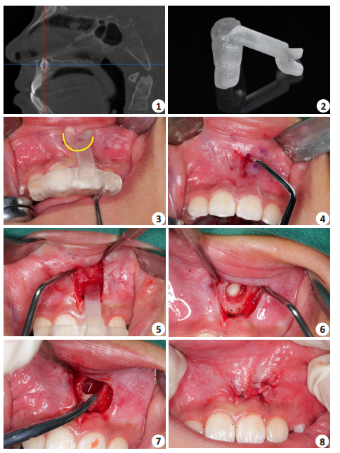
1.2.3 手术过程所有患者均采用全身麻醉,依据术前设计手术入路进行手术,手术定位导板就位,以导板定位部件的外缘为切口线(图 1中黄色标线),用龙胆紫标记,以移开定位导板,作黏膜弧形切口,翻起黏骨膜瓣,将定位导板再次就位,使用骨动力系统于开窗孔范围去骨,暴露多生牙,将其拔除,搔刮牙囊,生理盐水冲洗拔牙窝后缝合。典型病例见图 1。

|
图 1 数字化定位导板在埋伏多生牙拔除中应用 Fig.1 Application of digital positioning guide in extraction of impacted supernumerary teeth. 1: CT positioning; 2: Digital positioning guide; 3: Guide plate positioning; 4: Position determination; 5: Open flap, guide plate positioning again; 6 : Exposure of the supernumerary teeth; 7: Block removal of the supernumerary teeth; 8: Suture |
(1)术前设计时间:术前设计定位导板的时间;(2)找牙时间:从切开翻瓣至找到埋伏多生牙牙体组织需要的时间;(3)手术时间:从切开翻瓣至缝合结束需要的时间;(4)术中并发症:定位偏差、邻牙损伤、鼻底损伤。
1.2.5 统计学分析采用SPSS 18.0软件进行数据处理,结果用平均数和标准差表示,采用t检验进行统计学分析,P < 0.05为差异有统计学意义。
2 结果实验组术中定位导板均固位稳定,贴附良好,根据开窗孔去骨,迅速定位并暴露了多生牙,同时避免了术中损伤邻近重要解剖结构,迅速将其拔除。实验组平均术前设计时间为50±5 min,对照组为0 min;实验组平均找牙时间为5±2 min,对照组为10±3 min,实验组显著小于对照组(t=15.40,P < 0.01);实验组平均手术时间为25±4min,对照组为45±6 min,实验组显著小于对照组(t=35.50,P < 0.01);实验组未出现术中并发症,对照组出现一例定位稍偏差,正常恒牙根面暴露(多生牙与恒牙根未直接接触)。两组患者术后随访1个月,伤口均Ⅰ期愈合,未出现邻牙、鼻底等部位损伤的症状。
3 讨论微创、精确一直是牙槽外科医生追求的目标之一[24-25]。Hu等[26]利用手术导板配合超骨刀应用于埋伏多生牙的拔除,取得良好效果。刘培才等利用两段式导板分别定位切口线和多生牙,能快速精确定位并减少手术创伤,但两段式设计增加了制作和使用难度[27]。在导航系统辅助下完成埋伏多生牙拔除,明显缩短了拔牙时间,但术前增加导航系统安装时间,而且由于设备限制不便于在基层医院推广[28-29]。
本课题应用数字化定位导板,解决了影像定位无法准确转移到口内的问题,也简化了导板的结构,同时弥补了导航辅助拔牙的一些限制,提高了埋伏牙拔除过程准确性和安全性,且避免损伤邻近的重要解剖结构;首创以导板定位部件的外缘为切口线,一物两用,精准的定位也减少了翻瓣的面积,降低了术中的出血量,缩短了拔牙时间,减少患者术后不适反应和并发症的发生。牙支持式为定位导板提供了良好的固位,不易翘动。考虑到口腔环境复杂,患者情况不统一,以替牙期儿童居多,局麻术中患者配合程度不一[31],加之多生牙位置多样,解剖差异大,本研究严格限制纳入标准,只纳入了实验组、对照组各15例病例,并且均在全麻下进行。在临床应用中,实验组增加了术前设计时间,而且由于导板需要一定制作周期,一般为3 d,我们采取的措施是,在患者第1次前来预约床位时,收集好数据及制作导板,待患者正式入院时导板已到位。由于导板的使用,减小了翻瓣面积,缩短了找牙时间,也进一步缩短翻瓣时间和缝合时间,整个手术时间也了随着缩短。所以虽然实验组增加了术前准备时间,但简化了手术过程,是值得肯定的。
虽然本课题为了消除局麻下患者配合程度的影响,只纳入全麻病例,但笔者认为,数字化定位导板在门诊局麻下有更广阔的应用前景,在患者初诊时进行沟通,CBCT的拍摄,口腔模型的制取,待导板制作完毕,复诊进行手术,通过导板的使用,缩短手术时间,减少手术创伤,可减轻患者对拔牙过程的恐惧感,有利于提高医患关系。
本研究数字化定位导板在上颌唇侧埋伏多生牙拔除中的应用,取得了良好的临床效果,在后续的研究中,可将数字化定位导板的应用范围进一步扩大,如考虑进行牙瘤、内生性骨疣、根端囊肿等的定位与手术。本研究中,仅通过CT数据构建的导板与CT数据配合牙列模型构建的导板我们均有制作进行对比,发现在手术中的使用效果无异。虽然本文未提及腭侧入路的定位导板,但笔者也进行了一些尝试,发现由于腭侧软组织影响较大,仅通过CT数据构建的导板使用效果不稳定。所以在条件不充许时,唇侧入路的多生牙定位导板,可以考虑仅使用CT数据来构建,这在后续研究中将进一步阐明。
综上所述,应用数字化定位导板拔除上颌埋伏多生牙缩短了手术时间,减少了手术难度,减轻患者术后反应,值得临床推广。
| [1] |
Garvey MT, Barry HJ, Blake M. Supernumerary teeth--an overview of classification, diagnosis and management[J]. J Can Dent Assoc, 1999, 65(11): 612-6. |
| [2] |
Peter P, Jochen F, Sergio A, et al. Problems of supernumerary teeth, hyperdontia or dentes supernumerarii[J]. Ann Anat, 2006, 188(2): 163-9. |
| [3] |
Akgun OM, Fidan S, Altug A, et al. Non-syndrome patient with bilateral supernumerary teeth: Case report and 9-year follow-up[J]. Eur J Dent, 2013, 07(1): 123-6. |
| [4] |
Mason C, Azam N, Holt RD, et al. A retrospective study of unerupted maxillary incisors associated with supernumerary teeth[J]. Br J Oral Maxillofac Surg, 2000, 38(1): 62-5. |
| [5] |
Liu DG, Zhang WL, Zhang ZY, et al. Localization of impacted maxillary canines and observation of adjacent incisor resorption with cone-beam computed tomography[J]. Oral Surg Oral Med Oral Pathol Oral Radiol Endod, 2008, 105(1): 91-8. |
| [6] |
Bereket C, Çakir-Özkan N, Şener İ, et al. Analyses of 1100 supernumerary teeth in a nonsyndromic Turkish population: A retrospective multicenter study[J]. Niger J Clin Pract, 2015, 18(6): 731. |
| [7] |
Anthonappa RP, Rashied SO, Nigel MK. Characteristics of 283 supernumerary teeth in southern Chinese children[J]. Oral Surg Oral Med Oral Pathol Oral Radiol Endod, 2008, 105(6): e48-54. |
| [8] |
Parolia A, Kundabala M, Dahal M, et al. Management of supernumerary teeth[J]. J Conserv Dent, 2011, 14(3): 221-4. |
| [9] |
Kapila S, Conley RS, Harrell WE. The current status of cone beam computed tomography imaging in orthodontics[J]. Dentomaxillofac Radiol, 2011, 40(1): 24-34. |
| [10] |
司亚静, 任起辉, 张建丽, 等. CT三维重建技术在正畸埋伏牙诊治中的应用[J]. 中国CT和MRI杂志, 2018, 16(8): 40-2. |
| [11] |
Guerrero ME, Shahbazian M, Bekkering G, et al. The diagnostic efficacy of cone beam CT for impacted teeth and associated features: a systematic review[J]. J Oral Rehabil, 2011, 38(3): 208-16. |
| [12] |
Ali FA, Ata-Ali J, Penarrocha-Oltra D, et al. Prevalence, etiology, diagnosis, treatment and complications of supernumerary teeth[J]. J Clin Exp Dent, 2014, 6(4): e414-8. |
| [13] |
Liu DG, Zhang WL, Zhang ZY, et al. Three-dimensional evaluations of supernumerary teeth using cone-beam computed tomography for 487 cases[J]. Oral Surg Oral Med Oral Pathol Oral Radiol Endod, 2007, 103(3): 403-11. |
| [14] |
Nakajima A, Sameshima GT, Arai Y, et al. Two-and threedimensional orthodontic imaging using limited cone beam-computed tomography[J]. Angle Orthod, 2005, 75(6): 895-903. |
| [15] |
Katheria BC, Kau CH, Tate R, et al. Effectiveness of impacted and supernumerary tooth diagnosis from traditional radiography versus cone beam computed tomography[J]. Pediatr Dent, 2010, 32(4): 304-9. |
| [16] |
李志进, 郭家平, 石咏梅, 等. 锥形束CT在诊治阻生牙所致邻牙牙根外吸收中的应用[J]. 华西口腔医学杂志, 2013, 31(6): 588-91. |
| [17] |
龚二平. 微创技术在牙及牙槽外科中的应用价值分析[J]. 全科口腔医学电子杂志, 2016, 3(5): 93-4. |
| [18] |
Louvrier A, Marty P, Barrabé A, et al. How useful is 3D printing in maxillofacial surgery?[J]. J Stomatol Oral Maxillofac Surg, 2017, 118(4): 206-12. |
| [19] |
Prasad TS, Sujatha G, Muruganandhan J, et al. Three-dimensional printing in reconstructive oral and maxillofacial surgery[J]. J Contemp Dent Pract, 2018, 19(1): 1-2. |
| [20] |
Dawood A, Marti BM, Sauret-Jackson V, et al. 3D printing in dentistry[J]. Br Dent J, 2015, 219(11): 521-9. |
| [21] |
Azarmehr I, Stokbro K, Bell RB, et al. Surgical navigation: a systematic review of indications, treatments, and outcomes in oral and maxillofacial surgery[J]. J Stomatol Oral Maxillofac Surg, 2017, 75(9): 1987-2005. |
| [22] |
Demian N, Pearl C, Woernley TR, et al. Surgical navigation for oral and maxillofacial surgery[J]. Oral Maxillofac Surg Clin North Am, 2019, 31(4): 531-8. |
| [23] |
Chen X, Xu L, Sun Y, et al. A review of computer-aided oral and maxillofacial surgery: planning, simulation and navigation[J]. Expert Rev Med Devices, 2016, 13(11): 1043-51. |
| [24] |
薛洋, 胡开进. 中国牙及牙槽外科的现状与未来[J]. 中国实用口腔科杂志, 2019, 12(10): 600-3. |
| [25] |
黄志权, 张大明. 微创外科技术在口腔颌面外科中的应用[J]. 口腔疾病防治, 2018, 26(2): 75-82. |
| [26] |
Ying KH, Qian YX, Chi Y, et al. Computer-designed surgical guide template compared with free-hand operation for mesiodens extraction in premaxilla using "trapdoor" method[J]. Medicine (Baltimore), 2017, 96(26): e7310. |
| [27] |
刘培才, 王志兴. 手术定位导板在埋伏多生牙拔除中的应用[J]. 华西口腔医学杂志, 2019, 37(1): 58-61. |
| [28] |
吕坤, 杨荣涛, 周海华, 等. 导航下拔除上颌埋伏多生牙的临床分析[J]. 中华口腔医学杂志, 2018, 53(2): 103-6. |
| [29] |
Wang W, Somar M, Lv K. Safer alternative for extraction of impacted supernumerary teeth of a patient in the mixed dentition stage with the aid of an image-guided operating system[J]. Br J Oral Maxillofac Surg, 2017, 55(5): 551-3. |
| [30] |
吴中明, 李云波, 王维戚, 等. PET/MR在口腔颌面部恶性肿瘤外科手术中的应用价值[J]. 分子影像学杂志, 2020, 43(1): 41-4. |
| [31] |
曾桂琼, 邹海燕, 刘齐英, 等. 局部麻醉下儿童上颌埋伏多生牙拔除的护理配合[J]. 中外医疗, 2013, 32(34): 157-9. |
 2020, Vol. 40

