随着MRI在乳腺疾病诊断中的广泛应用,美国放射学会制定和发布的MRI乳腺影像报告和数据分析系统(BI-RADS),在乳腺疾病的MRI同质化诊断中得到了全球的广泛认可和应用[1-2]。然而,BI-RADS词典并没有提供进行诊断决策的具体方法。因此,使用BI-RADS词典进行疾病诊断时,阅片者之间的读图一致性良好,诊断的准确性差异较大[3-5]。为了协助放射科医师分析乳腺MRI特征,提高对乳腺病变良、恶性诊断的特异性,研究提出了一种鉴别乳腺良、恶性病变的树状流程图为“Kaiser评分”[6-7]。但是,目前有关乳腺病变Kaiser评分的研究报道极少。同时,对于乳腺非肿块性病变Kaiser评分的研究基本处于空白。
依据增强后乳腺病灶形态学上的不同,MRI BI-RADS将乳腺异常强化病灶分为3类:点状、肿块及非肿块强化;新版BI-RADS对于病变特征的描述趋于简单、实用,有效减少了不同研究者分析的主观差异性,为乳腺非肿块病变MRI研究的同质化和精确化提供了可能[8-9]。但目前国内外应用新版BI-RADS对非肿块病变的研究较少。本研究目的是评价Kaiser评分对乳腺非肿块强化病变的诊断效能,并与2013版MRI BI-RADS分类进行比较。
1 资料和方法 1.1 一般资料从PACS系统上选取2014年1月~2019年6月,在西安交通大学第二附属医院行术前乳腺3.0T MRI多模态扫描的患者,回顾性分析影像及临床病理资料,纳入标准为:MRI检查前未行有创性检查或任何有关乳腺癌的治疗;病变经手术或穿刺病理证实;MRI表现为非肿块强化。排除标准为:MRI表现为肿块型强化;病理结果不完善。
1.2 检查方法 1.2.1 设备和参数MRI检查采用3.0T MRI扫描仪,乳腺专用相控阵线圈,患者采取俯卧位进行扫描。MRI扫描序列包括:FSE T1WI(TR 680 ms,TE9 ms,矩阵320× 320),FSE T2WI脂肪抑制序列(TR 6700 ms,TE 37 ms,矩阵320×320)及T1WI动态增强(VIBRANT)序列(TR 5 ms,TE 2 ms,矩阵350×350)。动态增强序列包括增强前蒙片+6期动态增强,于造影剂注射后15 s开始扫描,每期64 s。造影剂采用Gd-DTPA,剂量0.2 mmol/kg,注射速率2.0 mL/s,再加15 mL生理盐水。
1.2.2 数据处理及测量形态学参数的获取:首先从乳腺背景实质强化中识别非肿块强化,然后获取病变的形态学指标,包括病变的位置、分布模式、边缘和内部强化模式等。动态增强曲线的获取:应用Functool软件,避开出血、坏死、囊变等区域,在病变最大层面中心区域放置感兴趣区(ROI),> 3个体素,自动获得时间信号曲线(TIC)。TIC分为3型:Ⅰ型为持续型,Ⅱ型为平台型,Ⅲ型为流出型。
1.3 诊断评估标准BI-RADS分类诊断参照美国放射学会的2013版BI-BADS词典[8],根据病灶的分布、内部强化特征,参考动态增强曲线,综合评估后做出最终分类诊断。本研究纳入病例不包括评估不全的0类、无异常发现的1类和活检已证实恶性的6类,将BI-RADS 2类和3类视为诊断良性,BI-RADS 4类和5类视为诊断恶性。
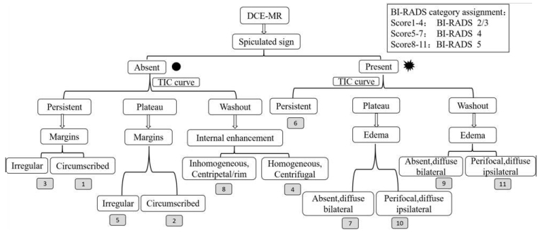
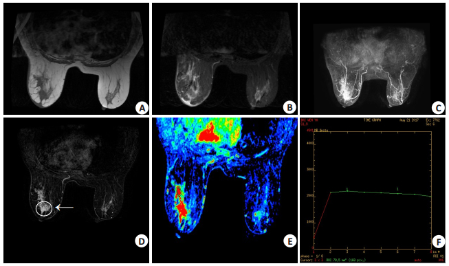
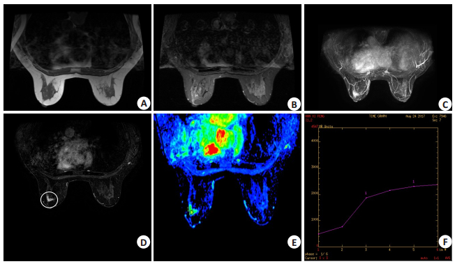
Kaiser评分参照Baltzer等的评分标准和Kaiser评分流程图进行[6-7],评分为1~11分(图 1),其中1~4分对应BI-RADS 2/3类,5~7分对应BI-RADS 4类,8~11分对应BI-RADS 5类(图 2、3)。

|
图 1 Kaiser评分流程图 Fig.1 Flow chart of Kaiser scoring. |

|
图 2 女,61岁,发现左乳肿块1年余 Fig.2 Female, 61 years old, found left breast mass for over 1 year. A: T1WI; B: T2WI; C: CE MRI-MIP; D: CE MRI; E, F: TIC. A non- mass enhancement lesion was found in the left breast, presenting with segmental distribution and clustered ring enhancement (arrow in D) with unclear and multiple spiculate margin (circle in D) and peritumoral edema (Ellipse B) on T2WI. The dynamic enhancement curve showed a flatform type. BI-RADS 5 category was considered, which corresponded to a Kaiser score of 10. The postoperative pathology showed left breast intraductal carcinoma with necrosis and local suspected infiltration. |

|
图 3 女,56岁,双侧乳腺胀痛1月 Fig.3 MRI findings in a 56-year-old female patient with complaint of bilateral breast swelling pain for one month. A: T1WI; B: T2WI; C: CE MRI-MIP; D: CE MRI; E, F: TIC. A non-mass enhancement lesion was found in the left breast (circle in D), showing a segmental distribution and heterogeneous enhancement with unclear margin and without spiculate margin or peritumoral edema. The dynamic enhancement curve showed persistent type. BI-RADS 4 category was considered, consistent with a Kaiser score of 3. The postoperative pathology showed left breast cystic hyperplasia with metaplasia of sweat glands. |
由2名中级职称影像科医师分别采用上述两种方法,在不知临床诊断和病理结果的情况下分别对所有患者MRI图像进行分析和评估,意见不一致时则由一名资深乳腺影像医师确定。
1.5 统计学方法采用SPSS20.0统计软件进行数据分析。以病理结果为金标准,计算2种方法诊断结果与病理结果的一致性采用Kappa分析:K≥0.8为一致性很好;0.4≤K < 0.8为中度一致性;K < 0.4为一致性较差。按BI-RADS>3和Kiaser评分>4的截断值分别计算两种诊断方法的敏感性、特异性、阳性预测值、阴性预测值和诊断准确性。两种诊断方法的诊断效能比较采用配对卡方检验。P < 0.05为差异有统计学意义。
2 结果 2.1 乳腺病变病理结果并良、恶性病变BI-RADS分类及Kaiser评分诊断结果本研究共纳入患者88例,均为女性,年龄22~75岁,平均47.1岁。88例共90个非肿块病变。双侧乳腺病变者共2例:1例双侧乳腺癌;1例一侧病变为浸润性导管癌,另一侧为囊性增生。良性病变28个,以腺病42.9%(12/28)及非哺乳期乳腺炎35.7%(10/28)最多见。恶性病变62个,以浸润性导管癌64.5%(40/62)和导管原位癌伴或不伴微浸润24.2%(15/62)最多见。乳腺良、恶性病变BI-RADS分类及Kaiser评分诊断结果具体见表 1。
| 表 1 乳腺良、恶性病变BI-RADS分类及Kaiser评分诊断结果 Tab.1 Diagnostic results of BI-RADS classification and Kaiser score in benign and malignant breast lesions |
BI-RADS分类及Kaiser评分结果与病理结果的对比分析具体见表 2。BI-RADS分类及Kaiser评分对乳腺非肿块强化病变的诊断敏感性、特异性、阳性预测值、阴性预测值及准确性具体见表 3。Kaiser评分诊断特异性为75%,明显高于BI-RADS分类诊断(46.4%),配对卡方检验结果显示差异有统计学意义(P=0.021)。
| 表 2 BI-RADS分类及Kaiser评分结果与病理结果的对比 Tab.2 Comparison of Kaiser score and BI-RADS classification results with pathology |
| 表 3 两种诊断方法效能比较 Tab.3 Comparison of the diagnostic efficacy of Kaiser score and BI-RADS classification |
临床上非肿块强化的发生率小于肿块,但是57%的临床无法触及的浸润性乳腺癌表现为非肿块强化,所以对非肿块强化的识别和诊断对乳腺MRI检查具有更为重要的意义[10]。目前国内外对于乳腺肿块型强化病变的研究已经较为成熟,MRI BI-RADS分类在预测其良恶性方面基本上形成了较统一和规范的诊断标准;而非肿块强化无论是在形态学还是血流动力学方面,良恶性病变相互重叠较多,临床容易漏诊或过度诊断[1, 11-15]。新版BI-RADS对于病变特征的描述趋于简单、实用,有效减少了不同研究者分析的主观差异性[16]。研究显示节段样分布、簇环形强化、TIC类型为Ⅲ型高度提示恶性病变[11]。Aydin[17]发现节段性分布、簇环形强化、TIC类型为Ⅱ型和扩散受限的存在与恶性肿瘤有关。有研究同样发现簇环状强化、节段性分布高度提示恶性肿瘤[18]。因此乳腺MRI BI-RADS具有较高的诊断敏感性和准确性,但诊断特异性较低[11]。本研究使用BI-RADS分类对非肿块乳腺病变诊断的特异性为46.4%,与上述研究结论一致。
有研究认为Kaiser评分系统有望提高乳腺MRI诊断的特异性[6, 19-20]。因为Kaiser评分是基于对恶性病变的影像学特征分析,从17个征象中选择了5个征象(根征、动态增强血流动力学、病灶边缘、内部增强模式和瘤周水肿)对每个病灶进行评分,来评估恶性肿瘤的可能性。此外,Kaiser评分系统仅需要简单的动态增强血流动力学和形态学特征,无需增加额外的成像序列。Marino等[19]研究认为Kaiser评分不仅适用于乳腺肿块性病变,也可以应用于非肿块性病变。Woitek等[20]研究显示使用Kaiser评分可以提高诊断特异性,避免了25%以上的活检率。本研究由经验尚浅的2名影像科医师对乳腺MR图像进行分析与评估,研究结果表明Kaiser评分对乳腺非肿块强化病变的诊断特异性明显高于BI-RADS分类,差异有统计学意义。此外,与病理结果的一致性也明显优于BI-RADS分类。实际工作中对于临床经验丰富的影像科医师,往往并不需要借助Kaiser评分系统评估病变良恶性。因此,本研究认为对于经验尚浅的影像科医师使用Kaiser评分能够明显提高其对乳腺非肿块强化病变的诊断特异性。
Kaiser评分及BI-RADS分类均能较全面的根据病灶的形态学及血流动力学特征分析乳腺病变。本研究90个非肿块样强化病变,73个病灶根据BI-RADS分类为恶性病变,有15例良性病变误诊为恶性,其中7例为乳腺囊性增生伴或不伴导管上皮增生,6例为非哺乳期乳腺炎。69个病灶根据Kaiser评分为恶性病变,有7例良性病变误诊为恶性,其中1例为囊性增生伴导管上皮增生,另6例均为非哺乳期乳腺炎。由于非哺乳期乳腺炎与非肿块强化乳腺癌形态学及血流动力学表现类似,故影响了本组病例的诊断准确性,诊断特异性也较文献报道略低[11, 20-21]。
本研究对乳腺非肿块强化病变进行了较大样本的研究,但良性病变数量较少,有待于进一步扩大样本量。实际临床工作中,如能够同时兼顾病灶的DWI特点,并结合乳腺X线摄影综合分析,可能会进一步提高乳腺MR非肿块强化病变的诊断准确性。
| [1] |
Sohns C, Scherrer M, Staab W, et al. Value of the BI-RADS classification in MR-Mammography for diagnosis of benign and malignant breast tumors[J]. Eur Radiol, 2011, 21(12): 2475-83. |
| [2] |
Aydin H. The MRI characteristics of non-mass enhancement lesions of the breast: associations with malignancy[J]. Br J Radiol, 2019, 92(1096): 20180464. |
| [3] |
Stoutjesdijk MJ, Futterer JJ, Boetes C, et al. Variability in the description of morphologic and contrast enhancement characteristics of breast lesions on magnetic resonance imaging[J]. Invest Radiol, 2005, 40(6): 355-62. |
| [4] |
Grimm LJ, Anderson AL, Baker JA, et al. Interobserver variability between breast imagers using the fifth edition of the BI-RADS MRI lexicon[J]. AJR Am J Roentgenol, 2015, 204(5): 1120-4. |
| [5] |
Lalonde L, El Khoury M, Mesurolle B, et al. Breast imaging reporting and data system (BI-RADS) lexicon for breast MRI: Interobserver variability in the description and assignment of BI-RADS category[J]. Eur J Radiol, 2015, 84(1): 71-6. |
| [6] |
Baltzer PA, Dietzel M, Kaiser WA. A simple and robust classification tree for differentiation between benign and malignant lesions in MR-mammography[J]. Eur Radiol, 2013, 23(8): 2051-60. |
| [7] |
Dietzel M, Baltzer P. How to use the Kaiser score as a clinical decision rule for diagnosis in multiparametric breast MRI: a pictorial essay[J]. Insights Imaging, 2018, 9(3): 325-35. |
| [8] |
Edwards SD, Lipson JA, Ikeda DM, et al. Updates and revisions to the BI-RADS magnetic resonance imaging lexicon[J]. Magnetic Res Imaging Clin North America, 2013, 21(3): 483-93. |
| [9] |
Shin K, Phalak K, Hamame A, et al. Interpretation of breast MRI utilizing the BI-RADS fifth edition lexicon: how are we doing and where are we headed?[J]. Curr Probl Diagn Radiol, 2017, 46(1): 26-34. |
| [10] |
Bartella L, Liberman L, Morris EA, et al. Nonpalpable mammographically occult invasive breast cancers detected by MRI[J]. AJR Am J Roentgenol, 2006, 186(3): 865-70. |
| [11] |
崔晓琳, 周纯武, 李静, 等. MRI乳腺影像报告和数据系统在非肿块强化病变中的诊断价值研究[J]. 临床放射学杂志, 2015, 34(4): 532-7. |
| [12] |
李晶英, 赵殿江. 乳腺MRI非肿块样强化病变的影像学诊断进展[J]. 中国医学影像学杂志, 2018, 26(7): 547-51. |
| [13] |
Baltzer PA, Benndorf M, Dietzel M, et al. False-positive findings at contrast-enhanced breast MRI: a BI-RADS descriptor study[J]. AJR Am J Roentgenol, 2010, 194(6): 1658-63. |
| [14] |
Gutierrez RL, DeMartini WB, Eby PR, et al. BI-RADS lesion characteristics predict likelihood of malignancy in breast MRI for masses but not for nonmasslike enhancement[J]. AJR Am J Roentgenol, 2009, 193(4): 994-1000. |
| [15] |
Benndorf M, Baltzer PA, Kaiser WA. Assessing the degree of collinearity among the lesion features of the MRI BI-RADS lexicon[J]. Eur J Radiol, 2011, 80(3): e322-4. |
| [16] |
Rao AA, Feneis J, Lalonde C, et al. A Pictorial review of changes in the BI-RADS fifth edition[J]. Radiographics, 2016, 36(3): 623-39. |
| [17] |
Aydin H. The MRI characteristics of non-mass enhancement lesions of the breast: associations with malignancy[J]. Br J Radiol, 2019, 92(1096): 20180464. |
| [18] |
Asada T, Yamada T, Kanemaki Y, et al. Grading system to categorize breast MRI using BI-RADS 5th edition: a statistical study of non-mass enhancement descriptors in terms of probability of malignancy[J]. Jpn J Radiol, 2018, 36(3): 200-8. |
| [19] |
Marino MA, Clauser P, Woitek R, et al. A simple scoring system for breast MRI interpretation: does it compensate for reader experience?[J]. Eur Radiol, 2016, 26(8): 2529-37. |
| [20] |
Woitek R, Spick C, Schernthaner M, et al. A simple classification system (the Tree flowchart) for breast MRI can reduce the number of unnecessary biopsies in MRI-only lesions[J]. Eur Radiol, 2017, 27(9): 3799-809. |
| [21] |
Chu AN, Seiler SJ, Hayes JC, et al. Magnetic resonance imaging characteristics of granulomatous mastitis[J]. Clin Imaging, 2017, 43: 199-201. |
 2020, Vol. 40

