2. 湖北医药学院附属太和医院创伤骨科,湖北 十堰 442000;
3. 南华大学附属第一医院创伤骨科,湖南 衡阳 421000;
4. 南方医科大学南方医院骨科,广东 广州 510515
2. Department of Orthpaedics and Traumatology, Taihe Hospital, Shiyan 442000, China;
3. Department of Traumatology and Orthopaedics, The First Affiliated Hospital of Nanhua University, Hengyang 421000, China;
4. Department of Orthopedics, Nanfang Hospital, Southern Medical University, Guangzhou 510515, China
骨盆或髋臼骨折相关的出血主要来自骶前和腰椎静脉丛,或直接来自骨折部位[1-3]。虽然骨盆或髋臼骨折引起的盆腔内主要大动脉损伤很少发生(15%~20%)[4-6],合并髂外动脉损伤的更为罕见,文献中只有少数病例系列报道或个案报告[4-5, 7-10];但这些损伤可导致75%~83%的死亡率[11-12],其中约60%患者死于创伤性失血性休克[6]。早期死亡原因最常见是无法控制的出血,晚期大部分为全身多器官功能衰竭。对于何种类型髋臼骨折容易并发髂外大血管损伤文献报道较少,近年来个别学者开始关注此损伤机制,他们试图找出骨盆骨折与髂外大血管损伤的特异性关系[4, 6, 10, 13-14]。但至今尚无明确阐述。在此,我们分享4例髋臼顶柱骨折[15]合并髂外动脉损伤病例外科治疗,并探讨该类型骨折引起髂外动脉损伤的机制。
1 资料和方法 1.1 一般资料回顾性分析2013年1月~2018年12月南方医科大学南方医院因髋臼顶柱骨折合并髂外动脉损伤住院治疗患者,研究通过南方医科大学南方医院伦理委员会审查(伦理编号:NFEC-2019-256),随访中患者本人或死亡患者的家属签署知情同意书。本研究共4例患者,其中男性3例,女性1例,年龄28~53岁,平均年龄42.8岁,受伤原因均为严重车祸。全部患者入院时存在外伤性失血性休克。左、右侧髋臼顶柱骨折各2例。闭合性髋臼顶柱骨折3例,开放性髋臼顶柱骨折1例。1例开放性损伤合并严重软组织挫伤,下腹壁及盆腔软组织广泛积气。2例合并耻骨骨折。3例患者入院查体足背动脉搏动消失,术前行增强血管CT检查(CTA)示髂外动脉远端均未显影,血管腔内血栓形成、闭塞。1例患者入院查体足背动脉搏动存在,术后行CTA检查示髂外动脉虽不显影,但臀下动脉与股深动脉存在侧支循环(表 1)。
| 表 1 4例髋臼顶柱骨折合并髂外动脉损伤患者资料表 Tab.1 General characteristics of 4 patients with acetabular roof column fractures and external iliac arterial injury |
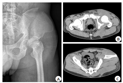
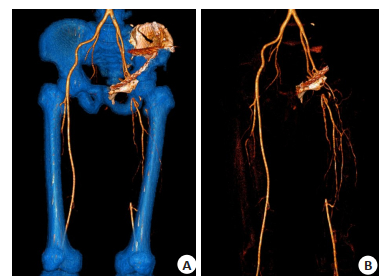
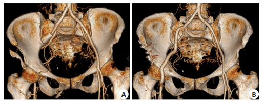
4例患者入院后予损伤性控制复苏,行补液、输血、维持循环等处理,急诊手术治疗。Case 1患者为右侧髋臼开放性顶柱骨折,右大腿中下段软组织大面积严重碾压伤,入院查体右下肢皮肤严重发绀,右足背动脉未触及。CTA检查示右侧髂外动脉管腔闭塞,充盈缺损。综合分析患者右侧髂外动脉损伤,考虑右大腿中下段软组织碾压伤,无法挽留患肢,遂行右侧大腿中下段截肢术。患者术后大腿残端出现脓性渗出伴恶臭,残端皮肤坏死,无法愈合,并发感染性休克。遂第2次行髋关节离断术及残端清创术,患者病情仍持续恶化,感染无法控制,继而出现败血症及弥漫性血管内凝血(DIC),最终因多脏器功能衰竭死亡。Case 2患者为左侧髋臼闭合性顶柱骨折,入院查体左下肢皮肤呈暗红色,皮温冰凉,左足背动脉未触及;术前CTA检查提示示左髂外动脉血栓行成,左下肢动静脉血管无显示,考虑患者左侧髂外动脉完全断裂。因该患者左下肢无血供近22 h,深浅静脉血栓已形成,患者最终行左髋关节离断术挽救生命,术后恢复良好。Case 3患者为左侧髋臼闭合性顶柱骨折(图 1),入院体格检查提示左下肢血运正常,足背动脉搏动存在;术中探查见髂腰肌和髂外动脉完全离断,结合查体考虑左下肢血供已形成侧支循环,遂单纯行髋臼骨折复位内固定术,髂外动脉损伤未予处理。患者术后恢复良好,术后CTA示左侧股动脉中上段大部分未见显示,考虑血栓栓塞,臀下动脉与股深动脉存在侧支循环(图 2)。Case 4患者为右侧髋臼闭合性顶柱骨折,术前CTA检查示右侧髂外动脉及其远端未显影,术中探查见右股总动脉及右股总动脉分叉以下约5 cm的股浅动脉无搏动,考虑右侧髂外动脉断裂,遂行骨盆骨折切开复位内固定+骨盆骨折外固定+右侧髂动脉、股动脉人工血管旁路术(图 3),吻合人工血管后,触摸股浅动脉搏动恢复。术后经抗凝、营养支持等对症治疗后,患者恢复良好,右下肢血运正常。

|
图 1 术前髋关节X线及CT检查 Fig.1 Preoperative radiographs and CT scan of the hip joint. A: Preoperative radiographs show a roof column acetabular fracture; B-C: CT scan reveals an acetabular fracture with roof wall and roof column. |

|
图 2 双下肢动脉CTA检查 Fig.2 CT angiography of lower limbs. A-B: CT angiography shows a filling defect of the left external iliac artery and collateral circulation of the lower gluteal artery and the deep femoral artery. |

|
图 3 髂外动脉术前术后CTA检查 Fig.3 Preoperative and postoperative CT angiography of the external iliac artery. A: CT angiography shows a filling defect of the right external iliac artery; B: Postoperative CT angiography shows the reconstruction of artificial blood vessels after right iliac artery and femoral artery bypass grafting. |
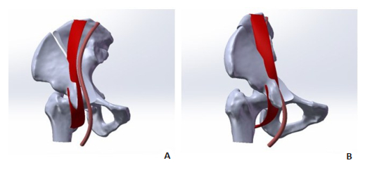
4例患者诊断为髋臼顶柱骨折合并髂外动脉损伤,入院后行损伤控制复苏,并采用不同手术方式治疗,包括截肢术、髋关节离断术、骨折切开复位和骨折内固定术及骨折外固定术、髂动脉和股动脉旁路移植术等。除1例患者死于多器官功能衰竭外,余患者恢复良好,术后均重返工作岗位。通过分析4例患者受伤经过及骨折类型,我们考虑髋臼顶柱骨折发生后,由于髂外动脉通过髂耻筋膜等软组织与髂腰肌有一定连接,髂外动脉可能随髂腰肌一起被牵拉入骨折端间隙,髂外动脉直接被骨折端割破,或患者在搬运及骨折复位过程中,骨折块刺破髂外动脉(图 4),从而导致创伤性失血性休克。

|
图 4 髋臼顶柱骨折后髂外动脉损伤示意图 Fig.4 Schematic diagram of external iliac artery injury after acetabular roof column fracture. A-B: The iliac vessels are connected to the iliopsoas muscle through soft tissues such as the iliac fascia. After the acetabular roof column fracture, the tension effect caused the iliopsoas muscle to fall between the fracture ends, and the external iliac artery was pulled into the fracture space together, causing damage to the external iliac artery. |
髋臼为典型的杵臼关节,其具有连接躯干和下肢、传导人体重力、维持正常生理活动等功能。髋臼骨折的受伤机制多为高能量暴力经股骨头作用于髋臼关节面,引起骨块移位及髋关节不稳。大部分髋臼骨折是股骨头侧方不同角度的撞击所致,这种类型骨折有时累及臼顶承重关节面和部分髂骨,但是影像学示髂耻线完整,真骨盆缘完整,Letournel[16-18]将这种类型归类为少见的后壁骨折类型,认为股骨头撞击髋臼顶骨折后继发髂骨骨折和髂骨外旋[16]。另有研究认为带有髋臼前上方关节面的髂前上棘以下骨折是前壁骨折[19]。谭国庆等[20]报道了12例该类型骨折治疗,认为这类骨折归属前柱骨折的范畴,是一种真骨盆缘完整的髋臼高位前柱骨折。侯志勇团队[15, 21-22]提出基于三柱构成理念的髋臼骨折改良分型,他们认为人发育成熟后,构成半骨盆的髂骨、耻骨和坐骨的骨质较厚,构成髋臼三个强有力的柱:顶柱、前柱和后柱,髋臼通过上方的顶柱与主骨支撑柱相连;将髂骨耻骨、髂骨坐骨和耻骨坐骨之间的移行薄弱区定义为前壁、后壁和内壁;其中简单顶柱骨折(A3.2)表现为髂耻线及髂坐线均完整。
本组4例患者为车祸高能量撞击伤所致,髋臼顶柱骨折块一般呈外旋畸形、向近端移位,所有病例均在髋部前外侧发现瘀斑。我们推测其受伤机制为暴力直接作用在骨盆前外侧挤压髂骨前部外旋所致。首先发生髂骨骨折继而累及髋臼臼顶区骨折,导致头臼不稳定,部分患者出现股骨头前上方脱位,由于暴力作用方向及对术者手术入路选择均在前方,按髋臼骨折三柱分型[15],我们认为这4例患者属于髋臼顶柱骨折,A3.2型。但这一损伤机制尚需进一步的生物力学研究加以证实。
骨盆一般来说更能抵抗侧向压缩力。作为一个直接的结果,对于侧方压力引起的骨盆骨折,与其他方向相比有着更低的血管损伤率及更好的止血效果[17-18]。按暴力方向对骨盆骨折进行的分型,血流动力学不稳定性是不太可能出现在侧方挤压损伤(LC骨折)[23]。相比之下,前部挤压损伤更容易导致临床显著的血管损伤,包括动脉和静脉出血或创伤性血管管腔的损伤(血栓形成,夹层)。通常的两种血管损伤机制是骨折块引起临近骨盆骨面的血管血栓形成或血管撕裂[3, 24-26]。
迄今为止,骨盆、髋臼骨折引起大动脉损伤的机制至今尚不明确。有报道发现有关检查骨盆环不同部位的骨折与血管损伤的关系(无断裂,前环,后环,和髋臼)[27],在429例患者中均未出现髂血管或股动脉损伤。另一项研究报道两例骨盆骨折合并髂股动脉损伤[14]。他们的损伤机制为骨盆前后压缩骨折,但这两个病人都有髋臼受累。因此,他们得出结论:髂股动脉损伤只有当骨盆骨折合并髋臼骨折时才会发生。
我们遇到这4例患者均为髋臼顶柱骨折合并髂外动脉损伤,有必要探讨一下髂血管受伤机制与该类型骨折的相关性。髂腰肌由髂肌和腰大肌组成。髂肌起自髂窝;腰大肌起自腰椎体侧面及横突。向下两肌相合,经腹股沟韧带深面,通过髂耻弓前缘(即髋臼前壁),止于股骨小转子。我们猜测这4例病人血管损伤的机制是,来自于前外侧的巨大暴力直接作用于髂骨翼,引起髂骨纵行断裂,即髋臼顶柱骨折,这种类型骨折骨折线大多终止于髋臼前方髂耻隆起处。当顶柱骨折块外旋移位时,带动股骨头同时外旋,小转子外旋牵拉髂腰肌,使髂腰肌张力增加,加上生理情况下髂腰肌通过髂耻弓前缘也会有一定张力。髋臼顶柱骨折发生后,张力作用使得髂腰肌陷入骨折断端之间。髂外动脉通过髂耻筋膜等软组织与髂腰肌有一定连接性,因而会被牵拉一起陷入移位明显的髂耻隆起骨折间隙处,锋利的骨折断端可直接割破髂外动脉,或患者在后期搬运及骨折复位过程中,骨折块刺破髂外动脉。但在其他一些髋臼顶柱骨折病例中,我们也观察到了髂腰肌的嵌顿受损,髂外动脉却未受损,这可能和髂外动脉与髂腰肌连接紧密程度有关。
这4例患者有相同的骨折类型及同样的髂外动脉损伤,但所采取的手术治疗方式却截然不同。采取截肢术或髋关节离断术的患者可能是因为髂外动脉断裂位置靠近近端,侧支循环尚未形成,下肢血供不足,从而引起肢体坏死。髋臼顶柱骨折合并髂外动脉损伤,血管不需处理的患者,考虑有髂内动脉的分支与股深动脉形成吻合支,侧支循环形成,能代偿提供下肢血运,最终预后良好。Case 4患者受伤后检查,首诊医师较早发现右侧足背动脉无搏动,立即行CTA检查发现右侧髂动脉损伤,发现较为及时,行右侧髂动脉、股动脉人工血管旁路术后,重新建立右下肢血运,患者得到及时、正确的治疗。
综上所述,虽然髋臼顶柱骨折合并髂外动脉损伤很少发生,但其损伤后果是灾难性的。临床医生遇到此类病人时,应高度警惕大血管损伤的可能性,必要时可通过CT血管三维重建或动脉介入血管造影术,早期干预、早期治疗以挽救肢体。
| [1] |
Marsh JL, Slongo TF, Agel J, et al. Fracture and dislocation classification compendium-2007-Orthopaedic Trauma Association classification, database and outcomes committee[J]. J Orthop Trauma, 2007, 21(10, S): S1-133. |
| [2] |
张奉琪, 潘进社, 张英泽. 骨盆骨折血管损伤的解剖学基础[J]. 中国临床解剖学杂志, 2004, 22(2): 116-9. DOI:10.3969/j.issn.1001-165X.2004.02.002 |
| [3] |
张奉琪, 潘进社. 骨盆骨折血管损伤的解剖学基础及临床[J]. 中国矫形外科杂志, 2003, 11(14): 985-7. DOI:10.3969/j.issn.1005-8478.2003.14.018 |
| [4] |
Pascarella R, Del Torto M, Politano R, et al. Critical review of pelvic fractures associated with external iliac artery lesion: a series of six cases[J]. Injury, 2014, 45(2): 374-8. DOI:10.1016/j.injury.2013.10.011 |
| [5] |
Carrillo EH, Wohltmann CD, Spain DA, et al. Common and external iliac artery injuries associated with pelvic fractures[J]. J Orthop Trauma, 1999, 13(5): 351-5. DOI:10.1097/00005131-199906000-00005 |
| [6] |
Porter SE, Schroeder AC, Dzugan SS, et al. Acetabular fracture patterns and their associated injuries[J]. J Orthop Trauma, 2008, 22(3): 165-70. DOI:10.1097/BOT.0b013e318165918b |
| [7] |
黄海生, 张谢安, 杨峰, 等. 髂外动脉损伤的治疗[J]. 中华创伤杂志, 1997, 13(4): 17. |
| [8] |
Ruotolo C, Savarese E, Khan A, et al. Acetabular fractures with associated vascular injury: a report of two cases[J]. J Trauma, 2001, 51(2): 382-6. DOI:10.1097/00005373-200108000-00028 |
| [9] |
Wolinsky PR, Johnson KD. Delayed catastrophic rupture of the external iliac artery after an acetabular fracture. A case report[J]. J Bone Joint SurgAm, 1995, 77(8): 1241-4. DOI:10.2106/00004623-199508000-00015 |
| [10] |
Langford JR, Trokhan S, Strauss E. External iliac artery thrombosis after open reduction of an acetabular fracture: a case report[J]. J Orthop Trauma, 2008, 22(1): 59-62. DOI:10.1097/BOT.0b013e31815affd2 |
| [11] |
Rothenberger DA, Fischer RP, Perry JF. Major vascular injuries secondary to pelvic fractures: an unsolved clinical problem[J]. Am J Surg, 1978, 136(6): 660-2. DOI:10.1016/0002-9610(78)90331-8 |
| [12] |
Perry JF. Pelvic open fractures[J]. Clin Orthop Relat Res, 1980(151): 41-5. |
| [13] |
Koelling E, Mukherjee D. Extrinsic compression of the external iliac artery following internal fixation of an acetabular fracture[J]. J Vasc Surg, 2011, 54(1): 219-21. DOI:10.1016/j.jvs.2010.11.123 |
| [14] |
Frank JL, Reimer BL, Raves JJ. Traumatic iliofemoral arterial injury: an association with high anterior acetabular fractures[J]. J Vasc Surg, 1989, 10(2): 198-201. DOI:10.1016/0741-5214(89)90356-X |
| [15] |
Zhang R, Yin Y, Li A, et al. Three-Column classification for acetabular fractures: introduction and reproducibility assessment[J]. J Bone Joint SurgAm, 2019, 101(22): 2015-25. DOI:10.2106/JBJS.19.00284 |
| [16] |
Letournel E, J R. Fractures of the acetabulum[M]. Heidelberg: Springer-Verlag, 1993.
|
| [17] |
Letournel E. Acetabulum fractures - classification and management[J]. Clin Orthop Relat Res, 1980, 15(1): 81-106. |
| [18] |
Letournel E. Acetabulum Fractures: Classification and Management[J]. J Orthop Trauma, 2019, 33(S2): S1-2. |
| [19] |
Lenarz CJ, Moed BR. Atypical anterior wall fracture of the acetabulum: case series of anterior acetabular rim fracture without involvement of the pelvic brim[J]. J Orthop Trauma, 2007, 21(8): 515-22. DOI:10.1097/BOT.0b013e31814612e5 |
| [20] |
谭国庆, 周东生, 王伯珉, 等. 真骨盆缘完整的髋臼高位前柱骨折的治疗[J]. 中华骨科杂志, 2011, 31(11): 1239-44. DOI:10.3760/cma.j.issn.0253-2352.2011.11.013 |
| [21] |
侯志勇, 张瑞鹏, 张英泽. 基于三柱构成理念的改良髋臼骨折分型[J]. 中华创伤杂志, 2018, 34(1): 6-10. DOI:10.3760/cma.j.issn.1001-8050.2018.01.003 |
| [22] |
王忠正.改良髋臼骨折分型系统的可信度与可重复性评价[D].石家庄: 河北医科大学, 2019: 35. http://cdmd.cnki.com.cn/Article/CDMD-10089-1019630401.htm
|
| [23] |
Young JW, Burgess AR, Brumback RJ, et al. Lateral compression fractures of the pelvis: the importance of plain radiographs in the diagnosis and surgical management[J]. Skeletal Radiol, 1986, 15(2): 103-9. DOI:10.1007/BF00350202 |
| [24] |
武兴国.骨盆骨折血管损伤与骨折类型相关性的临床研究[D].济南: 山东大学, 2006: 59. http://cdmd.cnki.com.cn/Article/CDMD-10422-2006166923.htm
|
| [25] |
张奉琪, 张英泽, 潘进社, 等. 骨盆前环后环骨折与骨盆动脉损伤的相关性研究[J]. 中国骨与关节损伤杂志, 2005, 20(8): 505-7. DOI:10.3969/j.issn.1672-9935.2005.08.001 |
| [26] |
李昶荣, 陈忠. X线、CT联合MRI在胫骨平台骨折中的诊断价值[J]. 分子影像学杂志, 2020, 43(1): 122-5. |
| [27] |
Klein SR, Saroyan RM, Baumgartner F, et al. Management strategy of vascular injuries associated with pelvic fractures[J]. J Cardiovasc Surg (Torino), 1992, 33(3): 349-57. |
 2020, Vol. 40

