2. Department of Critical Care Medicine, Nanfang Hospital, Southern Medical University, Guangzhou 510515, China
2. 南方医科大南方医院重症医学科, 广东 广州 510515
Central venous catheterization is required in many clinical settings, such as in the operating room, surgical intensive care unit, emergency department, and cardiovascular department for central venous pressure monitoring, rapid fluid resuscitation, and hemodialysis, etc. The common veins for puncture include the internal jugular vein (IJV), subclavian vein (SCV), and axillary vein (AV). The conventional cannulation (landmark method) of the central vein is performed using the anatomical landmarks as the guide and is associated with a high risk of mechanical complications including pneumothorax, hemothorax and arterial puncture[1]. A new safer and more efficient catheterization approach is thus urgently needed. Increasing evidence from studies has suggested that ultrasound-guided venipuncture can improve the success rate of puncture, shorten the operation time, reduce complications and lower the medical costs [2-4]. The current approaches of ultrasound-guidance central venous catheterization mainly include the long-axis (LAX) approach and short-axis (SAX) approach[5].
In recent years, a number of randomized controlled trials (RCTs) [6-12] were carried out to compare the advantages and disadvantages of these two approaches, but the conclusions derived from these studies are not consistent and can even be contradictory. To address the lack of high-quality systematic evaluation and meta-analysis based on human subjects, we searched the current databases and conducted a systematic review and meta-analysis of the RCTs comparing the efficacy, safety and complications of ultrasound-guided LAX and SAX approaches for central venous catheterization, aiming to provide reliable and convincing evidence to facilitate the choice of ultrasound-guided approach to central venous cannulation.
METHODS Study ethicsEthical approval and patient consent are waived because of the nature of this study, which is a systematic review and meta-analysis of previously published studies. The study was performed with close adherence to principles recommended by the Preferred Reporting Items for Systematic Reviews and Meta-Analyses (PRISMA) Statement[13].
Search strategy and study selectionTwo investigators independently searched the databases including PubMed, Cochrane Library, Science Direct, and Web of Science. The electronic search strategy combined terms related to ultrasound guide catheterization approach (including a keyword search using the words 'longitudinal', 'transverse', 'short-axis', 'long-axis ', 'in plane ' and 'out of plane or out-plane') and the terms related to the central vein (using the keywords 'femoral vein', 'axillary vein', 'subclavian vein', and 'jugular veins').
Inclusion and exclusion criteriaOnly prospective RCTs comparing ultrasound -guided LAX (in-plane) and SAX (out -of-plane) central venous catheterization in clinical adult patients requiring central venous cannulation were included in this study. The publications (1) that document systematic reviews or meta-analyses, (2) whose authors did not consent to the inclusion, (3) where the study subjects are not clinical patients, and (4) that are not published in English language were excluded from the analysis.
Data extraction and outcome measuresData were extracted independently by two investigators, and the discrepancies between the investigators were resolved by consensus. A piloted data-extraction sheet was applied, which covered the first author 's name, age of the patients, body mass index (BMI), paper source, experience of the operators, and ultrasound equipment used. The primary outcomes were the first-attempt success, mean attempts to success, and the incidence of complications. The secondary outcomes included the puncture time, number of repositioning and skin puncture times. The data obtained by two independent research evaluators were compared for homogeneity.
Assessment of bias and quality of evidenceAssessment of the risk of bias was performed in accordance with guidelines outlined in the Cochrane Handbook for Systematic Reviews of Interventions (Version 5.1.0)[14]. Two investigators reviewed all the studies and assigned a value from 'high', 'low', or 'unclear' to the following domains: random sequence generation; allocation concealment; blinding of participants and personnel; blinding of outcome assessment; incomplete outcome data; selective reporting; and other bias. Trials with a high risk of bias for one or more of these key domains were considered at high risk of bias. Trials with low risk of bias for all these domains were considered at low risk of bias. Otherwise, they were considered to have unclear risk of bias[14]. The overall quality of the evidence and the strength of the recommendations were evaluated using the Grading of Recommendations, Assessment, Development and Evaluation (GRADE) Working Group guidelines[15, 16]. Specifically, the evidence derived from the study is deemed to be (1) of high quality if further research is very unlikely to change our confidence in the estimate of effect; (2) of moderate quality if further research is likely to have an important impact on the confidence in the estimate of the effect and may change the estimate; (3) of low quality if further research is very likely to have an important impact on the confidence in the estimate of effect and is likely to change the estimate; and (4) of very low quality if it is very uncertain about the estimate.
Statistical analysisThe difference in the dichotomous data is expressed as the relative risk (RR) with 95% confidence interval (95% CI). The weighted mean difference (WMD) of the 95% CI was used for continuous outcomes. The Q test was used to calculate the I2 statistic to test the heterogeneity: P≤0.1 or I2 > 50% indicates significant statistical homogeneity[13, 17]. Whenever significant heterogeneity was present, we searched for potential sources of heterogeneity and a random effects model was applied. For example, if one study showed results that were completely out of range of the others, we searched for likely reasons explaining the difference and performed a sensitivity analysis excluding that study when deemed appropriate. We further carried out subgroup analysis according to the type of insertion (elective vs emergency). We estimated the difference between the estimates of the subgroups according to the tests for interaction. If not applicable, a fixed effect model was referred. Begg Test was calculated when needed, and Egger Test was used to evaluate the existence of publication bias[18, 19]. GRADE Profiler 3.6 was used to perform an overall quality assessment. A P value < 0.05 in the combined test was considered to indicate a statistically significant di-fference between the tested subgroups. RevMan (Version 5.2; Nordic Cochrane Center, Copenhagen, Denmark) and Stata 12.0 (Stata Corporation, College Station, TX, USA) were used for statistical analyses of the data.
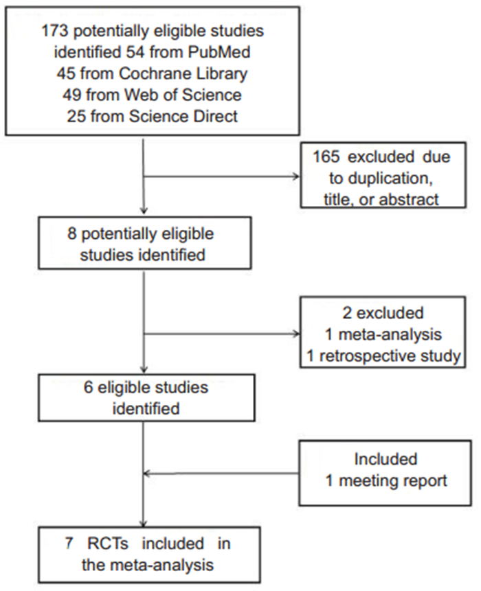
RESULTS Study selection and characteristicsBased on our search strategy, we identified a total of 173 potentially eligible RCTs from the databases (Fig. 1), among them 7 studies[6-12]involving a total of 729 patients met the inclusion criteria for the meta-analysis. The authors of these 7 studies were from the United States[7], Italy[6], Spain[8], Oman[9], Turkey[10], Ireland[11]or Egypt[12]. The settings of these studies were mostly operating room and surgical intensive care unit, and in a small part were the emergency department or kidney dialysis center. All the subjects receiving the puncture were adult patients (Tab. 1).

|
Fig.1 Flowchart illustrating the inclusion and exclusion of the studies identified from the databases. |
| Tab.1 Characteristics of the included RCTs |
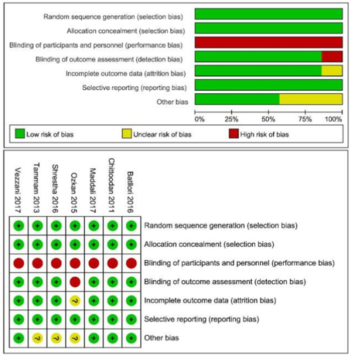
Cochrane tool was used to evaluate the risk of bias of the studies. All these studies were conducted with adequate randomization of sequence generation and allocation concealment. The participants and personnel in all these 7 studies were not blinded (Fig. 2). According to the GRADE Working Group criteria, the grades of evidence for the first-attempt success rate and overall success rate were moderate, and those of other outcome measures were low.

|
Fig.2 Assessment of risk of bias of the included studies. |
First-attempt success rate Six RCTs[6-11] reported the first-attempt success rate and were used to calculate the pooled estimate. Overall, the first-attempt success rate was higher with the SAX approach than with the LAX approach (RR=1.27; 95% CI: 1.11-1.46; P=0.0005, I2= 49%) (Fig. 3). Sensitivity analysis showed that the study by Shrestha et al[7] was a main source of heterogeneity, and the heterogeneity was significantly reduced after exclusion of this study[7](RR=1.31; 95%CI: 1.20-1.44; P < 0.00001, I2=0%) (Tab. 2). Subgroup analysis found significant heterogeneity in "patient characteristics", "number of puncture", and "operational department", while the "venous type", "patient number", and "publishing time" had small heterogeneities(Tab. 2).

|
Fig.3 Forest plot of the first-attempt puncture success rate. |
| Tab.2 Subgroup analysis of the first-attempt success rate |
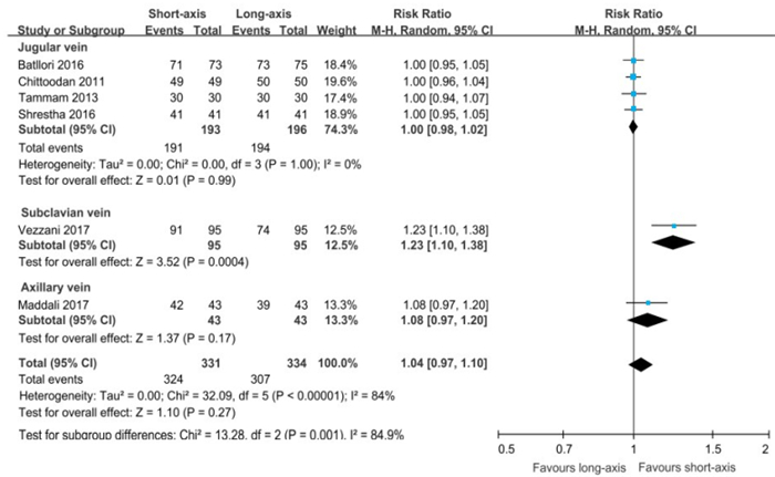
Overall puncture success rate Six RCTs [6, 9, 11, 12] reported the overall puncture success rate. Pooled analysis showed no significant difference in the overall puncture success rate between SAX and LAX approach (RR=1.04; 95% CI: 0.97-1.10; P=0.27; I2=84%) (Fig. 4). Subgroup analysis, however, showed that the overall puncture success rate was significantly higher with the SAX than with the LAX approach in the SCV (RR=1.23, 95% CI: 1.10-1.38; P=0.0004).

|
Fig.4 Forest plot of the overall puncture success rate. |
Complications Five RCTs reported the incidence of accidental arterial puncture. Pooled analysis showed that the arterial puncture rate was significantly higher in LAX than in SAX approach (RR=1.04; 95%CI: 1.01-1.08; P= 0.01, I2=30%) (Tab. 3). Sensitivity analysis indicated that the difference was more significant and the heterogeneity was reduced when the research by Tammam et al[12]was excluded (RR=1.06, 95%CI: 1.02-1.10; P=0.01, I2=7%).
| Tab.3 Summary of accidental arterial puncture, hematoma and secondary outcomes |
Four RCTs have reported the incidence of hematoma. Pooled analysis showed no differences in the incidence of hematoma between the two groups (RR= 0.67, 95% CI: 0.19-2.35;P=0.53, I2=0%) (Tab. 3). Subgroup analysis of venous type showed only insignificant heterogeneity, and the arterial puncture rate was significantly higher with the LAX than with the SAX approach in the SCV (RR=1.08, 95% CI: 1.02-1.15;P= 0.01).
Pooled analysis based on the SCV and the AV indicated that the SAX approach significantly increased the first-attempt success rate (RR=1.41; 95% CI: 1.08-1.85; P=0.01; I2=53%), and reduced the incidence of accidental arterial puncture (RR=0.15; 95% CI: 0.03-0.86; P=0.03; I2=0%) (Tab. 4).
| Tab.4 Summary of the outcomes of central venous cannulation through the SAX and LAX approaches in the subclavian vein and axillary vein |
No significant differences were found between the two approaches in the mean puncture time to success (WMD=-5.08; 95% CI: -12.76-2.61; P=0.20, I2=59%); repositioning of the needle (RR=-0.09;95%CI: -0.40-0.22; P=0.57, I2=77%) and the number of skin punctures (RR=-0.09, 95%CI : -0.23-0.05;P=0.19, I2=61%) (Tab. 3).
Publication biasAssessment of publication bias using Egger and Begg' test revealed no potential publication bias among the included trials (Egger test, P=0.82; Begg'test, P=0.85).
DISCUSSIONOur meta-analysis showed that the first-attempt success rate in central venous cannulation is higher when using a SAX approach than a LAX approach, and the SAX approach is also associated with a reduced incidence of such complications as accidental arterial puncture.
The conventional approach of central venous catheterization is time-consuming and has a low success rate, high incidences of complications, and a high medical cost. Ultrasound-guided puncture approaches can substantially remedy these problems. Currently researchers hold different opinions concerning the LAX approach (LAX) and SAX approach[20-22]. Recent studies by Gao et al[20] and Liu et al[21] suggested no significant differences between the LAX and SAX approaches in ultrasound-guided central venous catheterization, but Liu et al found that the SAX approach was superior to the LAX in the treatment of SCV and iliac vein catheterization under ultrasound guidance[21]; He et al[22] suggested that the LAX approach could increase the success rate of puncture in AV puncture. We noted a meeting report by Parlak et al[10], and after the inclusion of this trial, our study indicated that SAX was superior to LAX in central venous puncture, which differed from the previous results reported by Liu et al. But due to the lack of more rigorous peer review of the conference paper, more high-quality evidences are still needed to support this conclusion. Importantly, our conclusion based on this study is consistent with the recommendations of the international experts Frankel et al [23]. We thus recommend that the SAX approach be used to acquire high success rate and reduce the incidence of arterial puncture in central venous puncture.
According to the results of our analysis, the overall puncture success rate of a SAX approach was higher than that of a LAX approach in the SCV, which was different from the findings by Vezzani et al[6]. This discrepancy may arise from (1) the study design of Vezzani's, who limited the number of attempts to two for both techniques to limit the complication rate; (2) the large size and superficial location of the IJV, whose course along the neck does not vary much[24](thus allowing easier access to the short axis section of the IJV), as compared with the deeper location and smaller size of the SCV; and (3) the critical condition of the patients enrolled in Vezzani's study, who were more likely to have hemodynamic instabilities such as hypotension.
We found that the arterial puncture rate was higher with the LAX than with the SAX approach, which was especially obvious in the SCV. This may be due to the fact that the LAX approach does not allow visualization of the artery and the vein simultaneously as the SAX approach does. The deeper position and smaller size of the SCV also contribute to the higher arterial puncture rate associated with the LAX approach. No differences were observed in the incidence of hematoma between the two methods, for which a probable explanation is that the operators took extra attention when performing cannulation through the LAX approach, and provided timely treatment should arterial puncture occur.
No significant differences were found in the mean puncture time, number of repositioning and skin puncture times between the two approaches, but since significant heterogeneity was found in the combined results, this may have limited clinical significance. In particular, Vezzani et al[6]reported that the insertion time was shorter and the number of repositioning was smaller with the SAX approach than with the LAX approach; according to Batllori et al[8], the mean cannulation time was also longer in the LAX than in the SAX approach, and the patients in the LAX group required more needle passes (skin puncture times). But the study on human torso mannequin by Vogel et al [25] among resident physicians found that the long-axis view for the IJV was better than the short-axis view with fewer redirections. The long-axis view for subclavian central venous catheterization was also better with decreased time to cannulation and fewer redirections. But considering the different experiences of the operators, ultrasound equipment and the subjects receiving the puncture, this difference may need furthe study.
In addition, we found that the trial by Shrestha et al[7] is the source of heterogeneity regarding the first time success rate analysis, probably because of the heterogeneity in the age of the patients included and only one operator in the study. Our results supported the notion that the SAX approach can increase the first success rate and reduce the incidence of arterial puncture in the iliac vein and subclavian vein, as was consistent with the finding by Liu et al[21].
Due to the limited number of studies included, we did not use a funnel chart but employed the Begg's test and Egger' s Test to assess the publication bias. No publication bias was found in the studies included in this analysis. The sensitivity analysis and subgroup analysis suggested the sources of heterogeneity in this study may arise from (1) baseline characteristics of the population; (2) number of operators; and (3) the clinical operating environment (we did not assess the quality of the operators and the ultrasound equipment in emergency or non-emergency scenarios).
The limitations of this study lie in (1) the inclusion of only studies published in English language; (2) the small number of studies included, which may lead to overestimation of the conclusion; (3) failure of using double blinding in the included studies; (4) absence of studies involving special patients, such as children and obstetric patients; and (5) failure of assessment of satisfaction on the part of the operators and the patients. Further well-designed, more robust trials are needed to further validate our findings.
In recent years, new improvement approaches have been explored based on ultrasound-guided LAX and SAX approach. Dilisio et al[26]and Baidya et al[27]proposed an ultrasound -guided oblique-axis plane puncture approach combining the LAX and SAX approaches. Quan et al[28] proposed the ultrasound-guided development line approach and Tsuchiya et al [29] proposed the visual technology in plate. However, these techniques need to be validated in future clinical trials. We believed that the future improvement of ultrasound-guided puncture catheterization will attach greater importance to the success rate, reducing the operational complications, a more user-friendly operation interface and a simpler operative procedure.
| [1] |
Parienti JJ, Mongardon N, Megarbane B, et al. Intravascular complications of central venous catheterization by insertion site[J]. N Engl J Med, 2015, 373(13): 1220-9. DOI:10.1056/NEJMoa1500964 |
| [2] |
Calvert N, Hind D, Mcwilliams R, et al. Ultrasound for central venous cannulation: economic evaluation ofcost-effectiveness[J]. Anaesthesia, 2004, 59(11): 1116-20. DOI:10.1111/j.1365-2044.2004.03906.x |
| [3] |
Hind D, Calvert N, Mcwilliams R, et al. Ultrasonic locating devices for central venous cannulation: meta-analysis[J]. BMJ, 2003, 327(7411): 361-8. DOI:10.1136/bmj.327.7411.361 |
| [4] |
Gu WJ, Tie HT, Liu JC, et al. Efficacy of ultrasound-guided radial artery catheterization: a systematic review and meta-analysis of randomized controlled trials[J]. Crit Care, 2014, 18(3): 93-100. DOI:10.1186/cc13862 |
| [5] |
Chapman G, Johnson D, Bodenham A. Visualisation of needle position using ultrasonography[J]. Anaesthesia, 2006, 61(2): 148-58. DOI:10.1111/j.1365-2044.2005.04475.x |
| [6] |
Vezzani A, Manca T, Brusasco C, et al. A randomized clinical trial of ultrasound-guided infra-clavicular cannulation of the subclavian vein in cardiac surgical patients: short-axis versus long-axis approach[J]. Intensive Care Med, 2017, 43(11): 1594-601. DOI:10.1007/s00134-017-4756-6 |
| [7] |
Shrestha G, Gurung A, Koirala S. Comparison between long- and short-axis techniques for ultrasound-guided cannulation of internal jugular vein[J]. Ann Card Anaesth, 2016, 19(2): 288-92. DOI:10.4103/0971-9784.179629 |
| [8] |
Batllori M, Urra M, Uriarte E, et al. Randomized comparison of three transducer orientation approaches for ultrasound guided internal jugular venous cannulation[J]. Br J Anaesth, 2016, 116(3): 370-6. DOI:10.1093/bja/aev399 |
| [9] |
Maddali MM, Arora NR, Chatterjee N. Ultrasound guided out-of-plane versus in-plane transpectoral left axillary vein cannulation[J]. J Cardiothorac Vasc Anesth, 2017, 31(5): 1707-12. DOI:10.1053/j.jvca.2017.02.011 |
| [10] |
Parlak O, Ozkan G. Ultrasound-guided internal jugular vein access: comparison between short axis and long axis techniques[J]. Am J Cardiol, 2013, 24(4): S64. |
| [11] |
Chittoodan S, Breen D, O'Donnell B D, et al. Long versus short axis ultrasound guided approach for internal jugular vein cannulation: a prospective randomised controlled trial[J]. Med Ultrason, 2011, 13(1): 21-5. |
| [12] |
Tammam TF, El-Shafey EM, Tammam HF. Ultrasound-guided internal jugular vein access: comparison between short axis and long axis techniques[J]. Saudi J Kidney Dis Transpl, 2013, 24(4): 707-13. DOI:10.4103/1319-2442.113861 |
| [13] |
Moher D, Liberati A, Tetzlaff J, et al. Preferred reporting items for systematic reviews and meta-analyses: the PRISMA statement[J]. J Clin Epidemiol, 2009, 62: 1006-12. DOI:10.1016/j.jclinepi.2009.06.005 |
| [14] |
Higgins JPT, Altman DG, Gotzsche PC, et al. The Cochrane Co-llaboration's tool for assessing risk of bias in randomised trials[J]. BMJ, 2011, 343: d5928. DOI:10.1136/bmj.d5928 |
| [15] |
Higgins JP, Green S. Cochrane handbook for systematic reviews of interventions[M]. The Cochrane Collaboration, 2008. ISBN: 978-0-470-69951-5.
|
| [16] |
Brożek JL, Akl EA, Alonso-Coello P, et al. Grading quality of evidence and strength of recommendations in clinical practice guidelines[J]. Allergy, 2009, 64(5): 669-77. DOI:10.1111/j.1398-9995.2009.01973.x |
| [17] |
Brozek J L, Akl EA, Compalati E, et al. Grading quality of evidence and strength of recommendations in clinical practice guidelines part 3 of 3. The GRADE approach to developing recommendations[J]. Allergy, 2011, 66(5): 588-95. DOI:10.1111/j.1398-9995.2010.02530.x |
| [18] |
Begg C B, Mazumdar M. Operating characteristics of a rank correlation test for publication bias[J]. Biometrics, 1994, 50(4): 1088-101. DOI:10.2307/2533446 |
| [19] |
Higgins JPT, Thompson SG. Quantifying heterogeneity in a meta-analysis[J]. Stat Med, 2002, 21(11): 1539-58. DOI:10.1002/sim.1186 |
| [20] |
Gao Y, Yan J, Ma J, et al. Effects of long axis in-plane vs short axis out-of-plane techniques during ultrasound-guided vascular access[J]. Am J Emerg Med, 2016, 34(5): 778-83. DOI:10.1016/j.ajem.2015.12.092 |
| [21] |
Liu C, Mao Z, Kang HJ, et al. Comparison between the long-axis/in-plane and short-axis/out-of-plane approaches for ultrasound-guided vascular catheterization: an updated meta-analysis and trial sequential analysis[J]. Ther Clin Risk Manag, 2018, 14: 331-40. DOI:10.2147/TCRM.S152908 |
| [22] |
He Y, Zhong M, Wu W, et al. A comparison of longitudinal and transverse approaches to ultrasound-guided axillary vein cannulation by experienced operators[J]. J Thorac Dis, 2017, 9(4): 1133-9. DOI:10.21037/jtd.2017.03.137 |
| [23] |
Frankel HL, Kirkpatrick AW, Elbarbary M, et al. Guidelines for the appropriate use of bedside general and cardiac ultrasonography in the evaluation of critically Ill patients-Part Ⅰ[J]. Crit Care Med, 2015, 43(11): 2479-502. DOI:10.1097/CCM.0000000000001216 |
| [24] |
Breschan C, Platzer M, Jost R, et al. Size of internal jugular vs subclavian vein in small infants: an observational, anatomical evaluation with ultrasound[J]. Br J Anaesth, 2010, 105(2): 179-84. DOI:10.1093/bja/aeq123 |
| [25] |
Vogel JA, Haukoos JS, Erickson CL, et al. Is long-axis view superior to short-axis view in ultrasound-guided central venous catheterization[J]. Crit Care Med, 2015, 43(4): 832-9. DOI:10.1097/CCM.0000000000000823 |
| [26] |
Dilisio R, Mittnacht AJC. The"medial-oblique"approach to ul-trasound-guided central venous cannulation-maximize the view, minimize the risk[J]. J Cardiothorac Vasc Anesth, 2012, 26(6): 982-4. DOI:10.1053/j.jvca.2012.04.013 |
| [27] |
Baidya DK, Chandralekha, Darlong V, et al. Comparative sonoanatomy of classic "short axis" probe position with a novel "medial-oblique" probe position for ultrasound-guided internal jugular vein cannulation: a crossover study[J]. J Emerg Med, 2015, 48(5): 590-6. DOI:10.1016/j.jemermed.2014.07.062 |
| [28] |
Quan Z, Tian M, Chi P, et al. Modified short-axis out-of-plane ultrasound versus conventional long-axis in-plane ultrasound to guide radial artery cannulation[J]. Anesth Analg, 2014, 119(1): 163-9. DOI:10.1213/ANE.0000000000000242 |
| [29] |
Tsuchiya M, Mizutani K, Funai Y, et al. In-line positioning of ultrasound images using wireless remote display system with tablet computer facilitates ultrasound-guided radial artery catheterization[J]. J Clin Monit Comput, 2016, 30(1): 101-6. DOI:10.1007/s10877-015-9692-9 |
 2020, Vol. 40

