2. 广州医科大学附属第二医院设备科,广东 广州 510000;
3. 华南理工大学医学院,广东 广州 510000;
4. 南方医科大学南方医院胸外科,广东 广州 510515
2. Department of Medical Equipment, Second Affiliated Hospital of Guangzhou Medical University, Guangzhou 510000, China;
3. School of Medicine, South China University of Technology, Guangzhou 510000, China;
4. Department of Thoracic Surgery, Nanfang Hospital, Southern Medical University, Guangzhou 510515, China
介电特性是生物组织的固有属性,通常用介电常数和电导率表示[1-2]。对于人体组织,其介电特性与组织内蛋白质含量,离子浓度及结合水与自由水的比例等有关[3],当人体组织的生理或病理状态发生改变时,组织的介电特性也会发生变化。许多研究对人体组织介电特性进行了报道,结果表明人体癌症组织与对应的正常组织的介电特性往往存在较大差异[4-8]。基于这种差异发展出多种新型的诊断和治疗技术,包括磁共振介电特性断层成像(MR-EPT)[9-10]、射频/微波消融[11]、以及术中恶性组织边缘检测[12-13]等,因此研究人体组织的介电特性,建立人体组织介电特性数据库具有重要意义。有研究建立了一个10 Hz~100 GHz频率范围的人体组织介电特性数据资源网站[14],这是现在普遍使用的数据库,但其中关于淋巴结的数据并不全面。有学者测量了6个转移淋巴结样本,报道了10个频率点(50、100、200、300、400、500、600、700、800和900 MHz)的介电特性[15]。然而这项研究并没有分析非转移淋巴结的介电特性也没有指出测量的是人体哪个部位的淋巴结。有研究测量了27个压缩至1 mm的腋窝淋巴结的介电特性[16],还有研究测量了23个外表面和横断面的乳腺淋巴结的介电特性[17],结果都表明转移淋巴结和非转移淋巴结的介电特性具有明显差异。然而,现有研究中还未发现对肺癌转移和非转移淋巴结的介电特性数据的报道。
淋巴结转移是肺癌常见的转移途径,临床上主要采用在手术切除肿瘤的同时根据患者的实际情况进行淋巴结清扫,淋巴结是否转移与患者的预后和生存质量密切相关[18-19]。目前常用的淋巴结检测方法是通过对需检测的淋巴结组织的石蜡切片进行HE染色,但是此技术检测时间较长,检测程序繁琐且无法在术中实时使用,大大降低了手术效率。本研究使用的开端同轴探头技术是基于开端同轴探头终端与待测组织的阻抗不匹配,电磁波将在探头终端面上发生反射,通过测量反射系数并计算出组织的介电特性[20-21]。此技术具有简单、快速、非破坏性和可实现在体实时测量等优点。因此在手术过程中,如果能通过使用此技术快速测量肺癌淋巴结的介电特性,从而快速鉴别出淋巴结是否转移,使得实时指导手术清扫方案成为可能,这将具有重大临床意义。为了实现这一目标,首先需要建立淋巴结介电特性数据库。基于以上几点,本文使用开端同轴探头法,对肺癌转移和非转移淋巴结的介电特性进行了测量,获得了介电特性数据并对这些数据进行了对比分析。
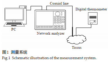
1 材料和方法 1.1 测量系统本研究采用开端同轴探头法[22-23],此方法操作简便且对样本没有破坏性。测量系统由一台网络分析仪(AV3680A)、一根特性阻抗为50 Ω的半刚性同轴线(UT141)和一台笔记本电脑组成(图 1)。探头通过BNC连接器连接到网络分析仪,网络分析仪通过USB线连接到笔记本电脑。笔记本电脑控制整个系统,包括设置网络分析仪的测量格式、测量频率点数和存储测量数据等。测量和存储的数据为网络分析仪校准面的反射系数,再根据探头终端平面等效模型和三参数计算方法[22-23]可求得组织的介电特性。本研究将测量频率点数设置为4000点,测量格式为Smith圆图。

|
图 1 测量系统 Fig.1 Schematic illustration of the measurement system. |
在测量淋巴结样本前,需要测量开路、短路和去离子水[22]的反射系数以求得系统参数。为验证系统的稳定性和准确性,测量已知介电常数和电导率的溶液(浓度为99.8%的甲醇、+浓度为99.8%的乙醇和生理盐水)[23-24]的反射系数,根据系统参数计算其介电常数和电导率。将测量值与标准值进行比较,控制误差在可接受的范围内则可以继续测量淋巴结的反射系数,否则调整系统重新求得系统参数。
待临床医生将淋巴结切除下来之后,需将淋巴结表面的脂肪组织清理干净,以免影响测试结果。测量时,探头垂直压在淋巴结表面,稍用力去除探头和淋巴结之间的空气间隙。选取淋巴结表面5个不同位置,依次进行测量,最后取这5个点的平均值来代表一个淋巴结的介电特性。最后测量了来自76名肺癌手术病人的219组淋巴结数据,根据病理切片检查结果将测量数据进行分类,其中41组为转移淋巴结,178组为非转移淋巴结。
1.3 介电特性数据分析依照每组淋巴结的病理检查结果将淋巴结分为转移淋巴结和非转移淋巴结。由于肺癌按组织学的细胞分化程度和形态特征主要分为非小细胞肺癌和小细胞肺癌,非小细胞肺癌约占所有肺癌的80%,其中鳞状细胞癌(鳞癌)和腺癌最多,所以将转移淋巴结又分为肺鳞癌转移淋巴结和肺腺癌转移淋巴结。将他们的介电特性分别进行对比。为了使数据易于应用,使用基于Debye方程的双极Cole-Cole方程拟合这些离散频率点的介电特性值并计算出拟合参数。Cole-Cole方程[25]是一种松弛模型,通常用于描述介电弛豫。在物理学中,介电弛豫是指介质对外部振荡电场的弛豫响应,这种响应通常用复介电常数作为频率的函数来描述。其次给出6个特定频率点(64、128、298、433、915、2450 MHz)的介电特性数据,其中64、128和2450MHz分别对应1.5、3和7T磁共振的拉莫尔频率,而频率433、915和2450 MHz为Industrial Scientific Medical(ISM)频带。由于一些数据不符合正态分布,所以本文使用Mann-Whitney U检验对这6个频率点的介电常数和电导率进行了统计学分析,P < 0.05为差异具有统计学意义。
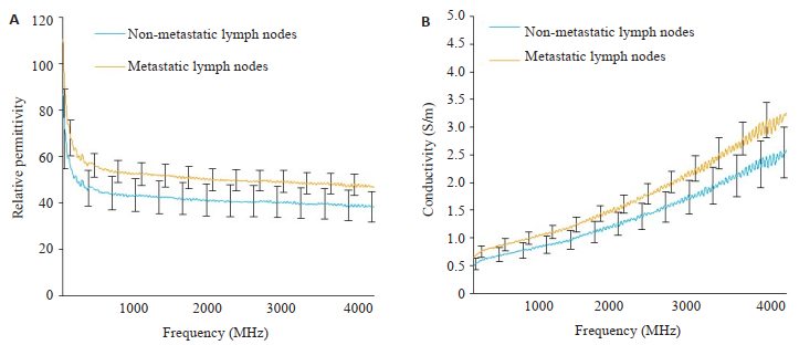
2 结果 2.1 转移和非转移淋巴结介电特性对比对41组转移淋巴结和178组正常淋巴结的介电特性值取均值后,对比曲线图显示,转移淋巴结的介电常数和电导率明显高于非转移淋巴结(图 2)。6个特定频率点下正常和转移淋巴结介电常数和电导率差异有统计学意义(P < 0.01,表 1)。

|
图 2 正常和转移淋巴结介电特性对比 Fig.2 Comparison of dielectric properties between metastatic and non-metastatic lymph nodes. A: Relative permittivity; B: Conductivity. |
|
表 1 6个特定频率点下的介电特性值
Tab.1 Dielectric property values at the 6 specific frequencies |
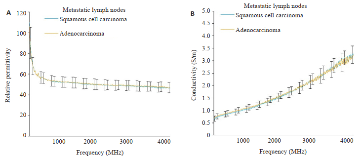
对比肺鳞癌和肺腺癌转移淋巴结的介电特性值,上述转移淋巴结数据中有20组肺腺癌转移淋巴结,18组肺鳞癌转移淋巴结(图 3)。结果显示肺鳞癌和肺腺癌转移淋巴结的介电特性值无明显差异,肺鳞癌转移淋巴结介电特性的标准差比肺腺癌转移淋巴结的标准差稍大。

|
图 3 肺鳞癌和肺腺癌转移淋巴结介电特性值对比 Fig.3 Comparison of dielectric properties between lymph node metastasis derived from squamous cell lung carcinoma and lung adenocarcinoma.A: Relative permittivity; B: Conductivity. |
本研究使用双极Cole-Cole方程拟合1 MHz~4 GHz频率范围内的测量值,双极Cole-Cole模型的表达式如下:
| $ \varepsilon _r^*(\omega ) = {\varepsilon _\infty } + \frac{{\Delta {\varepsilon _1}}}{{1 + {{(j\omega {\tau _1})}^{1 - {\alpha _1}}}}} + \frac{{\Delta {\varepsilon _2}}}{{1 + {{(j\omega {\tau _2})}^{1 - {\alpha _2}}}}} + \frac{{{\mathit{\sigma }_\mathit{s}}}}{{j\omega {\varepsilon _0}}} $ |
其中,ε0 = 8.854 × 10⁻¹² Fm-1是真空介电常数,ω是角频率且ω = 2πf,f为频率,εr*(ω)为复介电常数,ε∞, ∆ε1, ∆ε2, τ1, τ2, α1, α2和σs是Cole-Cole模型拟合参数,其中ε∞为高频介电常数,∆ε1和∆ε2分别为第1个和第2个弛豫过程的弛豫强度,α1和α2分别为第1个和第2个弛豫过程的分布参数,τ1和τ2分别为第1个和第2个弛豫过程的弛豫时间,σs为电介质材料的直流电导率。为方便计算,本文对4000个正常和转移淋巴结介电特性值等间隔取100个点的数据,计算得到的拟合参数值(表 2)。
| 表 2 双极Cole-Cole拟合参数 Tab.2 Parameters of double-pole Cole-Cole fitting |
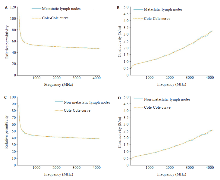
介电特性测量值和Cole-Cole拟合结果显示,转移淋巴结介电常数和电导率的Cole-Cole拟合结果(图 4A、B),正常淋巴结的介电常数和电导率的拟合结果(图 4C、D)。可见在1 MHz~4 GHz,使用双极Cole-Cole拟合方程对肺癌转移和非转移淋巴结的介电特性数据进行拟合,可以获得很好的拟合效果。

|
图 4 转移和非转移淋巴结介电特性Cole-Cole拟合结果 Fig.4 Cole-Cole fitting results of the dielectric properties of non-metastatic and metastatic lymph nodes. A: Relative permittivity of metastatic lymph nodes; B: Conductivity of metastatic lymph nodes; C: Relative permittivity of non-metastatic lymph nodes; D: Conductivity of non-metastatic lymph nodes. |
本研究验证了转移和非转移淋巴结的介电特性差异,观察到转移淋巴结的介电常数和电导率都明显高于正常淋巴结。有关研究表明介电常数的升高与肿瘤组织具有高的含水量密切相关[3]。随着频率增加到4 GHz,介电常数的差异会稍稍变小。本研究显示,在1 MHz~ 4 GHz频率范围内,转移淋巴结的介电常数的标准差比非转移淋巴结的低,这与其他文献在肝脏中、结肠组织中的报道[26-27]相同。本研究结果显示电导率差异随着频率增加而增加且转移淋巴结的标准差低于非转移淋巴结,6个特定频率点下的介电特性值差异均有统计学意义(P < 0.01)。然而,在研究肺鳞癌转移淋巴结和肺腺癌转移淋巴结的介电特性时,其差异不具有统计学意义。因此,可以初步认为这两种癌细胞转移至淋巴结时,对淋巴结介电特性影响程度的差异不明显。双极Cole-Cole方程拟合结果显示,使用两个弛豫模拟1 MHz~4 GHz频率范围内淋巴结的介电特性效果很好,说明本研究所测量的数据符合双极Cole-Cole模型。
转移和非转移淋巴结介电常数和电导率之间的差异是由许多因素引起的。例如,由于转移淋巴结血管增加从而血供增加可能会增加转移淋巴的介电特性;恶性肿瘤细胞的膜组成和膜通透性的变化、相关离子和蛋白质浓度的升高以及转移性淋巴结中水含量升高都可能会导致介电特性值升高[28-30];其他因素,如患者年龄、组织温度和肺癌分化程度等也可能会影响介电常数和电导率的值[31-32]。而对于肺鳞癌转移淋巴结和肺腺癌转移淋巴结的介电特性研究,可进一步增加样本数量和对于肺癌细胞分化程度进一步细分从而验证这一结论。
本文不仅报道了肺癌手术中获得的淋巴结的介电特性,而且给出了Cole-Cole拟合方程参数。研究结果表明在宽带频率下,转移和非转移淋巴结之间的介电特性存在显著差异。在单频点下,差异具有统计学意义,并且测量的数据符合双极Cole-Cole方程,取得很好的拟合效果。因此,此项研究可以为其他相关技术研究如术中组织良恶性鉴别技术,磁共振电特性断层成像技术等提供了基础数据,为术中鉴别淋巴结是否转移提供了一种新的可能。
| [1] |
Kremer F, Schönhals P. Broadband dielectric spectroscopy[D]. Springer: Berlin Heidelberg, 2003. http://www.springerlink.com/content/978-3-642-56120-7
|
| [2] |
Volkov AA, Prokhorov AS. Broadband dielectric spectroscopy of solids[J]. Radiophys Quantum Electron, 2003, 46(8/9): 657-65. DOI:10.1023/B:RAQE.0000024994.15881.c9 |
| [3] |
Pollacco DA, Farina L, Wismayer PS, et al. Characterization of the dielectric properties of biological tissues and their correlation to tissue hydration[J]. IEEE Transact Dielect Electr Insulat, 2018, 25(6): 2191-7. DOI:10.1109/TDEI.2018.007346 |
| [4] |
Guardiola M, Buitrago S, Fernández-Esparrach G, et al. Dielectric properties of colon polyps, cancer, and normal mucosa: Ex vivo measurements from 0.5 to 20 GHz[J]. Med Phys, 2018, 45(8): 3768-82. DOI:10.1002/mp.13016 |
| [5] |
Mirbeik-Sabzevari A, Ashinoff R, Tavassolian N. Ultra-wideband millimeter-wave dielectric characteristics of freshly-excised normal and malignant human skin tissues[J]. IEEE Trans Biomed Eng, 2017, 65(6): 1320-9. |
| [6] |
Li Z, Deng GH, Li Z, et al. A large-scale measurement of dielectric properties of normal and malignant colorectal tissues obtained from cancer surgeries at larmor frequencies[J]. Med Phys, 2016, 43(11): 5991-7. DOI:10.1118/1.4964460 |
| [7] |
周地福, 翟伟科, 孙颖, 等. 结直肠恶性组织黏膜面与浆膜面, 癌旁1, 3 cm以及正常组织黏膜面与浆膜面的介电特性差异[J]. 南方医科大学学报, 2018, 38(4): 434-42. |
| [8] |
张洒, 厉周, 辛学刚. 基于介电特性的人体恶性胃组织支持向量机辅助诊断方法[J]. 南方医科大学学报, 2017, 37(12): 1637-42. |
| [9] |
Ariturk G, Ider YZ. Optimal multichannel transmission for improved cr-MREPT[J]. Phys Med Biol, 2018, 63(4): 45001-12. DOI:10.1088/1361-6560/aaa732 |
| [10] |
Liu J, Shao Q, Wang Y, et al. In vivo imaging of electrical properties of an animal tumor model with an 8-channel transceiver array at 7 T using electrical properties tomography[J]. Magn Reson Med, 2017, 78(6): 2157-69. DOI:10.1002/mrm.26609 |
| [11] |
Sebek J, Bortel R, Prakash P. Broadband lung dielectric properties over the ablative temperature range: experimental measurements and parametric models[J]. Med Phys, 2019, 46(10): 4291-303. DOI:10.1002/mp.13704 |
| [12] |
Summers PE, Vingiani A, Di Pietro S, et al. Towards mm-wave spectroscopy for dielectric characterization of breast surgical margins[J]. Breast, 2019, 45(1): 64-9. |
| [13] |
Pappo I, Spector R, Schindel A, et al. Diagnostic performance of a novel device for real-time margin assessment in lumpectomy specimens[J]. J Surg Res, 2010, 160(2): 277-81. |
| [14] |
Italian National Research Council Institute for Applied Physics. An Internet resource for the calculation of the dielectric properties of body tissues in the frequency range 10 Hz-100 GHz[OL]. http://niremf.ifac.cnr.it/tissprop/[ 2018-09-10].
|
| [15] |
Joines WT, Zhang Y, Li C, et al. The measured electrical properties of normal and malignant human tissues from 50 to 900 MHz[J]. Med Phys, 1994, 21(4): 547-50. DOI:10.1118/1.597312 |
| [16] |
Choi JW, Cho J, Lee Y, et al. Microwave detection of metastasized breast cancer cells in the lymph node; potential application for sentinel lymphadenectomy[J]. Breast Cancer Res Treat, 2004, 86(2): 107-15. |
| [17] |
Cameron TR, Okoniewski M, Fear EC, et al. A preliminary study of the electrical properties of healthy and diseased lymph nodes[C]// international symposium on antenna technology and applied electromagnetics and the american electromagnetics conference, 2010: 1-3.
|
| [18] |
Torre LA, Siegel RL, Jemal A. Lung cancer statistics[J]. Adv Exp Med Biol, 2016, 893(12): 1-19. |
| [19] |
Jiang W, Chen X, Xi J, et al. Selective mediastinal lymphadenectomy without intraoperative frozen section examinations for clinical stage Ⅰ non-small-cell lung cancer: retrospective study of 403 cases[J]. World J Surg, 2013, 37(2): 392-7. DOI:10.1007/s00268-012-1849-9 |
| [20] |
邓官华, 蓝茂英, 冯健, 等. 开端同轴探头法原理及其在人体组织介电特性测量中的应用[J]. 中国医疗设备, 2016, 31(5): 12-4. DOI:10.3969/j.issn.1674-1633.2016.05.003 |
| [21] |
Bobowski JS, Johnson T. Permittivity measurements of biological samples by an Open-Ended coaxial line[J]. Prog Electromagn Res B, 2012, 40(4): 159-83. |
| [22] |
Ellison WJ. Permittivity of pure water, at standard atmospheric pressure, over the frequency range 0-25THz and the temperature range 0-100 ℃[J]. J Physical Chem Reference Data, 2007, 36(1): 1-18. DOI:10.1063/1.2360986 |
| [23] |
Gregory AP. Tables of the complex permittivity of dielectric reference liquids at frequencies up to 5 GHz[J]. Npl Report Cetm, 2001, 53(17): 2049-60. |
| [24] |
Stogryn A. Equations for calculating the dielectric constant of saline water[J]. IEEE Trans Microw Theory Tech, 1971, 19(8): 733-6. DOI:10.1109/TMTT.1971.1127617 |
| [25] |
Grosse C. A program for the fitting of Debye, Cole-Cole, ColeDavidson and Havriliak-Negami dispersions to dielectric data[J]. J Colloid Interface Sci, 2014, 419(4): 102-6. |
| [26] |
O'rourke AP, Lazebnik M, Bertram JM, et al. Dielectric properties of human normal, malignant and cirrhotic liver tissue: in vivo and ex vivo measurements from 0.5 to 20 GHz using a precision openended coaxial probe[J]. Phys Med Biol, 2007, 52(15): 4707-19. DOI:10.1088/0031-9155/52/15/022 |
| [27] |
Fornes-Leal A, Garcia-Pardo C, Frasson M, et al. Dielectric characterization of healthy and malignant colon tissues in the 0.5-18 GHz frequency band[J]. Phys Med Biol, 2016, 61(20): 7334-46. DOI:10.1088/0031-9155/61/20/7334 |
| [28] |
Chandra R, Zhou HY, Balasingham I, et al. On the opportunities and challenges in microwave medical sensing and imaging[J]. IEEE Trans Biomed Eng, 2015, 62(7): 1667-82. DOI:10.1109/TBME.2015.2432137 |
| [29] |
Peyman A, Kos B, Djokic M, et al. Variation in dielectric properties due to pathological changes in human liver[J]. Bioelectromagnetics, 2015, 36(8): 603-12. DOI:10.1002/bem.21939 |
| [30] |
Alshami AS, Tang JM, Rasco B. Contribution of proteins to the dielectric properties of dielectrically heated biomaterials[J]. Food Bioproc Technol, 2017, 10(8): 1548-61. DOI:10.1007/s11947-017-1920-5 |
| [31] |
Sebek J, Bortel R, Prakash P. Broadband lung dielectric properties over the ablative temperature range: experimental measurements and parametric models[J]. Med Physics, 2019, 46(10): 138-49. |
| [32] |
Fu FR, Xin SX, Chen WF. Temperature- and frequency-dependent dielectric properties of biological tissues within the temperature and frequency ranges typically used for magnetic resonance imagingguided focused ultrasound surgery[J]. Int J Hyperthermia, 2014, 30(1): 56-65. |
 2019, Vol. 39

