2008年国际口腔种植学会共识认为当上颌窦底剩余骨高度(RBH) < 5 mm时建议采用侧壁提升,当RBH > 4~5 mm时建议采用嵴顶提升[1]。由于侧壁提升手术创伤较大,患者术后反应重;经嵴顶提升明显创伤较少,术后反应较轻[2-3],所以近年来不断有学者挑战嵴顶提升的RBH,剩余骨高度不再被认为是决定侧壁提升与嵴顶提升的主要条件[4],然而对于RBH < 5 mm的一次性嵴顶提升被认为是高度复杂病例,存在一定的手术风险。因此,实现上颌后牙区剩余骨高度严重不足的患者手术微创并获得长期可靠的临床效果成为目前国内外研究方向。国外曾有研究报道中度骨高度不足(RBH≥3 mm且≤5 mm时)通过特殊新型器械行上颌窦提升的病例报道或类似提升后1~2年的短期临床观察[5-6],但目前国内外尚未见有关骨高度低于3 mm时行嵴顶提升的长期临床报道。本研究针对严重骨高度不足时使用常规器械微创下完成嵴顶提升并评估长期临床效果,旨在替代手术创伤较大的侧壁提升。因此,选取RBH≥1 mm且≤3 mm拟行种植修复的78例患者,采用经改良方法行二次提升、最终植入种植体并修复,随访5年,评估分析该改良方法的可行性及长期临床效果。
1 资料和方法 1.1 临床资料选取2012年3月~2014年12月在我院种植中心就诊的78名患者,本研究经过南方医科大学口腔医院伦理委员会审批。纳入标准为:上颌后牙缺失,上颌窦底剩余牙槽嵴垂直骨高度RBH≥1 mm且≤3 mm、牙槽嵴宽度≥5 mm,无上颌窦分隔,上颌窦无明显病变,患牙拔除3个月以上,软硬组织愈合较理想、全身无严重系统性疾病,无拔牙或种植手术禁忌症、无严重的咬合功能紊乱或磨牙症、无急性的上颌窦炎症、无未控制的邻牙根尖周疾病,无急性的局部感染、无进行性牙周炎或者牙周疾病,未接受全剂量放疗和静脉滴注双磷酸盐治疗,无长期吸烟病史。患者签字同意进行种植修复,其中男性46例,女性32例,平均年龄46.3±5.2岁,共植入148枚种植体。
1.2 主要仪器及材料Straumann种植系统和Straumann骨凿(Straumann)、Summers骨凿(Summers),天博齿固羟基磷灰石生物陶瓷(意化健科)。
1.3 手术方法术前常规拍摄CBCT及曲面断层片,测量缺牙区剩余牙槽嵴高度。术前常规预防性使用抗生素,常规消毒铺巾,局部浸润麻醉下于缺牙区牙槽嵴顶作切口,翻瓣,显露牙槽骨,用先锋钻钻入近上颌窦底1 mm处;Summers骨凿或Straumann骨凿轻轻敲击,击破窦底骨壁, 有落空感后缓缓向上轻推上颌窦底黏膜;植入适量骨粉,提升高度约6~9 mm,伤口缝合关闭。术中用“捏鼻鼓气法”检查窦底黏膜完整性。术后使用抗生素1周,10 d后拆线。一期术后6个月复查CBCT,检查成骨效果后行二阶段手术,于缺牙区牙槽嵴顶作切口,翻瓣, 显露牙槽骨,扩孔钻逐级扩孔至设计直径,然后用骨凿轻轻敲击,将第1次植入骨粉向上提升至拟植入高度,置入Straumann 4.1 mm×10.0 mm种植体101枚,4.8 mm× 10.0 mm种植体20枚,4.8 mm×8.0 mm植体27枚;使用共振频率分析仪测量种植体稳定性系数ISQ值,放置愈合基台后将种植体周围软组织严密缝合。术后抗炎及医嘱遵照第1次手术。手术均由同一术者完成,植入种植体术后6个月,转移印模,完成上部结构修复,修复前行共振频率分析仪测量种植体ISQ值,并行曲面断层片检查。若一期术中发生穿孔,则改为上颌窦外提升术,6个月后植入种植体。若种植体二期修复ISQ低于60,则待骨结合完成后再行修复。
1.4 临床评估标准(1)种植体稳定性系数应用共振频率分析仪对种植体进行共振频率分析(RFA),进一步转换成ISQ值(0~100)描述种植体稳定性。数值越大,稳定性越好。
(2)种植体存留率参照Buser [7]和Cochran [8]提出的标准:①种植体临床检查无动度;②无疼痛及任何主观感觉;③无复发的种植体周围炎;④X线检查种植体周围无连续的透射影。
(3)种植体探诊深度(PDi):0.25 N力量平行于种植体长轴对种植体周围龈沟利用种植体专用探针分别在颊侧、舌侧近中、中央、远中测量,每个牙位记录6各位点的探诊深度,取平均值记录,均由同一名医生完成。
(4)改良龈沟出血指数(mSBI):用约20 g的力控制种植体专用探针沿种植体龈缘探诊,记分标准:0=无出血;1=有分散出血;2=牙龈沟内有线状出血;3=重度自发性出血。每个牙位记录6各位点的出血指数,取平均值记录,均由同一名医生完成。
1.5 术后满意度调查、术中及术后临床评估及随访效果评估复诊询问患者术后有无任何疼痛,有无其他主观不适如鼻腔出血或上颌窦炎症。术中、术后及随访5年参照1.4标准行临床及影像学效果评估。
术后即刻、术后1年、5年复诊均拍摄CBCT,检查有无上颌窦异常,观察种植体骨结合情况并测量窦底新骨高度;拍摄曲面断层片比较边缘骨吸收情况。每次的CBCT图像均以种植体的横切面为基准,绿色切线平行于牙槽骨的长轴并通过种植体的中心得到种植体矢状面图像,红色切线垂直于牙槽骨并通过种植体的中心得到种植体冠状面图像最终三维显示种植体的近中、远中、颊侧及舌侧的牙槽骨,测量提升骨高度[9]。曲面断层片以种植体长度为参照排除X线片放大率的影响,测量边缘骨吸收[10]。利用图像处理软件Image Pro Plus 6.0测量,数据测量由同一名医生完成,反复测量3次,最终结果取平均值,数值精确到0.01 mm。
1.6 统计学分析采用SPSS21.0软件进行数据分析,计量资料用均数±标准差表示,计量资料符合正态分布时采用两独立样本t检验;不符合正态分布时,组间比较采用Wilconxon秩和检验。显著性水平取ɑ=0.05。
2 结果 2.1 种植体分布情况本研究中男性46例,女性32例,年龄46.3±5.2岁,植入148枚,种植牙位及种植体长度见表 1。
| 表 1 种植牙位的种植体数目、长度分布表 Tab.1 Number of implants and the length of implants depending on the area of surgery |
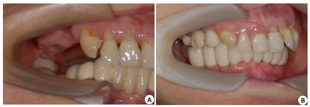
(1)78例患者中3例术中发生上颌窦穿孔、穿孔率为3.85%。该3例均同期改行上颌窦侧壁提升术,未发生上颌窦炎症,并最终修复。实际采用二次提升法患者共75例,共植入148枚植体。52例(69.33%)无头痛发热,无明显疼痛及肿胀反应,23例(30.67%)存在轻微疼痛。患者对手术术后反应总体感觉满意,对侧上颌后牙缺失患者均接受对侧再次行该术式。(2)随访率为100%。在愈合期及随访期内148枚种植体无一松动脱落,功能行使良好,患者无明显不适,种植体存留率达到100%。术前及术后随访照片如图 1。(3)种植体均采用基台埋入式愈合。种植术后即刻和术后半年种植体稳定性系数ISQ值分别为58.39±1.39,81.88±1.22,两组差异有统计学意义(t=-109.88,P < 0.05)。(4)临床检查种植体周软组织,术后1年、5年种植体探诊深度分别为3.21±0.06 mm、3.25±0.10 mm,两组差异无统计学意义(P > 0.05)。改良龈沟出血指数分别为0.78±0.19、0.87± 0.27,两组差异无统计学意义(P > 0.05)。(5)观察影像学资料,上颌窦底均可见新骨形成;经过不断骨吸收及骨改建,新骨形态良好且高度随时间增加略有减低,骨密度增加,形成新的上颌窦底骨板。绝大部分新骨形成且完全包绕种植体根尖,少部分新骨形成仅部分包绕种植体根尖,多见于上颌第2磨牙。术后第1年种植体周围骨吸收量明显高于术后2~5年。术后1年、术后5年的窦底提升高度分别为9.45±2.25 mm、8.81±2.56 mm,两组差异有统计学意义(P < 0.05)。术后1年、5年种植体周边缘骨吸收分别为0.81±0.04 mm、1.04±0.04 mm,两组差异有统计学意义(P < 0.05,表 2)。

|
图 1 术前及术后5年随访照片 Fig.1 Photographs taken before (A) and at 5 years after sinus lift (B). |
| 表 2 术后1年、术后5年数据统计表 Tab.2 Descriptive statistics at 1 year and 5 years after the operation (n=148, Mean±SD) |
本研究对上颌后牙区骨高度严重不足患者行二次提升法同期植入种植体长期随访病例75例,穿孔率为3.85%(3/78),与以往文献报道经嵴顶提升术的穿孔率类似[3, 11]。种植体植入半年后种植体稳定性系数ISQ值达到81.88±1.22,提示植入种植体后6个月种植体与周围牙槽骨达到良好骨结合。研究证明,剩余骨量并非影响种植体成功率的唯一因素,种植体与骨有效的接触表面是成功的关键[1, 12]。当RBH高度较低时,种植体-骨组织接触面积减小从而减低初稳[13];种植体突入上颌窦内部分增多导致窦黏膜张力增大从而加大了穿孔的风险;所以,随着嵴顶提升高度适应症扩大,术者的操作是导致窦膜穿孔的主要因素[14-15]。穿孔的风险随着提升的高度而增加,采用骨凿敲击形成骨刺、窦膜的厚薄程度、植骨材料的植入也是影响因素[16-17]。研究中第一阶段提升植骨后获得了新的RBH,提升高度无需达到种植所需种植体根方以上2 mm的理想位置[4, 18];实现一期微创植骨,为第二阶段提升同期植入创造良好条件。
种植体周围边缘骨吸收是影响种植体长期稳定性和成功率的主要因素之一[19-20]。研究中术后1年种植体周边缘骨吸收为0.81±0.04 mm,术后5年边缘骨吸收1.04±0.04 mm,术后1年边缘骨吸收与文献较为一致[11],主要发生在愈合期,随访期边缘骨吸收稳定。随访新骨生成情况,均有新骨形成,大部分病例全部包绕种植体根尖,少数部分包绕根尖。影像学分析发现,负载5年平均获得8.81±2.56 mm的新骨高度;术后5年较术后1年高度略有所降低,平均降低0.7 mm,这一现象与很多文献一致[21-23]。骨吸收程度可能与植入骨粉结构的改变、植骨材料的稳定性和吸收方式、种植体的受力以及上颌窦内压力有关[16, 24-25]。观察期内所有病例均无种植体周围疾病发生,术后1年与术后5年种植体探诊深度和改良龈沟出血指数两指标均无统计学差异。良好的种植体-骨结合是种植体行使功能的基础,而软组织良好的封闭对种植体周围骨组织的稳定起着重要作用,可避免种植体周围细菌侵入减少种植体周围疾病的发生[26-28]。吸烟因素也是上颌窦手术的危险因素之一[15, 30]。观察期内所有病例均无种植体周围疾病发生,术后1年与术后5年软组织指标均无统计学差异。这与患者的长期牙周维护、种植体周维护以及医生的口腔卫生宣教密切相关。
综上所述,上颌后牙区骨高度严重不足时行经牙槽嵴顶上颌窦底二次提升术取得了预期长期稳定的临床疗效,影像学证实了上颌窦底提升及成骨效果均良好。与侧壁开窗式上颌窦提升术相比,该术手术创伤小,患者不适感减轻明显,实现了较高难度手术的微创治疗,并且有效解决上颌后部牙槽骨度严重不足的问题。
| [1] |
Pommer B, Hof M, Fädler A, et al. Primary implant stability in the atrophic sinus floor of human cadaver maxillae:impact of residual ridge height, bone density, and implant diameter[J]. Clin Oral Implants Res, 2014, 25(2): e109-13. DOI:10.1111/clr.2014.25.issue-2 |
| [2] |
Zhao X, Gao W, Liu F. Clinical evaluation of modified transalveolar sinus floor elevation and osteotome sinus floor elevation in posterior maxillae:study protocol for a randomized controlled trial[J]. Trials, 2018, 19(1): 489. DOI:10.1186/s13063-018-2879-x |
| [3] |
Tan WC, Lang NP, Zwahlen M, et al. A systematic review of the success of sinus floor elevation and survival of implants inserted in combination with sinus floor elevation. Part Ⅱ:transalveolar technique[J]. J Clin Periodontol, 2008, 35(8 Suppl): 241-54. |
| [4] |
满毅. 经牙槽嵴顶上颌窦底提升术的应用研究进展[J]. 口腔疾病防治, 2018, 26(8): 477-83. |
| [5] |
Gatti F, Gatti C, Tallarico M, et al. Maxillary sinus membrane elevation using a special drilling system and hydraulic pressure:a 2-Year prospective cohort study[J]. Int J Periodontics Restorative Dent, 2018, 38(4): 593-9. DOI:10.11607/prd.3403 |
| [6] |
Krasny K, Krasny M, Kaminski A. Two-stage closed sinus lift:a new surgical technique for maxillary sinus floor augmentation[J]. Cell Tissue Bank, 2015, 16(4): 579-85. DOI:10.1007/s10561-015-9505-x |
| [7] |
Buser D, Mericske-Stern R, Bernard JP, et al. Long-term evaluation of non-submerged ITI implants. Part 1:8-year Life table analysis of a prospective multi-center study with 2359 implants[J]. Clin Oral Implants Res, 1997, 8(3): 161-72. DOI:10.1034/j.1600-0501.1997.080302.x |
| [8] |
Cochran DL, Buser D, Ten Bruggenkate CM, et al. The use of reduced healing times on ITI (R) implants with a sandblasted and acid-etched (SLA) surface:Early results from clinical trials on ITI (R) SLA implants[J]. Clin Oral Implants Res, 2002, 13(2): 144-53. DOI:10.1034/j.1600-0501.2002.130204.x |
| [9] |
Krasny K, Krasny M, Kaminski A. Two-stage closed sinus lift:a new surgical technique for maxillary sinus floor augmentation[J]. Cell Tissue Bank, 2015, 16(4): 579-85. DOI:10.1007/s10561-015-9505-x |
| [10] |
耿威, 林潇, 李晓光, 等. 下颌后牙区SLActive种植体3周早期负荷修复1年随访的临床观察[J]. 口腔医学研究, 2015, 23(5): 475-8. |
| [11] |
Corbella S, Taschieri S, Del Fabbro M. Long-term outcomes for the treatment of atrophic posterior maxilla:a systematic review of literature[J]. Clin Implant Dent Relat Res, 2015, 17(1): 120-32. DOI:10.1111/cid.2015.17.issue-1 |
| [12] |
周磊, 岳新新. All-on-Four技术在口腔种植领域中的应用进展[J]. 口腔疾病防治, 2017, 25(1): 1-7. |
| [13] |
Nedir R, Nurdin N, Khoury P, et al. Osteotome sinus floor elevation with and without grafting material in the severely atrophic maxilla. A 1-year prospective randomized controlled study[J]. Clin Oral Implants Res, 2013, 24(11): 1257-64. |
| [14] |
Yilmaz HG, Tozum TF. Are gingival phenotype, residual ridge height, and membrane thickness critical for the perforation of maxillary sinus[J]. J Periodontol, 2012, 83(4): 420-5. DOI:10.1902/jop.2011.110110 |
| [15] |
Tavelli L, Borgonovo AE, Ravida AA, et al. Analysis of forces applied during transalveolar sinus lift:a preliminary clinical study[J]. Implant Dent, 2018, 27(6): 630-7. DOI:10.1097/ID.0000000000000817 |
| [16] |
Cardoso CL, Curra C, Santos PL, et al. Current considerations on bone substitutes in maxillary sinus lifting[J]. Revista Clínica de Periodoncia, Implantología Y Rehabilitación Oral, 2016, 9(2): 102-7. DOI:10.1016/j.piro.2016.03.001 |
| [17] |
Janner SF, Caversaccio MD, Dubach PA, et al. Characteristics and dimensions of the Schneiderian membrane:a radiographic analysis using cone beam computed tomography in patients referred for dental implant surgery in the posterior maxilla[J]. Clin Oral Implants Res, 2011, 22(12): 1446-53. DOI:10.1111/clr.2011.22.issue-12 |
| [18] |
Trombelli L, Franceschetti G, Trisi PA. Incremental, transcrestal sinus floor elevation with a minimally invasive technique in the rehabilitation of severe maxillary atrophy. clinical and histological findings from a Proof-of-Concept case series[J]. J Oral Maxillofac Surg, 2015, 73(5): 861-88. DOI:10.1016/j.joms.2014.12.009 |
| [19] |
赖红昌, 史俊宇. 上颌窦提升术[J]. 口腔疾病防治, 2017, 25(1): 8-12. |
| [20] |
Lee CT, Tran D, Jeng MD, et al. Survival rates of hybrid rough surface implants and their alveolar bone level alterations[J]. J Periodontol, 2018, 89(12): 1390-9. DOI:10.1002/jper.2018.89.issue-12 |
| [21] |
Pjetursson BE, Lang NP. Sinus floor elevation utilizing the transalveolar approach[J]. Periodontol 2000, 2014, 66(1): 59-71. DOI:10.1111/prd.2014.66.issue-1 |
| [22] |
Si MS, Shou YW, Shi YT, et al. Long-term outcomes of osteotome sinus floor elevation without bone grafts:a clinical retrospective study of 4-9 years[J]. Clin Oral Implants Res, 2016, 27(11): 1392-400. DOI:10.1111/clr.12752 |
| [23] |
Bassi A, Pioto R, Faverani LP, et al. Maxillary sinus lift without grafting, and simultaneous implant placement:a prospective clinical study with a 51-month follow-up[J]. Int J Oral Maxillofac Surg, 2015, 44(7): 902-7. DOI:10.1016/j.ijom.2015.03.016 |
| [24] |
Del Fabbro M, Wallace SS, Testori T. Long-Term implant survival in the grafted maxillary sinus:a systematic review[J]. International Journal of Periodontics & Restorative Dentistry, 2013, 33(6): 773. |
| [25] |
Stefanski S, Svensson B, Thor A. Implant survival following sinus membrane elevation without grafting and immediate implant installation with a one-stage technique:an up-to-40-month evaluation[J]. Clin Oral Implants Res, 2017, 28(11): 1354-9. DOI:10.1111/clr.2017.28.issue-11 |
| [26] |
Menini M, Setti P, Pera P, et al. Peri-implant Tissue Health and Bone Resorption in Patients with Immediately Loaded, ImplantSupported, Full-Arch Prostheses[J]. International Journal of Prosthodontics, 2018, 31(4): 327-33. |
| [27] |
Monje A, Blasi G. Significance of keratinized mucosa/gingiva on peri- implant and adjacent periodontal conditions in erratic maintenance compliers[J]. J Periodontol, 2018 Nov 21. doi: 10.1002/JPER.18-0471.[Epubaheadofprint] https://www.researchgate.net/publication/329099028_Significance_of_keratinized_mucosagingiva_on_peri-implant_and_adjacent_periodontal_conditions_in_erratic_maintenance_compliers
|
| [28] |
Ng KT, Fan M, Leung MC, et al. Peri-implant inflammation and marginal bone level changes around dental implants in relation to proximity with and bone level of adjacent teeth[J]. Aust Dent J, 2018, 63(4): 467-77. DOI:10.1111/adj.2018.63.issue-4 |
| [29] |
Barbato L, Baldi N, Gonnelli A, et al. Association of smoking habits and height of residual bone on implant survival and success rate in lateral sinus lift:a retrospective study[J]. J Oral Implantol, 2018, 44(6): 432-8. DOI:10.1563/aaid-joi-D-17-00192 |
 2019, Vol. 39

 ,
,