下颌第一磨牙一直是临床上根管治疗的难点,因为其根管系统的复杂性,易导致治疗的失败,下颌第一磨牙近中第三根管的发现,使众多国内外学者对其做了大量研究[1-5]。大多数都以报告临床病例为主,同时也有用离体牙透明标本法、牙科显微镜等方法研究此根管形态。CBCT自带的计算机软件系统进行影像数据分析时,可在矢状面、冠状面和轴面同时显示,能更直观、更全面的了解根管系统,更有助于临床上治疗前对患牙根管生理形态的进一步观察[6-8]。
国内外有一些关于其他牙弯曲根管研究的报道[9-11]。但目前还缺乏针对下颌第一磨牙近中第三根管弯曲度的研究。锥形束CT(CBCT)与临床上其他的口腔根尖片,曲面体层摄影以及传统CT相比较而言,具有放射剂量更小、图像分辨率更高、模型的三维重建和操作更加简便等众多明显的优势。为了更适用于临床治疗的检测手段,本实验主要针对下颌第一磨牙近中第三根管进行研究观察其弯曲度及发生部位,探讨其与年龄的相关性,尝试为临床治疗提供科学可靠的理论依据,能在治疗牙髓病、尖周病的过程中,指导临床医生对其进行准确有效的根管预备,获得较高的成功率[12]。
1 资料和方法 1.1 基本资料自2016年1月起,收集在本院口腔影像科因口腔检查、种植、拔牙、正畸等原因拍摄的CBCT图像,截止2017年8月共获得1600名患者(年龄46.5±14.8岁)的2856颗下颌第一恒磨牙的CBCT图像作为研究对象,选取从中所获的含有近中第三根管的下颌第一恒磨牙的CBCT图像,共计168例。将其分为5组(A组:18~29岁;B组:30~39岁;C组:40~49岁;D组:50~59岁;E组:60~80岁)。图像均采用以下纳入标准:无根尖周病损;未进行过牙髓治疗及牙根切除术;根尖发育完好,无牙根吸收;扫描所得的CBCT,图像清晰无伪影。利用Planmeca Romexis Viewer软件对图像进行三维重建后再选择合适的位置,截取牙齿在近远中向和颊舌向的图像。釆用Schneider法对搜集到的图像进行解剖形态等相关参数的测量。统计下颌第一恒磨牙近中第三根管的发现率、根管弯曲度,及根管弯曲部位并做出相应的分析。
1.2 CBCT扫描本研究采用的Planmeca Romexis CBCT机为Planmeca Romexis。电压:84 kV;电流:14 mA;灰阶:15 bit;像素尺寸:200 μm×200 μm;影像分辨率:401× 401×401;最小层厚:0.2 mm;曝光时间:12 s;重建时间:30~150 s。探测器分辨率:1024×1024像素;像素尺寸:127 μm×127 μm。单次200°旋转进行影像釆集并拼接成像域:140 mm×105 mm×130 mm三维重建服务器使用Feldkamp型反投影重建算法。影像学资料采用DICOM格式导出、保存,以获取影像学资料。
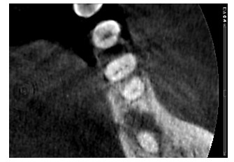
1.3 图像分析本研究纳入的所有图像均采用Planmeca Romexis CBCT机的图像重建系统进行三维重建并测量,沿轴向位观察重建影像牙齿的冠方-髓室底-根尖区,对横断面进行连续滚轴移动观察,同时调整矢状位和冠状位图像,使牙齿的根管系统和周围组织清楚可见。在暗室中对所有图像进行观察分析,分别观察矢状面、冠状面和横断面的CBCT图像3次,力求全面、详细的获得纳入牙齿的根管系统视图。本实验组2名高年资牙体牙髓专科医生进行标准一致性检验,合格后独立评价CBCT图像,确保结果真实准确。以下颌第一磨牙近中第三根管口轴位截(图 2)为例,利用CBCT可以清楚观察到各个面的根管形态。发现解剖复杂的根管或2位观察者无法达成共识时,由第三方(牙髓病专家)给予评估,最终确定根管解剖和形态,并将有争议的数据讨论后得出专业性结论。5组均统计其下颌第一恒磨牙近中第三根管的发现率、近远中向弯曲度、颊舌向弯曲度、根管弯曲部位等数据。

|
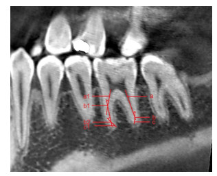
图 1 根管弯曲度测量方法 Figure 1 Method for measuring the curvature of root canal. a: Mesial root canal orifice; a1: Distal root canal orifice; b、b1: Linear inflection point; b2: The second linear inflection point; c: Distal root apical foramen; c1: Mesial root apical foramen. |

|
图 2 左侧下颌第一磨牙近中第三根管口轴位截图 Figure 2 Axial scan of the middle mesial canals of the left mandibular first molars. |
本研究采用改良的Schneider法对近中第三根管弯曲度、弯曲部位的弯曲度进行测量,操作方法为:使用Planmeca Romexis CBCT机的三维重建软件将根管口设置为a点,根尖孔为c点,自a点沿根管影像画一条直线,拐点处设为b点。ab、bc连线所组成的锐角则为根管弯曲度。将根管弯曲度分为3个等级:弯曲度≤10°记为轻度;弯曲度在10°~30°之间记为中度;弯曲度≥30°记为重度。根管弯曲位置测量:参考ab与bc的比值,如根管弯曲只有1处,设P=ab/bc(图 1左侧根管)。如有第2弯曲,则以b1点与b2点连线中偏离根管影像的一点记为b2点,根尖孔记为c1点。第1弯曲:P1= a1b1/b1b2+ b2c1,第2弯曲:P2= a1b1+b1b2/b2c1(图 1右侧根管)。依据P值计算出的结果将根管弯曲位置分为3类:P < 0.5为Ⅰ类,根管弯曲部位在根冠1/3处;0.5 < P < 2为Ⅱ类,弯曲部位在根中1/3处;P>2为Ⅲ类,弯曲部位在根尖1/3处。
1.5 统计学分析数据处理分析采用SPSS 19.0统计软件,计量资料以均数±标准差,计数资料以率(%)表示,正态分布资料采用t检验,计数资料比较采用χ2检验(格子数期望频数小于5时采用精确检验结果),两两比较用Bonferroni法校正检验水准,检验水准α=0.05,P < 0.05为差异有统计学意义。
2 结果 2.1 下颌第一磨牙近中第三根管发现率A组为37.5%;B组为17.86%;C组为14.89%;D组为19.64%;E组为10.12%。A组发现率显著高于其他各组(P < 0.01),B组发现率高于E组(P=0.041);D组发现率高于E组(P=0.187);其他各组之间比较,差异无统计学意义(P>0.05,表 1)。
| 表 1 不同年龄组下颌第一磨牙近中第三根管发现率 Table 1 Incidence rate of middle mesial canals of mandibular first molars (n, %) |
A组近远中向根管弯曲度中度大于其他各组(P < 0.05,图 2)。各组近远中向根管弯曲度差异有统计学意义(F=2.53, P=0.0426),各组颊舌向根管弯曲度差异无统计学意义(F=0.21, P=0.9312);5组近远中向根管弯曲度两两比较,结果显示:A组>C组(t=1.9922, P= 0.0495),A组>D组(t=2.4187, P=0.0175),A组>E组(t= 2.1455, P=0.0350),差异有统计学意义;其他各组相互比较,差异无统计学意义(P>0.05,表 2)。
| 表 2 下颌第一磨牙近中第三根管近远中向及颊舌向弯曲度 Table 2 Curvature in the mesiodistal direction and buccolingual direction of the middle mesial canals of mandibular first molars |
采用SPSS 19.0对数据进行统计分析,经χ2检验结果显示,近远中向和颊舌向弯曲部位在根中1/3处的发现率都显著高于根冠1/3处与根尖1/3处,差异有统计学意义(P < 0.01,表 3)。两两对比弯曲部位可知:近远中向根中1/3处高于根冠1/3处(P=0.000)和根尖1/3处(P= 0.000),近远中向根冠1/3处高于根尖1/3处(P=0.003);颊舌向根中1/3处高于根冠1/3处(P=0.000)和根尖1/3处(P=0.000);颊舌向根冠1/3处高于根尖1/3处(P= 0.006),差异均有统计学意义(P < 0.05,表 4)。
| 表 3 下颌第一磨牙近中第三根管近远中向及颊舌向弯曲部位 Table 3 Location of mesiodistal and buccolingual curvatures of the middle mesial canals of the mandibular first molars |
| 表 4 下颌第一磨牙近中第三根管近远中向及颊舌向弯曲部位发现率 Table 4 Detection rates of locations of mesiodistal and buccolingual curvatures of the middle mesial canals of the mandibular first molars (n, %) |
1981年美国人首次发现下颌第一磨牙近中第三根管,并对61颗下颌第一磨牙进行深入研究[13]。1984年另一美国学者也对下颌第一磨牙近中第三根管的根管系统进行观察分析,并提出了以其命名的Vertucci根管分类法[14]。研究发现,近中第三根管,被描述为一个额外的近中根管,是下颌第一磨牙的解剖变异[1-4]。近中第三根管的发现率/发生率存在相当大的异质性,从0.26% ~46.15%均有报导[1, 15-19]。这可能是由于样本来自不同的种族、年龄和性别的人群以及研究方法不同而造成。但有学者提出MMC可能不是真正独立的根管,推测是下颌第一磨牙近中颊和近中舌两主根管间的峡区[20-21]。研究发现下颌第一磨牙近中根峡区发生率在80%以上,对于根管治疗过程中,无论MMC是独立的根管还是峡区它都是一个需要清创和消毒的空间,否则将导致持续根管感染[22-24]。因此对MMC的研究对临床有重要的意义。目前国内关于近中第三根管的研究多为临床病例报告,而本团队利用CBCT图像对根管形态进行观察,属于国际前沿技术,具有先进性、可行性。本研究对2856颗下颌第一磨牙进行观察分析,发现168颗存在第三根管,MMC发现率为5.88%,研究结果位于上述研究结果区间。同时与Wang等[25]用CBCT研究中国西部人群下颌第一磨牙根管形态的结果基本相似,这也验证了本实验结果的科学、合理性。CBCT在还原牙体的形态结构方面有着很强的优势,尤其在根中和根尖1/3处具有优势,能更清晰地显示牙体结构,减少人为造成的误差,更有利于临床准确诊断,且辐射量不高[26]。虽然下颌第一磨牙近中第三根管(MMC)在临床上发生率不算很高,但是其根管形态复杂,若不能获取到清晰的根管系统视图、准确定位到根管口,将对临床治疗带来诸多不便。基于此,本实验将CBCT应用于下颌第一磨牙近中第三根管的研究分析,能获取到多个平面的清晰影像,为临床提供稳定可靠的影像学数据资料。另一方面,本团队率先对MMC的根管弯曲度进行深入探究,具有可靠有效的临床指导意义。
本研究结果显示下颌第一磨牙近中第三根管在年轻患者中很容易被发现,其发现率随年龄增长而下降,与其他研究结果一致[15-16],下颌第一磨牙近中牙根被证实有形态复杂的峡区存在,并且随着年龄增长,越近根尖部的峡区钙化越明显。年龄可能对是否存有MMC及其是否出现融合具有决定性作用,因而可以证明年龄越大,MMC发现率越低[27-28]。
牙周变性、牙齿磨耗,同时由于龋坏、创伤等外在损害导致了继发性牙本质及根管的钙化、牙骨质增厚、根部吸收及根部透明牙本质增加等无法避免的情况[29]。本研究结果显示,MMC近远中向及颊舌向弯曲度均值都在中、重度弯曲的范围内波动,而根管治疗具有一定的盲目性,弯曲度高为临床操作造成了较大的困难。且近远中向根管弯曲度随年龄增长而下降,这可能与随着年龄增长,根管的钙化程度增加有关。但5组弯曲度的平均值并没有太大差别,这可能是由于牙齿邻面磨耗程度随年龄增大而加重,牙冠有向近中移动以代偿邻面磨耗的趋势造成[30]。表 1~2均证明了MMC与年龄的相关性,因此,临床医生在治疗前可根据患者年龄设计不同的治疗方案。
本实验通过CBCT的观察,发现样本都有较高比例出现近远中向弯曲和颊舌向弯曲,与Perlea[31]所描述的一致。提示根管弯曲度变化常是三维立体的,临床医生进行根管治疗时应注意患牙近远中向、颊舌向弯曲的发生可能,不应只关注某一个方向。从表 3、4结果可见,MMC近远中向及颊舌向弯曲部位出现的频数皆为根中1/3>根冠1/3>根尖1/3。即下颌第一磨牙近中第三根管弯曲多发生在根中1/3处,故此部位是治疗过程应重点关注的。但根冠1/3及根尖1/3处的弯曲较大且较狭窄,根管预备时易造成根管器械的折断,因此在对其进行根管预备时,有必要根据术前X线诊断片的信息,针对不同类型的弯曲根管进而采取不同的抗弯曲措施,从而降低此牙位疑难根管治疗的风险,减少并发症的发生。而以往也有文献显示,相比根管上段和根管中段的弯曲,根管尖段的弯曲治疗难度最大,而且弯曲急的根管治疗难度也大于弯曲缓慢的根管,这证明了临床医生预先了解根管形态和弯曲程度可以提高根管治疗的成功率[10-11]。由于国内外较少学者研究MMC的弯曲度、弯曲部位,无法参考,但同时也说明本实验结果的必要性和重要性。
当然,本实验也存在一定的局限性。本研究采用的Schneider法,可以最大限度保留原有根管形态,减少了因破坏原始形态带来的偏差,这也是利用CBCT研究弯曲度的最大优势[32-34]。但在一定程度上无法避免人为因素带来的误差,如对于根管弯曲部分分布较广,尤其是当弯曲部位呈渐进性弯曲时,所测得角度与根管真实弯曲度之间会存在一定差异。这也是本项目组在往后的研究中将重点攻克及解决的问题。
综上所述,本实验进一步深化了CBCT在根管形态和牙体解剖结构方面的探索,弥补了国内在此方向研究的不足。实验结果可以指导临床医生更直观的掌握下颌第一磨牙近中第三根管的变化情况,从而在临床上处理复杂根管时,能借助CBCT获取到患牙更全面的信息,使根管治疗更加完善。对下颌第一磨牙根管发现率、根管弯曲度等情况进行分析,为临床医生寻找发现率低但意义重要的下颌第一磨牙近中第三根管提供了理论依据。CBCT的成像系统也为寻找根管提供了有效讯息,能显著避免临床医生行根管治疗时的盲目性,从而能对下颌第一磨牙近中第三根管进行充分的根管清理、预备及充填,达到提高根管治疗成功率的目的。
| [1] |
Kim SY, Kim BS, Woo J, et al. Morphology of mandibular first molars analyzed by cone-beam computed tomography in a Korean population: variations in the number of roots and canals[J].
J Endod, 2013, 39(12): 1516-21.
DOI: 10.1016/j.joen.2013.08.015. |
| [2] |
Banode AM, Gade V, Patil S, et al. Endodontic management of mandibular first molar with seven canals using cone-beam computed tomography[J].
Contemp Clin Dent, 2016, 7(2): 255-7.
DOI: 10.4103/0976-237X.183055. |
| [3] |
Sharma P, Shekhar R, Sharma A. Endodontic management of mandibular first molar with six canals using CBCT-report of a case[J].
J Clin Diagn Res, 2016, 10(8): ZJ12-3.
|
| [4] |
Martins JN, Anderson C. Endodontic treatment of the mandibular first molar with six Roots canals-two case reports and literature review[J].
J Clin Diagn Res, 2015, 9(4): ZD06-8.
|
| [5] |
Jabali AH. Middle mesial and middle distal canals in mandibular first molar[J].
J Contemp Dent Pract, 2018, 19(2): 233-6.
DOI: 10.5005/jp-journals-10024. |
| [6] |
Zahedi S, Mostafavi M, Lotfirikan N. Anatomic study of mandibular posterior teeth using cone-beam computed tomography for endodontic surgery[J].
J Endod, 2018, 44(5): 738-43.
DOI: 10.1016/j.joen.2018.01.016. |
| [7] |
Kiarudi AH, Eghbal MJ, Safi Y, et al. The applications of cone-beam computed tomography in endodontics: a review of literature[J].
Iran Endod J, 2015, 10(1): 16-25.
|
| [8] |
Zhang X, Xiong S, Ma Y, et al. A Cone-Beam computed tomographic study on mandibular first molars in a Chinese subpopulation[J].
PLoS One, 2015, 10(8): e0134919.
DOI: 10.1371/journal.pone.0134919. |
| [9] |
Gu Y, Lu Q, Wang P, et al. Root canal morphology of permanent threerooted mandibular first molars: Part Ⅱ-- measurement of root canal curvatures[J].
J Endod, 2010, 36(8): 1341-6.
DOI: 10.1016/j.joen.2010.04.025. |
| [10] |
Zheng QH, Zhou XD, Jiang Y, et al. Radiographic investigation of frequency and degree of canal curvatures in Chinese mandibular permanent incisors[J].
J Endod, 2009, 35(2): 175-8.
DOI: 10.1016/j.joen.2008.10.028. |
| [11] |
Fuentes R, Farfán C, Astete N, et al. Distal root curvatures in mandibular molars: analysis using digital panoramic X-rays[J].
Folia Morphol (Warsz), 2018, 77(1): 131-7.
DOI: 10.5603/FM.a2017.0066. |
| [12] |
Venskutonis T, Plotino G, Juodzbalys G, et al. The importance of cone beam computed tomography in the management of endodontic problems: a review of the literature[J].
J Endod, 2014, 40(12): 1895-901.
DOI: 10.1016/j.joen.2014.05.009. |
| [13] |
Pomeranz HH, Eidelman DL, Goldberg MG. Treatment considerations of the middle mesial canal of mandibular first and second molars[J].
J Endod, 1981, 7(12): 565-8.
DOI: 10.1016/S0099-2399(81)80216-6. |
| [14] |
Vertucci FJ. Root canal anatomy of the human permanent teeth[J].
Oral Surg Oral Med Oral Pathol, 1984, 58(5): 589-99.
DOI: 10.1016/0030-4220(84)90085-9. |
| [15] |
Nosrat A, Deschenes RJ, Tordik PA, et al. Middle mesial canals in mandibular molars: incidence and related factors[J].
J Endod, 2015, 41(1): 28-32.
DOI: 10.1016/j.joen.2014.08.004. |
| [16] |
Azim AA, Deutsch AS, Solomon CS. Prevalence of middle mesial canals in mandibular molars after guided troughing under high magnification: an in vivo investigation[J].
J Endod, 2015, 41(2): 164-8.
DOI: 10.1016/j.joen.2014.09.013. |
| [17] |
Versiani MA, Ordinola-Zapata R, Keleş A, et al. Middle mesial canals in mandibular first molars: A micro-CT study in different populations[J].
Arch Oral Biol, 2016, 61(3): 130-7.
|
| [18] |
Akbarzadeh N, Aminoshariae A, Khalighinejad N, et al. The association between the anatomic landmarks of the pulp chamber floor and the prevalence of middle mesial canals in mandibular first molars: an in vivo, analysis[J].
J Endod, 2017, 43(11): 1797-801.
DOI: 10.1016/j.joen.2017.07.003. |
| [19] |
Tahmasbi M, Jalali P, Nair MK, et al. Prevalence of middle mesial canals and isthmi in the mesial root of mandibular molars: an in vivo cone- beam computed tomographic study[J].
J Endod, 2017, 43(7): 1080-3.
DOI: 10.1016/j.joen.2017.02.008. |
| [20] |
Karapinar-Kazandag M, Basrani BR, Friedman S. The operating microscope enhances detection and negotiation of accessory mesial canals in mandibular molars[J].
J Endod, 2010, 36(8): 1289-94.
DOI: 10.1016/j.joen.2010.04.005. |
| [21] |
Mortman RE, Ahn S. Mandibular first molars with three mesial canals[J].
Gen Dent, 2003, 51(6): 549-51.
|
| [22] |
von Arx T, Steiner RG, Tay FR. Apical surgery: endoscopic findings at the resection level of 168 consecutively treated Roots[J].
Int Endod J, 2011, 44(4): 290-302.
DOI: 10.1111/iej.2011.44.issue-4. |
| [23] |
Mehrvarzfar P, Akhlagi NM, Khodaei F, et al. Evaluation of isthmus prevalence, location, and types in mesial Roots of mandibular molars in the Iranian Population[J].
Dent Res J (Isfahan), 2014, 11(2): 251-6.
|
| [24] |
Kim S, Jung H, Kim S, et al. The influence of an isthmus on the outcomes of surgically treated molars: a retrospective study[J].
J Endod, 2016, 42(7): 1029-34.
DOI: 10.1016/j.joen.2016.04.013. |
| [25] |
Wang Y, Zheng QH, Zhou XD, et al. Evaluation of the root and canal morphology of mandibular first permanent molars in a western Chinese population by cone-beam computed tomography[J].
J Endod, 2010, 36(11): 1786-9.
DOI: 10.1016/j.joen.2010.08.016. |
| [26] |
Mahajan P, Monga P, Goyal R, et al. Management of Independent middle mesial canal in mandibular first molar using cone beam computed tomography imaging as an adjunct: a case report[J].
J bagh college dentistry, 2016, 28(2): 26-9.
DOI: 10.12816/0028209. |
| [27] |
Gu L, Wei X, Ling J, et al. A microcomputed tomographic study of canal isthmuses in the mesial root of mandibular first molars in a Chinese population[J].
J Endod, 2009, 35(3): 353-6.
DOI: 10.1016/j.joen.2008.11.029. |
| [28] |
de Pablo OV, Estevez R, Péix Sánchez M, et al. Root anatomy and canal configuration of the permanent mandibular first molar: a systematic review[J].
J Endod, 2010, 36(12): 1919-31.
DOI: 10.1016/j.joen.2010.08.055. |
| [29] |
Jaju PP, Jaju SP. Clinical utility of dental cone-beam computed tomography: current perspectives[J].
Clin Cosmet Investig Dent, 2014, 6(6): 29-43.
|
| [30] |
王美青, 王军, 吴尧平, 等. 牙合面磨耗对下颌第一磨牙牙体长轴倾斜度影响的解剖学测量研究[J].
牙体牙髓牙周病学杂志, 2001, 11(2): 67-9.
|
| [31] |
Perlea P, Nistor CC, Imre M, et al. Middle mesial canal of the permanent mandibular first molars: an anatomical challenge directly related to the outcome of endodontic treatment[J].
Rom J Morphol Embryol, 2017, 58(3): 1083-9.
|
| [32] |
de Alencar AH, Dummer PM, Oliveira HC, et al. Procedural errors during root canal preparation using rotary NiTi instruments detected by periapical radiography and cone beam computed tomography[J].
Braz Dent J, 2010, 21(6): 543-9.
DOI: 10.1590/S0103-64402010000600011. |
| [33] |
Estrela C, Holland R, Estrela CR, et al. Characterization of successful root canal treatment[J].
Braz Dent J, 2014, 25(1): 3-11.
DOI: 10.1590/0103-6440201302356. |
| [34] |
Mamede-Neto I, Borges ÁH, Alencar AHG, et al. Multidimensional analysis of curved root canal preparation using continuous or reciprocating nickel-titanium instruments[J].
Open Dent J, 2018, 12(3): 32-45.
|
 2018, Vol. 38

