2. 南方医科大学珠江医院普外科,广东 广州 510280
2. Department of General Surgery, Zhujiang Hospital, Southern Medical University, Guangzhou 510280, China
组织介电特性是生物组织的固有属性,可以反映生物组织的一些生理和病理的变化。一系列研究发现良性和恶性组织的介电特性普遍存在一定的差异[1-6],基于良恶性组织的介电特性差异而发展出多种新型的诊断和治疗技术,包括肿瘤组织的射频/微波消融[7-10]、磁共振介电特性断层成像(MREPT)[11-15]以及术中恶性组织边缘检测[16-18]等技术。有文献报道恶性肿瘤组织对电磁能的吸收率普遍高于正常组织[4, 19-20],而生物组织对电磁能的特定吸收率取决于其介电特性,所以对肿瘤组织和周围组织的介电特性研究是至关重要的,这将有助于肿瘤组织射频/微波消融技术及相关设备的发展。良恶性组织介电特性差异研究对于磁共振介电特性断层成像(MREPT)也是不可或缺的,能够为MREPT提供拉莫尔频率1.5T(64 MHz)、3T(128 MHz)、7T(298 MHz)下组织介电特性的实测数据。此外,良恶性组织介电特性差异研究的一项直接临床应用是术中恶性组织边缘检测技术,该技术的基本原理是将术中实时测量的组织介电特性与事先已建立的对应组织的良恶性介电特性数据库比对,以判断被测部位是否为恶性,但目前该技术仅应用于乳腺组织。此技术今后若要应用于其他组织,则必须预先获取和分析各种恶性组织及其周围组织介电特性参数并建立相应的数据库。
至于结直肠介电特性的研究,Gabriel等[21-23]研究了正常结直肠组织的介电特性,但Gabriel研究的标本仅来源于羊的正常结直肠组织。Joines等[4]针对人体的正常和恶性结直肠组织的介电特性进行了研究,其测量频段仅限于50~900 MHz。此外,Fornes-Leal等[24]在0.5~18 GHz频率范围内对20例结直肠癌患者的正常和恶性结直肠组织的介电特性进行了研究。我们课题组及合作单位在进行结直肠介电特性的相关研究,这些研究包括:结直肠正常和恶性组织介电特性的测量[25],结直肠标本的癌组织黏膜面和切缘黏膜面介电特性的比较[26],对结直肠的粘液腺癌组织和腺癌组织介电特性差异的研究[27],以及不同分期的结直肠癌介电特性的研究[28-29],但由于我们前期的研究只是针对磁共振拉莫尔频率,故频率范围仅限于500 MHz以下。目前国内外缺乏针对结直肠恶性组织,癌旁1、3 cm以及正常组织介电特性差异的完整性研究,以及未见同时针对恶性组织黏膜面和浆膜面,正常组织黏膜面和浆膜面的介电特性差异性对比研究的报道。结直肠正常组织和恶性组织介电特性的测量研究对以电磁场为基础的诊断和治疗技术的发展具有重要的作用,尤其是MREPT和肿瘤热消融技术研究。此外,结直肠癌旁1、3 cm介电特性差异的研究为术中恶性组织边缘检测技术及设备的开发提供判断依据,因此对结直肠正常组织和恶性组织及癌旁组织介电特性的研究是非常必要的。恶性组织黏膜面和浆膜面,正常组织黏膜面和浆膜面的介电特性测量研究有助于实现结直肠癌组织的术中在体非破坏性测量,进而使得结直肠恶性组织边缘的术中在体检测成为可能,有着重要的临床应用价值。本研究对恶性组织,癌旁1、3 cm以及正常组织介电特性差异,以及恶性组织黏膜面和浆膜面、正常组织黏膜面和浆膜面的介电特性差异进行了详细的对比研究。
本研究在50 MHz~3 GHz频率范围内利用开端同轴探头法对结直肠恶性组织黏膜面和浆膜面,癌旁1、3 cm和正常组织黏膜面和浆膜面介电特性分别进行测量,分析比较其在全频段及6个特定频率(64、128、298、433、915和2450 MHz)点处的介电特性差异性。
1 材料和方法 1.1 生物组织介电特性介电特性常用相对复介电常数εr*表示:
| $ \varepsilon _{r}^{*}=\varepsilon _{r}^{'}-j\varepsilon _{r}^{''} $ | (1) |
其中εr'为相对介电常数,εr"是与电导率相关的介电损耗因子,即电导率为σ= -ωε0εr",ε0为真空介电常数(8.854 × 10⁻¹²F/m)。在相关研究中常以相对介电常数εr'和电导率σ两个参数来描述组织介电特性。
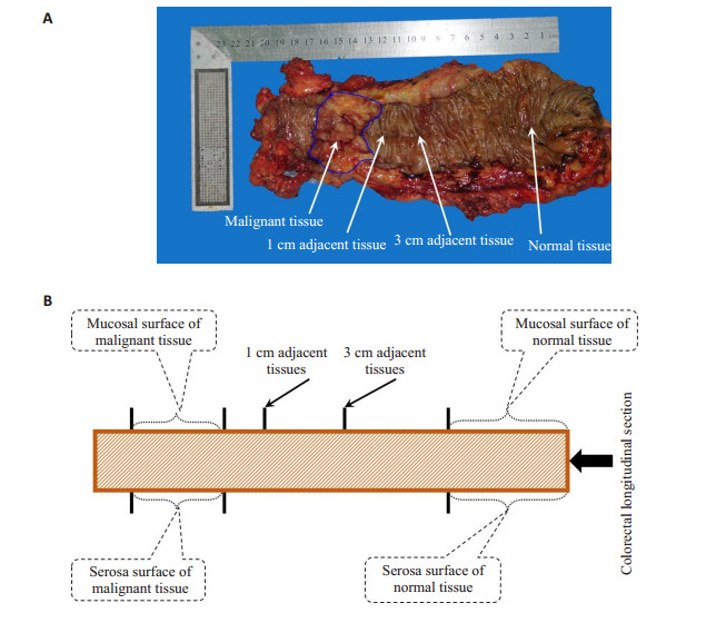
1.2 样品准备实验中所有结直肠标本均来源于南方医科大学珠江医院普外科,整个测量过程都在手术室内进行,一共测量了39个病例的新鲜离体结直肠组织,图 1 A展示了一结直肠组织标本(图中仅展示了黏膜面),图 1 B为结直肠的测量示意图。所有测量都在室温下进行并且在标本离体后0.5 h之内完成,结直肠标本的详细信如表 1所示。
|
图 1 结直肠标本(A)及测量示意图(B) Figure 1 Colorectal specimens (A) and measurement diagram (B). |
| 表 1 结直肠标本的相关信息 Table 1 The relevant information of colorectal samples |
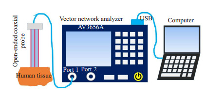
本研究采用操作简便,对样本具有非破坏性,适用于宽频测量的开端同轴探头法。测量系统由一根特征阻抗为50 Ω、外径3.58 mm的半刚性开端同轴探头(UT-086)连接到一台网络分析仪(AV3656A)组成,笔记本电脑用于网络分析仪的控制和数据处理,测量系统如示意图 2所示,并在50 MHz~3 GHz频率范围内测量探头反射系数,然后进一步转换成相对介电常数和电导率[30]。
|
图 2 测量系统 Figure 2 Measurement system. |
在标本切下之前需要对测量系统进行参数校准和误差分析,系统参数校准通常利用开路、短路和去离子水三种标准负载来完成,随后通过一系列标准物如甲醇(浓度99.8%)、乙醇(浓度99.8%)、乙二醇(浓度99.5%)来分析测量系统的准确性。将测量系统的校准结果与文献[31]中提供的参考数据进行比较。对于相对介电常数,甲醇、乙醇和乙二醇与参考值在整个频段内的最大误差分别为:1.9001 F/m、1.9548 F/m和1.4627 F/m;而电导率最大误差误差分别为:0.0852、0.0453和0.0753 S/m。通过上述的校准结果可知本测量系统具有较高的准确性和可靠性,进而为人体组织的测量提供可靠依据。
标本切除下来之后送往测量平台,在测量前需要用纱布将组织表面的血液和粘液擦干净,以免影响测量结果。再由外科医生的确认肿瘤组织黏膜面和浆膜面,正常组织黏膜面和浆膜面及癌旁1、3 cm黏膜面所在的位置。每次测量的时候将探头开端处的平整面垂直压在被测组织上面,可以稍稍施加一些压力以便减小探头和被测组织面之间的空气间隙。对肿瘤组织黏膜面和浆膜面,正常组织黏膜面和浆膜面及癌旁1、3 cm(本研究中的癌旁仅测量黏膜面)分别测量5个不同的点。测量完成之后紧接着用数字温度计(apuhua TM-902C)测量组织的温度。
1.4 测量数据统计分析依据常规病理检查结果对被测标本进行分类处理,并计算恶性组织,癌旁1、3 cm及正常组织的均值和相应的标准差。列出6个频率点64、128、298、433、915和2450 MHz的相对介电常数和电导率的数据,其中64、128和2450 MHz分别对应磁振共常用的拉莫尔频率1.5、3和7 T。而频率433、915和2450 MHz为ISM频带。
采用Mann-Whitney U检验分别对恶性组织,癌旁1、3 cm以及正常组织在频率点64、128、298、433、915和2450 MHz处的相对介电常数和电导进行统计学检验,其中P<0.01表示差异有统计学意义,所有统计检验均在IBM SPSS20中完成的。
1.5 测量数据拟合为了使测量数据易于应用,且将离散频率点的介电特性测量数据转换成连续频率介电特性。本研究采用了该领域常用的Cole-Cole模型对测量数据进行拟合。为了提高拟合精度,本研究采用二级Cole-Cole模型[32]进行拟合,如下:
| $ {\varepsilon ^*}(\omega ) = {\varepsilon _\infty } + \frac{{\Delta {\varepsilon _1}}}{{1 + {{(j\omega {\tau _1})}^{1 - {\alpha _1}}}}} + \frac{{\Delta {\varepsilon _2}}}{{1 + {{(j\omega {\tau _2})}^{1 - {\alpha _2}}}}} + \frac{{{\sigma _s}}}{{j\omega {\varepsilon _0}}} $ | (2) |
其中,ω是角频率且ω = 2πf,f为频率,ε*(ω)为复介电常数,是频率的函数,ε∞,Δε1,Δε2,τ1,τ2,α1,α2和σs是通过实验数据拟合得到的Cole-Cole模型参数。
2 结果 2.1 恶性组织、癌旁1、3 cm及正常组织黏膜面介电特性比较本研究结果以相对介电常数和电导率两个参数来分析结直肠组织介电特性。图 3为全频段下恶性组织,癌旁1、3 cm及正常组织黏膜面的介电参数均值(曲线表示)和标准差(误差棒表示),图 3为相对介电常数和电导率。显然恶性组织,癌旁1 cm,癌旁3 cm以及正常组织的相对介电常数和电导率均值依次减小。其中恶性组织相对介电常数和电导率均值高于癌旁1 cm,但二者误差棒出现小部分重叠。然而恶性组织相对介电常数和电导率均值明显高于癌旁3 cm和正常组织,并且恶性组织的误差棒与癌旁3 cm及正常组织明显分离。癌旁1、3 cm及正常组织的相对介电常数和电导率较为接近,且三者的误差棒相互交叠。此外还可以发现恶性组织的标准差(误差棒)小于癌旁1、3 cm及正常组织。

|
图 3 恶性组织,癌旁1、3 cm及正常组织黏膜面的全频段介电特性比较 Figure 3 Full-band dielectric properties of the mucosal surface of malignant tissue, adjacent tissues at 1 cm and 3 cm and normal tissue. Error bars represent standard deviation. A: Relative permittivity; B: Conductivity. |
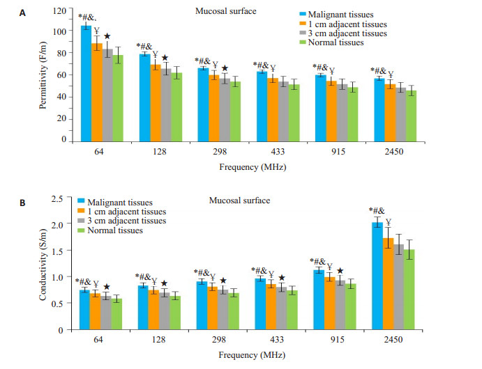
图 4给出了恶性组织和癌旁1、3 cm及正常组织黏膜面在6个特定频率点处的相对介电常数和电导率,显然恶性组织和癌旁1、3 cm及正常组织的相对介电常数和电导率在6个频率点处均依次减小,且恶性组织和癌旁1、3 cm及正常组织的介电参数在6个频率点处都存在显著性差异(P<0.01),癌旁1、3 cm黏膜面介电参数在6个频率点处依然存在显著性差异(P<0.01)。癌旁3 cm与正常组织的相对介电常数在64、128和298 MHz有显著性差异(P<0.01),在433、915和2450 MHz不存在显著性差异(P>0.01);癌旁3 cm与正常组织的电导率在64、128、298、433和915 MHz有统计学差异(P<0.01),而在2450 MHz无统计学意义(P>0.01)。
|
图 4 恶性组织,癌旁1、3 cm及正常组织在6个离散频率点的介电特性比较 Figure 4 Dielectric properties of the mucosal surface of malignant tissue, adjacent tissues at 1 cm and 3 cm and normal tissue at the discrete frequency. Error bars represent standard deviation. A: Relative permittivity; B: Conductivity. *P < 0.01 malignant vs 1 cm adjacent tissue; #P < 0.01 malignant vs 3 cm adjacent tissue; &P < 0.01 malignant vs normal tissue; ¥P < 0.01 1 cm adjacent tissue vs 3 cm adjacent tissue; ★P < 0.01 3 cm adjacent tissue vs normal tissue. |
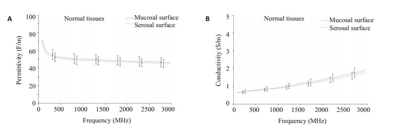
为了研究结直肠的内外壁介电特性,则分别测量了结直肠恶性组织与正常组织的黏膜面和浆膜面。恶性组织黏膜面的相对介电常数和电导率高于其浆膜面,但二者的误差棒(标准差)基本重叠,恶性组织的浆膜面的数据波动比黏膜较大,并且从误差棒可以发现浆膜面的数据波动涵盖了黏膜面数据(图 5)。恶性黏膜面与浆膜面介电特性在2450 MHz频率点处没有显著差异(P>0.01),其余的5个频率点依然有显著差异(P<0.01,图 6)。对于正常组织而言,黏膜面和浆膜面的介电特性差异非常小(图 7),正常组织黏膜面和浆膜面的介电特性在6个特定频率点处均不存在显著性差异(P>0.01,图 8)。

|
图 5 恶性组织黏膜面和浆膜面介电特性比较 Figure 5 Comparison of dielectric properties between mucosal and serosal surfaces in malignant tissues. Error bars represent standard deviation. A: Relative permittivity; B: Conductivity. |

|
图 6 恶性组织黏膜面和浆膜面在6个离散频率点的介电特性比较 Figure 6 Comparison of dielectric properties between mucosal and serosal surfaces in malignant tissues at six discrete frequencies. Error bars represent standard deviation. A: Relative permittivity; B: Conductivity. *P < 0.01 mucosal vs serosal. |
|
图 7 正常组织黏膜面和浆膜面介电特性比较 Figure 7 Comparison of dielectric properties between mucosal and serosal surfaces in normal tissues. Error bars represent standard deviation. A: Relative permittivity; B: Conductivity. |

|
图 8 正常组织黏膜面和浆膜面在6个离散频率点的介电特性比较 Figure 8 Comparison of dielectric properties between mucosal and serosal surfaces in normal tissues at 6 discrete frequencies. Error bars represent standard deviation. A: Relative permittivity; B: Conductivity. |
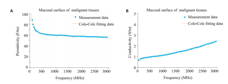
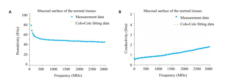
为了使数据可利用以及能够计算任意频率下介电参数,则对常见恶性组织和正常组织黏膜面介电特性数据进行两级Cole-Cole拟合。拟合结果如图 9、图 10所示,其中图 9A图 10A分别为恶性组织和正常组织黏膜面的相对介电常数拟合结果,图 9B图 10B分别为恶性组织和正常组织黏膜面的电导率数拟合结果,表 2给出了对应的拟合参数。
|
图 9 恶性组织黏膜面介电特性Cole-Cole拟合结果 Figure 9 Cole-Cole fitting results of dielectric parameters of malignant tissues mucosal surface. A: Relative permittivity; B: Conductivity. |
|
图 10 正常组织黏膜面介电参数的Cole-Cole拟合结果 Figure 10 Cole-Cole fitting results of dielectric parameters of normal tissues mucosal surface. A: Relative permittivity; B: Conductivity. |
| 表 2 恶性组织和正常组织黏膜面的Cole-Cole拟合参数 Table 2 Cole-Cole fitting parameters for malignant and normal tissues |
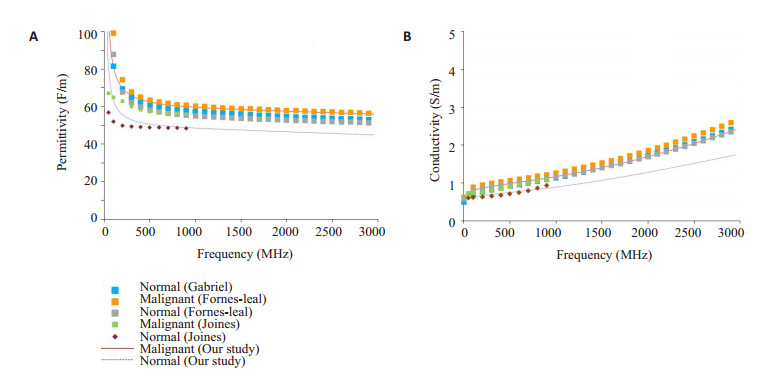
为了验证结果,将本研究中的结果与文献进行比较(图 11)。这里选取本次测量研究中的介电特性最大的恶性黏膜和介电特性最小的正常组织黏膜的结果与文献比较。首先,从相对介电常数和电导率的频率变化趋势来看,本研究的结果与文献保持一致。对于恶性组织,本研究的恶性组织相对介电常数和Fornes-Leal[24]的结果非常相似,两条曲线几乎重合,而电导率略低于Fornes-Leal[24]的结果。本研究中的相对介电常数和电导率均高于Joines[4]的结果。至于正常组织,我们的正常组织黏膜面介电特性均低于Fornes-Leal[24]的结果。在Gabrie[21]研究中仅测量了羊的结直肠正常组织介电特性,并且Gabrie[21]的结果高于本研究中正常组织的黏膜面的介电特性。本研究的正常组织黏膜面的相对介电常数与Joines[4]的结果较为接近,尤其是在500 MHz以上,而两者电导率则更为相似。
|
图 11 恶性组织和正常组织黏膜面介电特性与文献对比 Figure 11 Relative permittivity (A) and conductivity (B) of malignant and normal tissue mucosal surface measured in this study and reported in literature. |
组织介电特性作为生物组织的固有属性,其变化通常暗示着组织的生理、病理状态的改变,本研究针对结直肠癌标本介电特性进行了较为细致的研究。结果中恶性组织介电特性高于正常组织(P<0.01),从宏观方面可以解释为恶性组织水含量高于正常组织[33],尤其是结合水,并且有研究表明结合水介电特性高于自由水[34],这与生理上恶性组织伴随血管增生及扩张[35],血液含量相对较高的现象相符合。微观层面,细胞癌变后物质成分及含量发生改变,此外,细胞癌变后细胞膜的通透性的增加使得钠、钾等离子积聚到癌细胞中,致使癌组织具有更高的介电常数和电导率[3, 36]。另外,Sugitani[37]在研究乳腺癌组织介电特性的变异性时发现癌细胞体积分数与电导率、相对介电常数之间存在相关性——癌细胞密度越高则介电特性越大,癌细胞密度越低介电特性越小,这一发现可以很好的解释恶性组织,癌旁1、3 cm及正常组织介电特性的依次减小的现象,由于癌组织具有向周围组织生长和侵袭的能力,所以越靠近癌组织区域癌细胞的体积分数越高,即相对介电常数和电导率也越大,反之则越小。因此恶性组织,癌旁1、3 cm及正常组织介电特性的差异在一定程度上可以反映癌细胞向周围组织逐渐浸润及生长的过程。至于恶性组织的标准差(误差棒)小于癌旁1、3 cm及正常组织,这说明恶性组织的介电特性的波动较小,在O'rourke[6]和FornesLeal[24]的研究中也出现类似的现象。但无论是恶性组织、癌旁组织还是正常组织的介电特性均存在一定的波动性(图 3和图 4中的误差棒),这种波动现象可以归于以下原因:病人之间的个体差异;被测标本相对湿度的小幅度变化(标本离体到测量完成时间间隔相当的情况下);结直肠癌的类型[38];病人的年龄差异[39]。但这些影响因素相对于组织的生理和病理性质的变化都只是轻微的。
至于恶性组织黏膜面的介电特性高于浆膜面,这是因为恶性组织主要生长在黏膜面,然后再向浆膜面侵袭,显然黏膜面癌细胞的密度高于浆膜面,即黏膜面的相对介电常数和电导率高于浆膜面。对于浆膜面介电特性波动(图 5和图 6中的误差棒)比黏膜面大,甚至浆膜数据波动范围涵盖了黏膜面数据的波动(图 5中的误差棒重叠),这些现象源于恶性组织对浆膜浸润深度的影响,当恶性组织突破浆膜层时浆膜面介电特性与黏膜面相当,即此时的浆膜面介电特性较大,然而当恶性组织未突破或浸润浆膜层时浆膜面介电特性则较小,这也正是造成了浆膜面介电变化较大的主要原因。关于恶性组织浸润/突破浆膜面对浆膜面介电特性的影响在王卫卫等的研究中有较为详细的描述[29]。正常组织黏膜面和浆膜面介电特性无显著差异说明正常组织黏膜面介电特性与其浆膜面极为相似。图 6、图 8中的恶性组织浆膜面的介电特性高于正常组织浆膜也可以归因于恶性组织对其浆膜层的浸润而使得恶性组织浆膜面高于正常组织浆膜面[29]。
对于测量数据的拟合结果可见拟合效果很好,这反映了本研究采集的介电特性数据符合Cole-Cole模型[32]。本研究的恶性黏膜面的介电特性与Fornes-Leal[24]的恶性组织介电特性测量一致性较好。而正常组织黏膜面介电特性比Fornes-Leal[24]的正常组织介电特低,分析原因有以下几个方面:一是Fornes-Leal[24]对正常组织介电特性测量没有说明与恶性组织的距离,也没有指明正常组织是黏膜面还是浆膜面;二是结直肠的正常组织的介电特性也可能存在人种差异。Gabrie[21]的正常组织介电特性高于本研究的正常组织黏膜面介电特性,其原因可能是Gabrie[21]的标本来源于羊组织。本研究的恶性、正常组织黏膜面介电特性与Joines[4]的结果存在差异,很可能是因为Joines[4]的标本测量是在标本离体后1~2 h完成,导致标本水分散失,而本研究标本测量是在标本离体后0.5 h内完成。
通过本研究,对于术中恶性组织边缘检测/良恶性鉴别技术及设备的开发有直接性的临床应用价值,而对于磁共振电特性断层成像技术的发展以及肿瘤组织的热消融技术的发展有着基础性的作用。
| [1] |
Martellosio A, Pasian M, Bozzi MA, et al. Dielectric properties characterization from 0.5 to 50 GHz of breast cancer tissues[J].
IEEE Trans Microw Theory Tech, 2017, 65(3): 998-1011.
DOI: 10.1109/TMTT.2016.2631162. |
| [2] |
Mirbeik-Sabzevari A, Ashinoff R, Tavassolian N. Ultra-wideband millimeter-wave dielectric characteristics of freshly-excised normal and malignant human skin tissues[J]. IEEE Trans Biomed Eng, 2017, [Epub ahead of print].
|
| [3] |
Peyman A, Kos B, Djokic M, et al. Variation in dielectric properties due to pathological changes in human liver[J].
Bioelectromagnetics, 2015, 36(8): 603-12.
DOI: 10.1002/bem.21939. |
| [4] |
Joines WT, Zhang Y, Li C, et al. The measured electrical properties of normal and malignant human tissues from 50 to 900 MHz[J].
Med Phys, 1994, 21(4): 547-50.
DOI: 10.1118/1.597312. |
| [5] |
Cheng Y, Fu M. Dielectric properties for differentiating normal and malignant thyroid tissues[J].
Med Sci Monit, 2018, 24: 1276-81.
DOI: 10.12659/MSM.908204. |
| [6] |
O'rourke AP, Lazebnik M, Bertram JM, et al. Dielectric properties of human normal, malignant and cirrhotic liver tissue: in vivo and ex vivo measurements from 0.5 to 20 GHz using a precision open-ended coaxial probe[J].
Phys Med Biol, 2007, 52(15): 4707-19.
DOI: 10.1088/0031-9155/52/15/022. |
| [7] |
Possebon R, Jiang Y, Mulier S, et al. A piecewise function of resistivity of liver: determining parameters with finite element analysis of radiofrequency ablation[J].
Med Biol Eng Comput, 2018, 56(3): 385-94.
DOI: 10.1007/s11517-017-1699-6. |
| [8] |
Converse M, Bond EJ, Van Veen BD, et al. A computational study of ultra-wideband versus narrowband microwave hyperthermia for breast cancer treatment[J].
IEEE Trans Microw Theory Tech, 2006, 54(5): 2169-80.
DOI: 10.1109/TMTT.2006.872790. |
| [9] |
Etoz S, Brace CL. Analysis of microwave ablation antenna optimization techniques[J].
Int J RF Microwave Comp Engineer, 2018, 28(3, SI): e21224.
DOI: 10.1002/mmce.v28.3. |
| [10] |
Yu J, Liang P, Yu X, et al. A comparison of microwave ablation and bipolar radiofrequency ablation both with an internally cooled probe: results in ex vivo and in vivo porcine livers[J].
Eur J Radiol, 2011, 79(1): 124-30.
DOI: 10.1016/j.ejrad.2009.12.009. |
| [11] |
Ariturk G, Ider YZ. Optimal multichannel transmission for improved cr-MREPT[J].
Phys Med Biol, 2018, 63(4): 045001.
DOI: 10.1088/1361-6560/aaa732. |
| [12] |
Liu J, Shao Q, Wang Y, et al. In vivo imaging of electrical properties of an animal tumor model with an 8-channel transceiver array at 7 T using electrical properties tomography[J].
Magn Reson Med, 2017, 78(6): 2157-69.
DOI: 10.1002/mrm.v78.6. |
| [13] |
Katscher U, Voigt T, Findeklee C, et al. Determination of electric conductivity and local SAR via B1 mapping[J].
IEEE Trans Med Imaging, 2009, 28(9): 1365-74.
DOI: 10.1109/TMI.2009.2015757. |
| [14] |
Tha KK, Katscher U, Yamaguchi S, et al. Noninvasive electrical conductivity measurement by MRI: a test of its validity and the electrical conductivity characteristics of glioma[J].
Eur Radiol, 2018, 28(1): 348-55.
DOI: 10.1007/s00330-017-4942-5. |
| [15] |
Bulumulla SB, Lee SK, Yeo D. Conductivity and permittivity imaging at 3.0 T[J].
Concepts Magn Reson Part B Magn Reson Eng, 2012, 41B(1): 13-21.
DOI: 10.1002/cmr.b.v41b.1. |
| [16] |
Pappo I, Spector R, Schindel A, et al. Diagnostic performance of a novel device for real-time margin assessment in lumpectomy specimens[J].
J Surg Res, 2010, 160(2): 277-81.
DOI: 10.1016/j.jss.2009.02.025. |
| [17] |
Allweis TM, Kaufman Z, Lelcuk S, et al. A prospective, randomized, controlled, multicenter study of a real-time, intraoperative probe for positive margin detection in breast-conserving surgery[J].
Am J Surg, 2008, 196(4): 483-9.
DOI: 10.1016/j.amjsurg.2008.06.024. |
| [18] |
Dumitru D, Douek M, Benson JR. Novel techniques for intraoperative assessment of margin involvement[J].
Ecancermedicalscience, 2018, 12: 795.
|
| [19] |
Joines WT. Frequency-dependent absorption of electromagnetic energy in biological tissue[J].
IEEE Trans Biomed Eng, 1984, 31(1): 17-20.
|
| [20] |
Joines WT, Jirtle RL, Rafal MD, et al. Microwave power absorption differences between normal and malignant tissue☆[J].
Int J Radiat Oncol Biol Phys, 1980, 6(6): 681-7.
DOI: 10.1016/0360-3016(80)90223-0. |
| [21] |
Gabriel C, Gabriel S, Corthout E. The dielectric properties of biological tissues: Ⅰ. Literature survey[J].
Phys Med Biol, 1996, 41(11): 2231-49.
DOI: 10.1088/0031-9155/41/11/001. |
| [22] |
Gabriel S, Lau RW, Gabriel C. The dielectric properties of biological tissues: Ⅱ. Measurements in the frequency range 10 Hz to 20 GHz[J].
Phys Med Biol, 1996, 41(11): 2257-69.
|
| [23] |
Gabriel S, Lau RW, Gabriel C. The dielectric properties of biological tissues: Ⅲ. Parametric models for the dielectric spectrum of tissues[J].
Phys Med Biol, 1996, 41(11): 2271-93.
DOI: 10.1088/0031-9155/41/11/003. |
| [24] |
Fornes-Leal A, Garcia-Pardo C, Frasson M, et al. Dielectric characterization of healthy and malignant colon tissues in the 0.5-18 GHz frequency band[J].
Phys Med Biol, 2016, 61(20): 7334-46.
DOI: 10.1088/0031-9155/61/20/7334. |
| [25] |
Li Z, Deng GH, Li Z, et al. A large-scale measurement of dielectric properties of normal and malignant colorectal tissues obtained from cancer surgeries at larmor frequencies[J].
Med Phys, 2016, 43(11): 5991-7.
DOI: 10.1118/1.4964460. |
| [26] |
魏珊珊, 冯晓创, 符芳翔, 等. 开端同轴线射频鉴别系统检测的结肠癌组织、癌周正常组织介电常数和电导率比较[J].
山东医药, 2016, 56(27): 80-2.
DOI: 10.3969/j.issn.1002-266X.2016.27.028. |
| [27] |
王卫卫, 厉周, 林世明, 等. 101例手术切除的结直肠癌组织介电特性的探讨[J].
生物医学工程研究, 2016, 35(4): 270-3.
|
| [28] |
Li Z, Wang WW, Cai Z, et al. Variation in the dielectric properties of freshly excised colorectal cancerous tissues at different tumor stages[J].
Bioelectromagnetics, 2017, 38(7): 522-32.
DOI: 10.1002/bem.v38.7. |
| [29] |
王卫卫, 蔡寨, 韩帅, 等. 应用开端同轴线仪器检测结直肠癌组织和评估肿瘤浸润深度[J].
实用医学杂志, 2016, 32(20): 3376-9.
DOI: 10.3969/j.issn.1006-5725.2016.20.026. |
| [30] |
Bobowski JS, Johnson T. Permittivity Measurements of biological samples by an open -ended coaxial line[J].
Prog Electromagnet Res, 2012, 40(40): 159-83.
|
| [31] |
Gregory AP, Clarke RN.
Tables of the complex permittivity of dielectric reference liquids at frequencies up to 5 GHz[M]. 2009.
|
| [32] |
Grosse C. A program for the fitting of Debye, Cole-Cole, ColeDavidson, and Havriliak-Negami dispersions to dielectric data[J].
J Colloid Interface Sci, 2014, 419(4): 102-6.
|
| [33] |
Schepps JL, Foster KR. The UHF and microwave dielectric properties of normal and tumour tissues: variation in dielectric properties with tissue water content[J].
Phys Med Biol, 1980, 25(6): 1149-59.
DOI: 10.1088/0031-9155/25/6/012. |
| [34] |
Pethig R. Dielectric properties of biological materials: biophysical and medical applications[J].
Electr Insulat IEEE Transact, 1984, 19(5): 453-74.
|
| [35] |
Raza A, Franklin MJ, Dudek AZ. Pericytes and vessel maturation during tumor angiogenesis and metastasis[J].
Am J Hematol, 2010, 85(8): 593-8.
DOI: 10.1002/ajh.v85:8. |
| [36] |
Chandra R, Zhou HY, Balasingham I, et al. On the opportunities and challenges in microwave medical sensing and imaging[J].
IEEE Trans Biomed Eng, 2015, 62(7): 1667-82.
DOI: 10.1109/TBME.2015.2432137. |
| [37] |
Sugitani T, Kubota SI, Kuroki SI, et al. Complex permittivities of breast tumor tissues obtained from cancer surgeries[J].
Appl Phys Lett, 2014, 104(25): 253702.
DOI: 10.1063/1.4885087. |
| [38] |
杨军, 郭睿, 康安静, 等. 一种新的结直肠癌组织学分型、分级-评分方案[J].
南方医科大学学报, 2014, 34(2): 169-73.
|
| [39] |
Peyman A, Rezazadeh AA, Gabriel C. Changes in the dielectric properties of rat tissue as a function of age at microwave frequencies[J].
Phys Med Biol, 2001, 46(6): 1617-29.
DOI: 10.1088/0031-9155/46/6/303. |
 2018, Vol. 38

