2. 南方医科大学中医药学院,广东 广州 510515
2. School of Traditional Chinese Medicine, Southern Medical University, Guangzhou 510515, China
高血压病是当今世界最主要的流行病、慢性病之一。动脉粥样硬化(AS)是一种慢性炎症性疾病,也是高血压靶器官损害的重要中间坏节。AS是血管壁对各种损伤的异常反应[1],往往无特异性症状,多为相关器官受累后出现相应的临床表现,在诸多AS中,颈动脉粥样硬化(CAS)较常见,是心肌梗死、缺血性脑卒中等急性心脑血管疾病的病理基础[2-3]。最新数据显示,我国脑卒中亚型中,约60%~80%为缺血性脑卒中[4],其中由CAS导致的缺血性脑卒中约占20% [5]。现代医学对高血压的治疗,主要以药物控制为主,虽然都能有效控制血压,但同时也存在一些副作用[6-9]。而有CAS的高血压患者更应该慎重选择高血压药物。祖国医学并没有提及CAS的有关概念,不过多数学者认为CAS属于中医“淤血”、“痰浊”的范畴。常见的中医药疗法包括重要复方治疗[10-13]、中药提取物[14-15]。然而,CAS高血压的药物治疗几乎伴随患者终身,积极探讨非药物疗法对高血压病的防治具有重要意义。
推拿疗法经诸多临床研究证明是防治高血压的有效方法之一[16-17],其中又以推桥弓手法[18]尤为突出。桥弓穴的相关记载,可以最早追溯到《黄帝内经》时期,但是,其确切方法的提出,则属于现代。桥弓穴是由民间医家经验总结的一个推拿要穴,现代人将其运用到治疗高血压[19]。虽然推桥弓手法治疗高血压疗效显著,但仍可能伴发诸多并发症,如脑血管意外等[20]。由于推拿手法使用不当,如操作不规范、用力过大等,可能改变CAS斑块的生物力学性能[21],导致CAS斑块破裂[22-23],进而引发缺血性脑卒中等急性心脑血管疾病。相关研究报道,颈动脉血流动力学变化是CAS斑块破裂的关键因素之一。如局部血流动力学改变可能使血流类型由层流变为湍流,对管壁冲击力增加,产生较大的剪切应力,从而破坏斑块的稳定性,导致斑块脱落或破裂[24]。故检测推桥弓手法对CAS血流动力学的影响对提高推桥弓手法的安全性具有重要意义。
目前,推桥弓手法的临床治疗作用得到一些研究的肯定,但是,在手法操作过程中是否会影响CAS斑块稳定性,及血流动力学变化,甚至加重CAS,引起斑块脱落,危及生命,尚未有明确定论,相关研究也鲜有报道。因此,本研究对推桥弓手法干预CAS模型血流动力学的影响进行深入研究具有一定临床意义。
1 材料和方法 1.1 主要试剂、耗材及仪器Zoletil 50麻醉剂购自法国Virbac公司。碘伏、无菌镜器械包、止血带等均购自北京中杉金桥公司。高脂饲料(含2%胆固醇、10%猪油和88%普通颗粒饲料)由云南英茂公司配制提供。彩色多普勒超声诊断系统产自荷兰Philips公司(型号:L15-7io)。
1.2 实验动物及分组选取SPF级正常食蟹猴9只,均为雄性,体质量6~7 kg,年龄4~5岁,动物饲养和动物实验均在云南英茂公司实验中心完成,动物许可证号:SYXK(滇) 2009-0003。所有程序通过了该公司的动物实验伦理审查委员会的批准。将9只食蟹猴随机分为3组,每组3只,分别是:推桥弓组、模型对照组、空白对照组。先进行分组,然后再对推桥弓组和模型对照组进行CAS造模,空白组不做特殊干预。
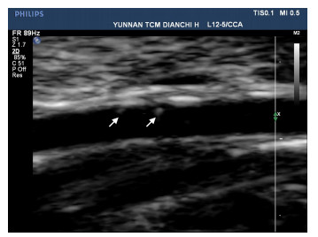
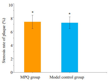
1.3 模型建立将推桥弓组和模型对照组食蟹猴进行CAS造模,每只食蟹猴只造一侧颈总动脉。造模方法:给予Zoletil 50 (5 mg/kg)麻醉后,常规消毒、铺巾,在胸锁乳突肌和喉结之间的间隙分离出颈总动脉,用注射器针头穿刺入动脉,并在动脉内壁上反复刮檫。拔出针头,用纱布按压进针点,待无明显渗血后,冲洗伤口,逐层缝合,术毕。手术均有同一组操作人员完成。术后注意观察伤口及动物吞咽、进食等情况。术后3 d分别给予盐酸左氧氟沙星氯化钠注射液(8 mg/kg)静脉滴注预防感染,同时给予注射用盐酸曲马多(2 mg/kg)肌注减轻术后疼痛。术后即给予高脂饲料喂养,8周后,在麻醉状态下行颈部彩超检测,可见颈总动脉内壁上有CAS斑块形成(图 1)。CAS模型斑块狭窄率在(7.28±0.82) %,根据2003年北美放射学会超声会议制定的标准[25],属于轻度狭窄。其中,推桥弓组的斑块狭窄率为(7.35±0.98) %,模型对照组的斑块狭窄率为(7.22±0.85) %,两组对比差异无统计学意义(P>0.05),说明两组具有可比性。
|
图 1 颈动脉彩超验证CAS斑块情况 Figure 1 CAS plaques (white arrows) observed by color Doppler ultrasound. |
造模成功后,将推桥弓组食蟹猴固定在固定椅上,沿着左右颈动脉各推20次,1次/d,共30 d。模型对照组、空白组不做特殊干预。所有手法操作均由1名主治医师完成。
1.5 检查方法给予Zoletil 50 (5 mg/kg)麻醉后,取仰卧位,头微后仰,充分暴露颈部。检查时,彩超探头检查范围距离颈总动脉分叉处1.37±0.10 cm,主要检测颈总动脉血管和斑块情况,指标包括:血管横切面面积、斑块横切面面积、斑块狭窄率(斑块横切面面积/血管横切面面积× 100%)。同时,检测颈总动脉血流动力学,指标包括:收缩期峰值血流速度(PSV)、舒张末期血流速度(EDV)、时间平均血流速度(TAV)、阻力指数(RI)[(PSV-EDV)/ PSV]、搏动指数(PI)[(PSV-EDV)/TAV]。
1.6 统计学方法使用SPSS 20.0软件进行统计分析,计量数据以均数±标准差表示,组间比较应用单因素方差分析(LSD-t检验)进行统计,检验水准α=0.05。当P<0.05认为差异有统计学意义。
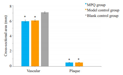
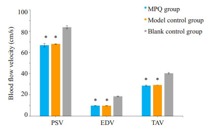
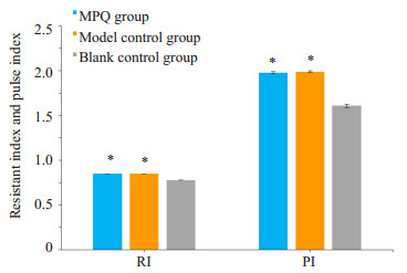
2 结果在彩超检查中,推桥弓组、模型对照组可见斑块形成,同时,在血管横切面面积、斑块横切面面积、斑块狭窄率方面,与空白对照组比较,推桥弓组、模型对照组差异均有统计学意义(P<0.05),而推桥弓组和模型对照组比较,差异无统计学意义(P>0.05)(图 2、3)。在各项血流动力学指标中(PSV、EDV、TAV、RI、PI),与空白对照组对比,推桥弓组、模型对照组差异均有统计学意义(P<0.05),而推桥弓组和模型对照组比较,差异无统计学意义(P>0.05,图 4、5)。
|
图 2 3组在血管横切面面积、斑块横切面面积的对比情况 Figure 2 Comparison of vascular cross-sectional area and plaque cross-sectional area among the 3 groups. *P < 0.05 vs blank control group. |
|
图 3 3组在斑块狭窄率方面的对比情况 Figure 3 Comparison of stenosis rate of plaque among the 3groups. *P < 0.05 vs blank control group. |
|
图 4 3组PSV、EDV和TAV的对比情况 Figure 4 Comparison of PSV, EDV, and TAV among the 3 groups. *P < 0.05 vs blank control group. |
|
图 5 3组RI和PI的对比情况 Figure 5 Comparison of RI and PI among the 3 groups. *P < 0.05 vs blank control group. |
CAS是动脉内膜表面隆起的斑块,动脉内膜增厚是CAS的早期指征,而粥样斑块形成是其最具特征性的病变[26-29],因此,我们通过针刺划痕法联合高脂饲养的CAS造模方法对动物进行造模,在术后8周,通过颈部彩超检测,可见颈总动脉内壁上有CAS斑块形成。CAS模型斑块狭窄率在(7.28±0.82) %,属于轻度狭窄。在造模过程中未发生动物死亡的不良事件,这说明我们的造模方法安全有效,可以有效的建立轻度CAS模型,为后续的实验提供了研究基础。
AS破裂是一个复杂的过程,往往是多因素联合作用的结果:(1)血流动力学因素。局部低壁面切应力和高剪切震荡指数是粥样硬化斑块形成、破裂的高危因素[30-33]。(2)斑块易损性。斑块的形态、大小、成分、生物学活性等与斑块稳定性密切相关[34-36]。斑块易损性与纤维帽厚度呈负相关,而与炎症细胞数量、脂质核心的大小呈正相关[37-38]。斑块内巨噬细胞的数量是决定斑块稳定性的重要因素,巨噬细胞数量越多, 斑块越不稳定[39-40]。同时,血流动力学与斑块易损性相关[41]。(3)机械应力。颈部推拿由于手法操作不规范、过度用力或违背解剖学原则等,可能损伤颈动脉、撕裂神经纤维,AS斑块受到高应力,引起脱落、破裂等,造成缺血性脑卒中等急性脑血管疾病[42-44]。
在诸多推拿著作及临床实验中,均指出推桥弓手法能起到很好的即刻降压效应,具有良性、双向调节和无毒副作用等优点。而血流动力学的异常可促进CAS的形成、发展[45-47]。因此,观察推桥弓手法对食蟹猴轻度CAS斑块模型血流动力学的影响具有重要的意义。
在本研究中,在颈总动脉横切面面积、斑块横切面面积、斑块狭窄率方面,与空白对照组比较,推桥弓组、模型对照组差异均有统计学意义(P<0.05),而推桥弓组和模型对照组比较,差异无统计学意义(P>0.05),这说明推桥弓组和模型对照组均有斑块形成,且推桥弓手法在短期内不会影响斑块的稳定性。在各项血流动力学指标中,推桥弓组、模型对照组PSV、EDV、TAV均低于空白对照组,说明斑块的形成会影响颈动脉的血流速度,从而进一步影响血供,同时,RI、PI均高于正常组(P<0.05),说明在CAS中,血管壁负荷增大。但推桥弓组和模型对照组比较,PSV、EDV、TAV、RI、PI差异均无统计学意义(P>0.05),这说明推桥弓手法在短期内不会加重血流动力学的影响。
本研究在实施过程中,由于客观原因和条件的限制,存在以下不足:(1)本研究只针对轻度CAS,对中、重度CAS有待进一步的研究。(2)出于伦理和安全性的考虑,实验对象选择食蟹猴,而不是直接作用于人体,手法对两者的影响是否有所不同,需待相关研究进一步探讨。
综上所述,AS斑块对颈动脉的血流动力学会产生影响,但推桥弓手法在短期内不会影响斑块的稳定性,也不会加重血流动力学的影响。
| [1] | De Gaetano M, Crean D, Barry M, et al. M1- and M2-Type macrophage responses are predictive of adverse outcomes in human atherosclerosis[J]. Front Immunol, 2016, 7: 275. |
| [2] | Chen ZL, Wang F, Zheng YS, et al. H-type hypertension is an important risk factor of carotid atherosclerotic plaques[J]. Clin Exp Hypertens, 2016, 38(5): 424-8. DOI: 10.3109/10641963.2015.1116547. |
| [3] | Hurtubise J, Mclellan K, Durr K, et al. The different facets of dyslipidemia and hypertension in atherosclerosis[J]. Curr Atheroscler Rep, 2016, 18(12): 82. DOI: 10.1007/s11883-016-0632-z. |
| [4] | 李海林, 贾晓雁, 王晋鹏, 等. 急诊独立模式溶栓缩短急性缺血性脑卒中患者入院-溶栓时间探讨[J]. 中华急诊医学杂志, 2016, 25(9): 1180-3. |
| [5] | Ammirati E, Moroni F, Norata GD, et al. Markers of inflammation associated with plaque progression and instability in patients with carotid atherosclerosis[J]. Mediators Inflamm, 2015, 2015: 718329. |
| [6] | Mancia G, De Backer G, Dominiczak A, et al. 2007 Guidelines for the management of arterial hypertension: The Task Force for the Management of Arterial Hypertension of the European Society of Hypertension (ESH) and of the European Society of Cardiology (ESC)[J]. Eur Heart J, 2007, 28(12): 1462-536. |
| [7] | Bangalore S, Parkar S, Grossman E, et al. A meta-analysis of 94, 492 patients with hypertension treated with beta blockers to determine the risk of new-onset diabetes mellitus[J]. Am J Cardiol, 2007, 100(8): 1254-62. DOI: 10.1016/j.amjcard.2007.05.057. |
| [8] | Matchar DB, Mccrory DC, Orlando LA, et al. Systematic review: comparative effectiveness of angiotensin-converting enzyme inhibitors and angiotensin Ⅱ receptor blockers for treating essential hypertension[J]. Ann Intern Med, 2008, 148(1): 16-29. DOI: 10.7326/0003-4819-148-1-200801010-00189. |
| [9] | Law MR, Moris JK, Wald HJ, et al. Use of blood pressure lowering drugs in the prevention of cardiovascular disease: meta-analysis of 147 randomised trials in the context of expectations from prospective epidemiological studies[J]. BMJ, 2009, 338: b1665. DOI: 10.1136/bmj.b1665. |
| [10] | 王春兰, 杨焕斌. 冠心Ⅱ号治疗颈动脉粥样硬化斑块的临床研究[J]. 光明中医, 2008, 23(8): 1148-50. |
| [11] | 陈俊发. 大黄虫丸治疗颈动脉粥硬化斑块50例[J]. 山东中医杂志, 2001, 16(6): 331-2. |
| [12] | 余虹, 万青, 石欣, 等. 调脂胶囊治疗颈动脉粥样硬化风块37例临床观察[J]. 中医杂志, 2003, 44(9): 664-5. |
| [13] | 党毓起, 吴青霞, 刘军, 等. 健脾补肾消瘀祛毒法治疗颈动脉粥样硬化40例临床研究[J]. 实用中医内科杂志, 2006, 20(6): 654-5. |
| [14] | 段明福, 宋梅, 孙秋红. 碟脉灵治疗颈与脑动脉硬化斑块的疗效观察[J]. 中华实用中西医杂志, 2003, 16(5): 631. |
| [15] | 何国厚, 艾志兵, 刘勇, 等. 小檗碱对颈动脉粥样硬化中内膜增生和巨噬细胞趋化作用的影响[J]. 中风与神经疾病杂志, 2006, 23(1): 94-6. |
| [16] | Field T. Massage therapy research review[J]. Complement Ther Clin Pract, 2014, 20(4): 224-9. DOI: 10.1016/j.ctcp.2014.07.002. |
| [17] | Nelson NL. Massage therapy: understanding the mechanisms of action on blood pressure. A scoping review[J]. J Am Soc Hypertens, 2015, 9(10): 785-93. DOI: 10.1016/j.jash.2015.07.009. |
| [18] | 娄晓峰, 廖品东. 头面部推拿与推桥弓辅助治疗高血压病临床疗效比较[J]. 时珍国医国药, 2009, 20(10): 2623-4. DOI: 10.3969/j.issn.1008-0805.2009.10.121. |
| [19] | 冯跃, 杨洁, 杨馨. 桥弓穴源流简考[J]. 吉林中医药, 2010, 30(6): 542-3. |
| [20] | Bowler N, Shamley D, Davies R. The effect of a simulated manipulation position on internal carotid and vertebral artery blood flow in healthy individuals[J]. Man Ther, 2011, 16(1): 87-93. DOI: 10.1016/j.math.2010.07.007. |
| [21] | Thomas LC, Mcleod LR, Osmotherly PG, et al. The effect of endrange cervical rotation on vertebral and internal carotid arterial blood flow and cerebral inflow: A sub analysis of an MRI study[J]. Man Ther, 2015, 20(3): 475-80. DOI: 10.1016/j.math.2014.11.012. |
| [22] | Riou LM, Broisat A, Ghezzi C, et al. Effects of mechanical properties and atherosclerotic artery size on biomechanical plaque disruption -mouse vs. human[J]. J Biomech, 2014, 47(4): 765-72. DOI: 10.1016/j.jbiomech.2014.01.020. |
| [23] | Vergallo R, Papafaklis MI, Yonetsu T, et al. Endothelial shear stress and coronary plaque characteristics in humans: combined frequency-domain optical coherence tomography and computational fluid dynamics study[J]. Circ Cardiovasc Imaging, 2014, 7(6): 905-11. DOI: 10.1161/CIRCIMAGING.114.001932. |
| [24] | He F, Hua L, Gao LJ. Computational analysis of blood flow and wall mechanics in a model of early atherosclerotic artery[J]. J Mech Sci Tech, 2017, 31(2): 1015-20. DOI: 10.1007/s12206-017-0154-9. |
| [25] | Paolo P, Napoli A, Anzidei M, et al. Comparison between dualenergy CT-angiography, MR-angiography and digital substraction angiography for the evaluation of carotid artery stenosis: A prospective study[J]. BMC Vet Res, 2015, 11(1): 1-5. DOI: 10.1186/s12917-014-0312-6. |
| [26] | Quillard T, Libby P. Molecular imaging of atherosclerosis for improving diagnostic and therapeutic development[J]. Circ Res, 2012, 111(2): 231-44. DOI: 10.1161/CIRCRESAHA.112.268144. |
| [27] | Li J, Ley K. Lymphocyte migration into atherosclerotic plaque[J]. Arterioscler Thromb Vasc Biol, 2015, 35(1): 40-9. DOI: 10.1161/ATVBAHA.114.303227. |
| [28] | Skagen K, Skjelland M, Zamani M, et al. Unstable carotid artery plaque: new insights and controversies in diagnostics and treatment[J]. Croat Med J, 2015, 57(4): 311-20. |
| [29] | Giannoglou GD, Antoniadis AP, Koskinas KC, et al. Flow and atherosclerosis in coronary bifurcations[J]. EuroIntervention, 2010, 6(Suppl J): J16-23. |
| [30] | Pedrigi RM, Mehta VV, Bovens SM, et al. Influence of shear stress magnitude and direction on atherosclerotic plaque composition[J]. R Soc Open Sci, 2016, 3(10): 160588. DOI: 10.1098/rsos.160588. |
| [31] | Chatzizisis YS, Jonas M, Coskun AU, et al. Prediction of the localization of high-risk coronary atherosclerotic plaques on the basis of low endothelial shear stress: an intravascular ultrasound and histopathology natural history study[J]. Circulation, 2008, 117(8): 993-1002. DOI: 10.1161/CIRCULATIONAHA.107.695254. |
| [32] | Assemat P, Siu KK, Armitage JA, et al. Haemodynamical stress in mouse aortic arch with atherosclerotic plaques: Preliminary study of plaque progression[J]. Comput Struct Biotechnol J, 2014, 10(17): 98-106. DOI: 10.1016/j.csbj.2014.07.004. |
| [33] | Kafi O, Khatib NE, Tiago J, et al. Numerical simulations of a 3D fluid-structure interaction model for blood flow in an atherosclerotic artery[J]. Math Biosci Eng, 2017, 14(1): 179-93. |
| [34] | Honda S, Miyamoto T, Watanabe T, et al. A novel mouse model of aortic valve stenosis induced by direct wire injury[J]. Arterioscler Thromb Vasc Biol, 2014, 34(2): 270-8. DOI: 10.1161/ATVBAHA.113.302610. |
| [35] | Naghavi M, Libby P, Falk E, et al. From vulnerable plaque to vulnerable patient: a call for new definitions and risk assessment strategies: Part Ⅱ[J]. Circulation, 2003, 108(15): 1772-8. DOI: 10.1161/01.CIR.0000087481.55887.C9. |
| [36] | Mughal MM, Khan MK, Demarco JK, et al. Symptomatic and asymptomatic carotid artery plaque[J]. Expert Rev Cardiovasc Ther, 2011, 9(10): 1315-30. DOI: 10.1586/erc.11.120. |
| [37] | Galaz R, Pagiatakis C, Gaillard E, et al. A parameterized analysis of the mechanical stress for co-ronary plaque fibrous caps[J]. J Biomed Sci Engin, 2013, 6(12A): 38-46. |
| [38] | Edsfeldt A, Grufman H, Asciutto G, et al. Circulating cytokines reflect the expression of pro-inflammatory cytokines in atherosclerotic plaques[J]. Atherosclerosis, 2015, 241(2): 443-9. DOI: 10.1016/j.atherosclerosis.2015.05.019. |
| [39] | Liberale L, Dallegri F, Montecucco F, et al. Pathophysiological relevance of macrophage subsets in atherogenesis[J]. Thromb Haemost, 2017, 117(1): 7-18. |
| [40] | Chinetti-Gbaguidi G, Colin S, Staels B. Macrophage subsets in atherosclerosis[J]. Nat Rev Cardiol, 2015, 12(1): 10-7. DOI: 10.1038/nrcardio.2014.173. |
| [41] | Sanyal A, Han HC. Artery buckling affects the mechanical stress in atherosclerotic plaques[J]. Biomed Eng Online, 2015, 14(Suppl 1): S4. DOI: 10.1186/1475-925X-14-S1-S4. |
| [42] | Cassidy JD, Boyle E, Côté P, et al. Risk of carotid stroke after chiropractic care: a Population-Based Case-Crossover study[J]. J Stroke Cerebrovasc Dis, 2017, 26(4): 842-50. DOI: 10.1016/j.jstrokecerebrovasdis.2016.10.031. |
| [43] | Biller J, Sacco RL, Albuquerque FC, et al. Cervical arterial dissections and association with cervical manipulative therapy: a statement for healthcare professionals from the American heart association/American stroke association[J]. Stroke, 2014, 45(10): 3155-74. DOI: 10.1161/STR.0000000000000016. |
| [44] | Cassidy JD, Bronfort G, Hartvigsen J. Should we abandon cervical spine manipulation for mechanical neck pain? No[J]. BMJ, 2012, 344: e3680. DOI: 10.1136/bmj.e3680. |
| [45] | Drucaroff L, Ramirez A, Sanchez R, et al. Assessment of arterial stiffness by 24-hour ambulatory blood pressure monitoring in nocturnal hypertensive or normotensive subjects[J]. Integra Med Int, 2015, 1(3): 129-34. |
| [46] | Magdas A, Szilagyi L, Belenyi B, et al. Ambulatory monitoring derived blood pressure variability and cardiovascular risk factors in elderly hypertensive patients[J]. Biomed Mater Eng, 2014, 24(6): 2563-9. |
| [47] | Cuspidi C, Sala C, Tadic M, et al. Untreated masked hypertension and carotid atherosclerosis: a meta-analysis[J]. Blood Press, 2015, 24(2): 65-71. DOI: 10.3109/00365521.2014.992185. |
 2017, Vol. 37

