2. 广州医科大学 病理生理学教研室,广东 广州 511436;
3. 广州市第一人民医院泌尿外科,广东 广州 510180
2. Department of Pathophysiology, Guangzhou Medical University, Guangzhou 511436, China;
3. Department of Urology, Guangzhou First People's Hospital, Guangzhou 510180, China
前列腺癌是男性常见的恶性肿瘤之一,早期局限性前列腺癌的治疗一般采用手术及局部放疗的方式,但由于前列腺癌早期常无症状,大多数发现时已属中晚期,错过了根治性治疗的时机,中晚期癌症患者主要采取睾丸切除联合内分泌治疗,在治疗过程中极易发展成为雄激素非依赖型前列腺癌,目前,对于雄激素非依赖型前列腺癌还没有非常有效的治疗方法[1-3]。桑根酮C是从桑科植物中提取的黄酮类化合物,主要存在于桑白皮中[4-6]。桑白皮为桑科植物桑的干燥根皮,具有抗癌、抗炎、降血压、降血糖、和抗动脉粥样硬化作用,临床主要用于治疗前列腺癌、高血压,高血脂和糖尿病等[7-11]。近年研究发现从桑科植物分离纯化的黄酮类化合物(quercetin-3-O-β-D-glucopyranoside和quercetin-3-7-diO-β-D-glu copyranoside可抑制人早幼粒白血病细胞HL60的生长并诱导其分化,表达CD66B和CD14抗原[12]。桑白皮的甲醇提取物morusin可通过抑制NF-ΚB的衰减诱导人宫颈癌HeLa细胞的凋亡,并能靶向杀灭肿瘤干细胞[13]。桑根酮C是桑白皮黄酮类化合物中最有活性的成分,它可以作为一个候选抗癌剂来使用。目前对于桑根酮C的生物学作用实验研究非常少,多停留在纯化提取方面,鲜有在抗氧化和抗炎等方面作用的报道[14-15]。目前尚未有研究探讨桑根酮C对前列腺癌的作用,本研究以雄激素非依赖型前列腺癌细胞PC3细胞为研究对象,通过观察桑根酮C对PC3细胞的增殖和凋亡的影响,并探讨相关机制,以期为桑根酮C的抗癌治疗提供新的实验依据。
1 材料和方法 1.1 材料PC3细胞株购于中国科学院上海生命科学研究院细胞库;桑根酮C购自上海经科化学科技有限公司;甲基噻唑蓝(MTT)、DMEM F12培养基、胰蛋白酶、胎牛血清、二甲基亚砜(DMSO)均购自Gibco;流式细胞检测试剂盒、Ac-DEVD-AMC、z-IETD-fmk、z-DEVD-fmk及z-LEHD-fmk均购于BioVision;caspase 3检测试剂盒购自中国碧云天公司。
1.2 方法 1.2.1 细胞培养人前列腺癌细胞PC3在含10%胎牛血清的DMEM F12培养基中,置于37 ℃、CO2体积分数为5%的饱和湿度培养箱中培养,隔天换液,融合度达80%时传代。
1.2.2 MTT法检测细胞抑制率细胞按5000/孔的密度接种于96孔板,培养24 h后,分别加入不同浓度的桑根酮C(0、1、5、20、50、100 μmol/L),其中对照组只加DMSO,每组设5个复孔。加药培养24 h后每孔加MTT(5 g/L)20 μL继续培养4 h后每孔加入150 μL DMSO,置摇床上低速振荡10 min后在酶标仪A490 nm处测量各孔的吸光值A,计算抑制率(%)=[1-(药物组吸光度值/对照组吸光度值)]×100%,各药重复试验3次。
1.2.3 流式细胞仪检测细胞凋亡细胞按5×10⁸/L的密度接种在6孔板,对数生长期加入20 μmol/L的桑根酮C,分别培养12、24、48 h。离心收集细胞(1~5)× 105。加入300 μL Binding buffer悬浮细胞。再加入5 μL FITC标记的Annexin V,室温混匀并避光30 min,然后加入PI,避光反应5 min后。上流式细胞仪进行检测。
1.2.4 Caspase 3活性检测桑根酮C 20 μmol/L作用PC3细胞,分别在时间点0、6、12、18、24 h收集细胞。取1×10⁶个细胞,加入100 L细胞裂解液,反复吹打并震荡混匀,室温作用30 min后离心取上清,加入AC-DEVD AMC 10 μL(浓度为1 g/L)和900 μL Hepes缓冲液,37 ℃水浴1 h。用荧光分光光度计检测荧光强度(激发波长360 nm、发射波长440 nm),实验重复3次。
1.2.5 Caspase抑制剂对桑根酮C抑制PC3细胞增殖的影响PC3细胞接种于96孔板,培养24 h后分别加入20 μmol/L z-DEVD-fmk(caspase-3抑制剂)、z-LEHD fmk(caspase-8抑制剂)及z-IETD-fmk(caspase-9抑制剂),1 h后各孔加入20 μmol/L的桑根酮C,每组设5个复孔,作用24 h后按1.2.2项方法进行MTT检测。
1.3 统计学处理所有实验结果数据均以均数±标准差表示,采用SPSS17.0统计软件对数据进行处理,分析方法采用方差分析,组间两两比较用LSD法,以P<0.05为差异有统计学意义。
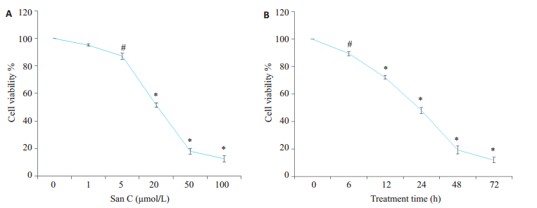
2 结果 2.1 桑根酮C对PC3细胞生长的抑制作用阴性对照组PC3细胞生长活跃,经1、5、20、50、100 μmol/L桑根酮C处理24 h后,PC3细胞呈不同程度的生长抑制,抑制率为(4.86 ± 0.88)%、(12.92 ± 2.13)%、(52.34 ± 1.88)%、(82.05 ± 2.08)%、(87.45 ± 2.16)%,各组间比较除对照组和1 μmol/L组及50 μmol/L与100 μmol/L组之间P>0.05外,其他组间比较差异均有统计学意义,半数有效抑制浓度为:18.76±1.53 μmol/L。20 μmol/L桑根酮C作用PC3细胞6、12、24、48、72 h后,抑制率分别为:(10.57 ± 1.54)%、(27.09 ± 1.35)%、(51.88±2.21)%、(80.73±2.96)%、(87.99±2.14)%,组间比较除48 h与72 h组间没有明显差异外,其他均有统计学意义。结果表明随着剂量的增加、时间的延长,抑制率明显增加(图 1A,B)。

|
图 1 桑根酮C对PC3细胞增殖抑制作用 Figure 1 Inhibitory effect of Sanggenon C on the proliferation of PC3 cells detected by MTT assay. A: PC-3 cells treated with indicated concentrations of Sanggenon C for 24 h; B: PC-3 cells treated with 20 μmol/L Sanggenon C for up to 72 h. #P < 0.05, *P < 0.01 vs control group. |
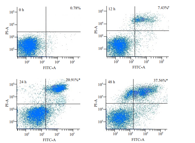
桑根酮C 20 μmol/L作用PC3细胞12、24、48 h后,流式细胞仪检测细胞凋亡率分别为:(7.43±0.97)%、(20.91±2.34)%、(37.56±1.03)%,与阴性对照组(0.78± 0.23)%间比较差异有统计学意义(P<0.05),各组间比较差异有统计学意义(P<0.05,图 2)。

|
图 2 20 μmol/L桑根酮C在不同时间点诱导PC3细胞凋亡作用 Figure 2 Apoptotic rates of PC-3 cells treated with 20 μmol/L Sanggenon C for up to 48 h measured with flow cytometry. #P < 0.05, *P < 0.01 vs control group. |
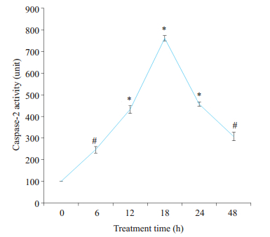
桑根酮C 20 μmol/L作用PC3细胞后,分别在6、12、18、24 h检测活性,发现随着时间的延长caspase 3的活性先增加,在18 h达高峰后逐渐下降(图 3)。

|
图 3 桑根酮C对PC3细胞caspases 3活性的影响 Figure 3 Effect of Sanggenon C on caspases 3 activity in PC3 cells. #P < 0.05, *P < 0.01 vs control group. |
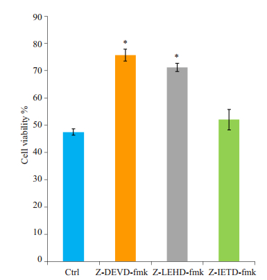
Caspase 3抑制剂z-DEVD-fmk及Caspase 9抑制剂z-LEHD-fmk,能有效逆转桑根酮C对PC3细胞的增殖抑制作用,细胞生长抑制率由(52.48±1.17)%分别降至(24.29±2.24)%及(28.81±1.52)%(P<0.05);Caspase 8抑制剂z-IETD-fmk的几乎没有作用,细胞生长抑制率由52.48%降至(47.95±3.73)%(P>0.05)。
3 讨论前列腺癌是西方国家男性发病率最高、死亡率居第2位的实体肿瘤[16]。我国近年来前列腺癌的发病率呈“井喷式”增长,在上海地区发病率甚至排在男性泌尿系统恶性肿瘤的第1位[2]。中晚期前列腺癌多数发展成为雄激素非依赖型前列腺癌,主要以内分泌治疗为主,当前常规使用的抗癌药物不仅治疗效果不佳而且毒副作用大[17-18]。桑白皮是一种可食用的中草药,几乎无毒副作用,临床中药方剂中常用来治疗前列腺癌[5]。桑白皮可通过线粒体或死亡受体途径,减少抗凋亡蛋白Survivin的表达,上调促凋亡蛋白Bax的表达来诱导人白血病细胞HL60及人乳腺癌细胞MCF-7及MDA-MB-231的凋亡[19-20]。但目前还没有研究探讨其抗前列腺癌的机制。

|
图 4 Caspase抑制剂对桑根酮C抑制PC3细胞增殖的影响 Figure 4 Effects of the caspase inhibitors on Sanggenon C-induced inhibition of PC3 proliferation. *P < 0.01 vs control. |
桑根酮C是桑白皮中最有活性的黄酮类化合物,本实验室既往研究发现其可抑制蛋白酶体的活性引起小鼠肝癌细胞H22、小鼠白血病细胞P388及人慢性髓系白血病K562细胞的死亡,以坏死为主,也有一部分凋亡[21]。本研究发现桑根酮C可抑制雄激素非依赖型前列腺癌PC3细胞的增殖活性,呈时间、剂量依赖性,作用24 h后半数有效抑制浓度IC50为18.76 μmol/L;同时发现桑根酮C可引起PC3细胞的死亡,以凋亡为主,考虑其引起PC3细胞死亡的机制可能与之前不同。凋亡经典途径包括内源性线粒体细胞色素释放途径和外源性死亡受体途径,现有研究表明这两种通路并非独立,而是相互关联交错的,而这两种途径最终都将激活凋亡终末效应酶caspase 3[22-25]。本实验发现20 μmol/L桑根酮C作用PC3细胞12 h后细胞开始凋亡(凋亡率为7.43%,以早期凋亡为主),同时检测caspase 3的活性较对照组增加了4.3倍,且随着时间推移,caspase 3的活性逐渐增强,在18 h时达高峰增加了7.6倍,随后在24 h后活性逐渐减弱,考虑为18 h后细胞在桑根酮C的诱导下逐渐转入凋亡晚期或消亡期所致(与本实验凋亡检测结果相符)。为探讨其凋亡的机制,应用caspase抑制剂发现caspase 9及caspase 3抑制剂可明显逆转桑根酮C对PC3细胞的生长抑制作用;而caspase 8抑制剂几乎没有作用。其中caspase 8是外源性凋亡途径的执行者,caspase 9是内源性凋亡途径的执行者[26-29]。
综上所述,桑根酮C能有效抑制PC3细胞的增殖,并诱导其凋亡;这无疑显示了它在中晚期前列腺癌治疗中的潜在应用价值,目前只初步探讨了其诱导凋亡的信号转导通路是以caspase 9激活的线粒体通路为主,其他具体的分子机制以及药物作用的靶点有待深入探讨。
| [1] | Adeola HA, Blackburn JM, Rebbeck TR, et al. Emerging pro teomics biomarkers and prostate cancer burden in Africa[J]. Oncotarget, 2017, 8(23): 37991-8007. |
| [2] | Cheng HH, Lin DW, Yu EY. Advanced clinical states in prostate cancer[J]. Urol Clin North Am, 2012, 39(4): 561. DOI: 10.1016/j.ucl.2012.07.011. |
| [3] | 徐亚文, 郑少波, 陈玢屾, 等. 苯丁酸钠对多烯紫杉醇耐药前列腺癌细胞株的增值抑制和凋亡诱导作用[J]. 南方医科大学学报, 2017, 37(1): 130-4. |
| [4] | Kwon DH, Cheon JM, Choi EO, et al. The immunomodulatory activity of Mori folium, the leaf of morus alba L, in RAW 264.7 macrophages in vitro[J]. J Cancer Prev, 2016, 21(3): 144-51. DOI: 10.15430/JCP.2016.21.3.144. |
| [5] | 高学敏. 中药学[M]. 北京: 中国中医药出版社, 2007: 23-8. |
| [6] | Kuk EB, Jo AR, Oh SI, et al. Anti-Alzheimer's disease activity of compounds from the root bark of morus alba L[J]. Arch Pharm Res, 2017, 40(3): 338-49. DOI: 10.1007/s12272-017-0891-4. |
| [7] | Ann JY, Eo H, Lim Y. Mulberry leaves (Morus alba L.) ameliorate obesity-induced hepatic lipogenesis, fibrosis, and oxidative stress in high-fat diet-fed mice[J]. Genes Nutr, 2015, 10(6): 46. DOI: 10.1007/s12263-015-0495-x. |
| [8] | Guo H, Xu Y, Huang W, et al. Kuwanon G preserves LPS-Induced disruption of gut epithelial barrier in vitro[J]. Molecules, 2016, 21(11): 1597-611. DOI: 10.3390/molecules21111597. |
| [9] | Qin J, Fan M, He J, et al. New cytotoxic and anti-inflammatory compounds isolate-ed from Morus alba L[J]. Nat Prod Res, 2015, 29(18): 1711-8. DOI: 10.1080/14786419.2014.999333. |
| [10] | Nguyen TD, Binh Xuan Phung Thi, Le Thi Phuong Quynh, et al. Cytotoxic prenylated flavonoids from Morus alba[J]. Fitoterapia, 2010, 81(8): 1224-7. DOI: 10.1016/j.fitote.2010.08.006. |
| [11] | Eo HJ, Park JH, Park GH, et al. Anti-inflammatory and anti-cancer activity of mulberry (Morus alba L.) root bark[J]. BMC Complement Altern Med, 2014, 14(1): 200. DOI: 10.1186/1472-6882-14-200. |
| [12] | Kim SY, Gao JJ, Kang HK. Two flavonoids from the leaves of Morus alba induce differentiation of the human promyelocytic leukemia (HL-60) cell line[J]. Biol Pharm Bull, 2000, 23(4): 451-5. DOI: 10.1248/bpb.23.451. |
| [13] | Kondylis V, Kumari S, Vlantis K, et al. The interplay of IKK, NF-κB and RIPK1 signaling in the regulation of cell death, tissue homeostasis and inflammation[J]. Immunol Rev, 2017, 277(1): 113-27. DOI: 10.1111/imr.2017.277.issue-1. |
| [14] | Li LC, Shen F, Hou Q, et al. Inhibitory effect and mechanism of action of sanggenon C on human polymorphonuclear leukocyte adhesion to human synovial cells[J]. Acta Pharmacol Sin, 2002, 23(2): 138-42. |
| [15] | Dat NT, Binh PT, Quynh le TP, et al. Sanggenon C and O inhibit NO production, iNOS expression and NF-κB activation in LPS induced RAW264.7 cells[J]. Immunopharmacol Immunotoxicol, 2012, 34(1): 84-8. DOI: 10.3109/08923973.2011.580755. |
| [16] | Siegel RL, Miller KD, Jemal A. Cancer statistics, 2017[J]. CA Cancer J Clin, 2017, 67(1): 7-30. DOI: 10.3322/caac.21387. |
| [17] | Rivera M, Ramos Y, Rodríguez-Valentín M, et al. Targeting multiple pro-apoptotic signaling pathways with curcumin in prostate cancer cells[J]. PLoS One, 2017, 12(6): e0179587. |
| [18] | Deng X, Zhou P, Wei X, et al. Plasma proteasomal chymotrypsin like activity correlates with prostate cancer progression[J]. Tumour Biol, 2015, 36(6): 4115-21. DOI: 10.1007/s13277-015-3045-7. |
| [19] | Kikuchi T, Nihei M, Nagai H, et al. Albanol a from the root bark of morus alba L. induces apoptotic cell death in HL60 human leukemia cell line[J]. Chem Pharm Bull (Tokyo), 2010, 58(4): 568-71. DOI: 10.1248/cpb.58.568. |
| [20] | Kang S, Kim EO, Kim SH, et al. Morusin induces apoptosis by regulating expression of Bax and Survivin in human breast cancer cells[J]. Oncol Lett, 2017, 13(6): 4558-62. |
| [21] | Huang H, Liu N, Zhao K, et al. Sanggenon C decreases tumor cell viability associated with proteasome inhibition[J]. Front Biosci (Elite Ed), 2011, 3(3): 1315-25. |
| [22] | Duclos C, Lavoie C, Denault JB. Caspases rule the intracellular trafficking cartel[J]. FEBS J, 2017, 284(10): 1394-420. DOI: 10.1111/febs.14071. |
| [23] | Qiu M, Chen L, Tan G, et al. JS-K promotes apoptosis by inducing ROS production in human prostate cancer cells[J]. Oncol Lett, 2017, 13(3): 1137-42. |
| [24] | Kim YS, Kim SK, Park SJ. Apoptotic effect of demetho xyfumitremorgin C from Marine fungus Aspergillus fumigatus on PC3 human prostate cancer cells[J]. Chem Biol Interact, 2017, 269(269): 18-24. |
| [25] | Lee JC, Shin EA, Kim B, et al. Auraptene induces apoptosis via myeloid cell leukemia 1-Mediated activation of caspases in PC3 and DU145 prostate cancer cells[J]. Phytother Res, 2017, 31(6): 891-8. DOI: 10.1002/ptr.5810. |
| [26] | Derakhshan A, Chen Z, Van Waes C. Therapeutic small molecules target inhibitor of apoptosis proteins in cancers with deregulation of extrinsic and intrinsic cell death pathways[J]. Clin Cancer Res, 2017, 23(6): 1379-87. DOI: 10.1158/1078-0432.CCR-16-2172. |
| [27] | Yuan J, Najafov A, Py BF. Roles of caspases in necrotic cell death[J]. Cell, 2016, 167(7): 1693-704. DOI: 10.1016/j.cell.2016.11.047. |
| [28] | Cao YM, Jiang Z, Zeng Z, et al. Bcl-2 silencing attenuates hypoxia-induced apoptosis resistance in pulmonary microvascular endothelial cells[J]. Apoptosis, 2016, 21(1): 69-84. DOI: 10.1007/s10495-015-1184-3. |
| [29] | Patmanathan SN, Wang W, Yap LF, et al. Mechanisms of sphingosine 1-phosphate receptor signalling in cancer[J]. Cell Signal, 2017, 34(34): 66-75. |
 2017, Vol. 37

