Hemangioma of the small intestine is a rare disease among gastrointestinal tumors, accounting for 5%-10% of all small intestinal benign tumors[1]. Based on the histological features of the vascular endothelium, small intestinal hemangiomas are categorized into capillary, cavernous and mixed hemangiomas, among which cavernous hemangioma is the most common[1]. The most frequent manifestation of small intestinal hemangioma is gastrointestinal bleeding. Bleeding caused by a capillary hemangioma can be occult, but cavernous hemangioma often causes sudden hematemesis or melena, and obscure gastrointestinal bleeding due to cavernous hemangioma is unusual[2, 3]. In the report, we describe a case of small intestinal cavernous hemangioma that caused occult gastrointestinal bleeding and review the documentation of this condition in literature.
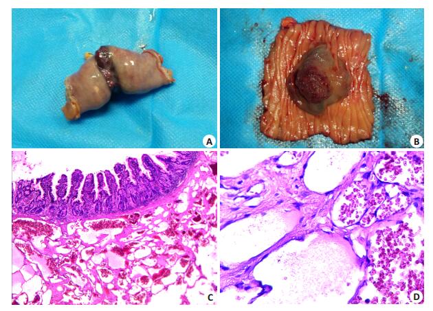
CASE REPORTA 44-year-old man was hospitalized for the complaint of weakness and dizziness for 2 years, which aggravated in the recent month before admission accompanied by intermittent melena. He denied a history of familial diseases or tuberculosis. Physical examination showed normal pallor and bowel sounds without intestinal patterns or abdominal masses. Laboratory tests revealed leukopenia (WBC 2.81 × 10⁹/L), microcytic and hypochromic anemia (HGB 41 g/L, MCV 64.5 × 10⁻⁶ L and MCHC 262 g/L), decreased ferritin (2.70 × 10⁻⁶ g/L), increased folate ( > 20.00 × 10⁻⁶ g/L) and normal tumor markers. Bone marrow puncture showed iron-deficiency anemia (IDA). Computed tomography of the abdomen demonstrated signs of anemia, and subsequent upper gastrointestinal endoscopy and colonoscopy detected ischemic mucosa but without active bleeding lesion. Capsule endoscopy was performed on the following day, which detected bleeding lesions and a prominent neoplasm in the jejunum. Hemostatic measures and blood transfusion failed to improve the symptoms of the patient. For a definite diagnosis and correcting anemia, the patient underwent an exploratory laparotomy with segmental resection of the jejunum. During the operation, a circular, irregular and polypoid neoplasm 100 cm distal to the ligament of Treitz was found (Fig. 1 A and B). Pathological examination identified the neoplasm as a cavernous hemangioma of the small intestine with a pinkish-grey and soft section composed of congestive vascularized structures and flimsy vascular veins (Fig. 1C and D). The patient recovered uneventfully after the operation and experienced no further symptoms during the follow-up for 11 months.

|
Figure 1 Intraoperative findings of the cavernous hemangioma in the small intestine and pathological examination of the lesion. A: Intraoperative examination of the resected intestinal segment revealed a fuchsia, circular and irregular vascularized neoplasm in the jejunum; B: A polypoid neoplasm was found after surgical resection of the small intestine; C: Pathological examination of the resected specimen revealed polypoid cavernous hemangioma with a pinkish-grey and soft section (HE staining, original magnification: × 20); D: Pathological examination revealed congestive vascular structures with flimsy vascular veins and fewer interstitial tissues (HE staining, ×40). |
Small intestinal hemangioma is a rare and congenital vascular hamartomatous tumor[4, 5] with single or multiple lesions involving most commonly the jejunum and rarely the duodenum[4, 6]. The tumor sizes range from a few millimeters to several centimeters (in the case of giant tumors). In terms of gross features, the tumors are nodular, polypus or diffusely infiltrative in shape. The tumors are often confined to the submucosa, resulting from the submucosal vascular plexuses. Subserosal vasculature is rarely observed. In addition, these tumors can also invade the muscular layer and affect the mesentery or retroperitoneum [7]. Small intestinal hemangiomas have been reported in patients almost at all ages, ranging from 2 months to 79 years old[6], but commonly in young patients around 30 years of age without obvious sexual differences [4].
We searched the PubMed database and retrieved 39 cases of small intestinal cavernous hemangioma accompanied by gastrointestinal hemorrhage reported since 1990. Of these cases, only 25.6% were diagnosed preoperatively. It is difficult for clinicians to detect specific symptoms or obvious lesions in these patients before surgery. In addition to the common manifestation of gastrointestinal bleeding, other rare manifestations of this condition include intestinal obstruction, intussusception, perforation, intramural hematoma or platelet sequestration [7]. Therefore, hemangiomas need to be distinguished from gastrointestinal erosions and ulcers, colorectal mucosal lesions, intestinal tuberculosis, inflammatory bowel diseases and nosohemia. In cases of obscure gastrointestinal bleeding, such examinations are essential as computed tomography, gastroscopy, colonoscopy or bone marrow puncture, and if necessary, capsule endoscopy, double-balloon enteroscopy and multidetector computed tomography can be considered [8]. When a definitive diagnosis can not be reached after all these examinations, a laparoscopic exploration or exploratory laparotomy should be performed immediately. In our case, when computed tomography, gastroscopy and colonoscopy demonstrated signs of anemia and capsule endoscopy revealed small intestinal erosions, bleeding lesions and prominent neoplasms, an exploratory laparotomy was performed immediately, and the cavernous hemangioma was confirmed by intraoperative frozen biopsy and postoperative pathological examination. Pathological examination still remains the gold standard for diagnosis of this disease.
Surgery is the optimal choice of treatment for a hemangioma of the small intestine. Segmental resection is appropriate for solitary small intestinal hemangioma, and for multiple lesions, careful detection should be performed during the operation. For patients with other comorbidities who can not tolerate surgery, local treatments such as electrocoagulation and laser or sclerotherapy at endoscopy can be good alternatives. Nevertheless, the recurrence rate of hemangioma is high after these surgical interventions [9]. According to Kirkham et al, thalidomide could be effective in the treatment of a bleeding hemangioma[10]. In the study, the patient underwent segmental resection of the jejunum and recovered smoothly after the operation.
This patient had a cavernous hemangioma of the small intestine with occult bleeding and severe anemia (HGB < 60 g/L) for a long time at the age of 44 years. This case demonstrated that a small intestine neoplasm may cause chronic bleeding, and alleviation of the discomforts would provide benefits to the patient. As cavernous hemangioma of the small intestine is a rare condition without specific symptoms, its early diagnosis can be difficult before operation. The diagnostic examinations and treatments of this disease need to be further explored.
| [1] | Kuo LW, Chuang HW, Chen YC. Small bowel cavernous hemangioma complicated with intussusception: report of an extremely rare case and review of literature[J]. Indian J Surg, 2015, 77(Suppl 1): 123-4. |
| [2] | Ersoy O, Akin E, Demirezer A, et al. Cavernous haemangioma of small intestine mimicking gastrointestinal stromal tumour[J]. Arab J Gastroenterol, 2013, 14(3): 139-40. DOI: 10.1016/j.ajg.2013.08.008. |
| [3] | Fernandes D, Dionisio I, Neves S, et al. Cavernous hemangioma of small bowel: a rare cause of digestive hemorrhage[J]. Rev Esp Enferm Dig, 2014, 106(3): 214-15. |
| [4] | Ruiz AR, Ginsberg AL. Giant mesenteric hemangioma with small intestinal involvement: an unusual cause of recurrent gastrointestinal bleed and review of gastrointestinal hemangiomas[J]. Dig Dis Sci, 1999, 44(12): 2545-51. DOI: 10.1023/A:1026659710815. |
| [5] | Huber A, Abdel Samie A, Kychenko D, et al. A rare cause of recurrent iron-deficiency anemia: cavernous hemangioma of the small intestine[J]. J Gastrointestin Liver Dis, 2012, 21(4): 343. |
| [6] | Garvin PJ, Herrmann V, Kaminski DL, et al. Benign and malignant tumors of the small intestine[J]. Curr Probl Cancer, 1979, 3(9): 1-46. DOI: 10.1016/S0147-0272(79)80037-9. |
| [7] | Pera M, Marquez L, Dedeu JM, et al. Solitary cavernous hemangioma of the small intestine as the cause of long-standing iron deficiency anemia[J]. J Gastrointest Surg, 2012, 16(12): 2288-90. DOI: 10.1007/s11605-012-1991-6. |
| [8] | Akazawa Y, Hiramatsu K, Nosaka T, et al. Preoperative diagnosis of cavernous hemangioma presenting with melena using wireless capsule endoscopy of the small intestine[J]. Endosc Int Open, 2016, 4(3): 249-51. DOI: 10.1055/s-00025476. |
| [9] | Chaves DM, Sakai P, Oliveira CV, et al. Watermelon stomach: clinical aspects and treatment with argon plasma coagulation[J]. Arquivos De Gastroenterologia, 2006, 43(3): 191-5. DOI: 10.1590/S0004-28032006000300007. |
| [10] | Kirkham SE, Lindley KJ, Elawad MA, et al. Treatment of multiple small bowel angiodysplasias causing severe life-threatening bleeding with thalidomide[J]. J Pediatr Gastroenterol Nutr, 2006, 42(5): 585-7. DOI: 10.1097/01.mpg.0000215308.86287.98. |
 2017, Vol. 37
