卵圆孔未闭(PFO)是一种先天性房间隔发育缺损,约占正常人群的25% [1-2]。以往认为PFO是一种解剖学变异很少导致严重后果。近年来,越来越多的研究表明PFO与隐源性脑卒中[3-5]、短暂性脑缺血发作[6]、减压病[7-9]、偏头痛[10-11]等疾病的发生存在密切相关性。PFO引起疾病的主要原因是左右心系统之间异常的血液循环途径即心脏右向左分流(RLS)。目前认为PFO是引起RLS最常见的病因,约占所有RLS的95%。常用的诊断PFO的方法包括经食道超声心动图、经胸超声心动图及经颅多普勒超声(c-TCD)。其中经食道超声心动图因与尸检结果一致性较高,被认为是诊断PFO的金标准[12-13]。但因其费用较高,有创性及完成标准的Valsalva动作(VM)困难等特点,临床工作中经食道超声心动图检查并未得到广泛应用。研究证实,c-TCD具有价格低廉、安全性高、无创性等优点,并且有较高的灵敏度及特异性,被认为是诊断PFO的替代方法[14-15]。但是关于c-TCD检查过程中使用的造影剂(CA)目前尚未完全统一,主要包括六氟化硫微泡、半乳糖空气微泡、生理盐水-气体混合液(AS)和生理盐水-气体-血混合液(ASb)。前两种CA因价格高、稳定性差等问题现已较少使用。目前临床上使用最多的就是AS与ASb,但是两者诊断PFO的优劣性尚无明确定论。本文目的即比较AS和ASb这两种造影剂在诊断PFO阳性率、分流程度及第1个微泡出现时间的差异。
1 资料和方法 1.1 研究对象选取2015年11月~2016年1月于西安交通大学第二附属医院神经内科TCD室行c-TCD检查的门诊及住院患者248例。所有患者签署知情同意书后,通过掷硬币法决定AS或ASb这两种造影剂使用的先后顺序。本研究除外已知肺动静脉分流、颞窗缺乏、严重凝血功能障碍、严重心肺功能障碍、严重认知障碍或其它疾病导致配合不佳或影响VM完成的患者。该研究获得西安交通大学第二附属医院伦理委员会批准。
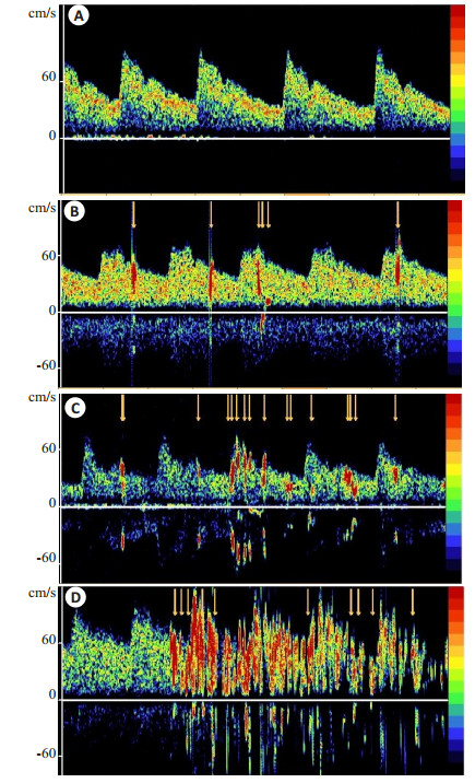
1.2 研究方法本试验采用美国Natus的SONARA TEK型c-TCD检测仪。检查时患者取仰卧位姿势,使用频率为2 MHz的探头,以50~55 mm的深度监测右侧大脑中动脉血流速度,通过仪器屏幕可以观察血流速度变化曲线。当大脑中动脉血流速度一过性下降≥25%时,确定该VM有效。检查前嘱患者反复练习VM,待患者熟练掌握后,经肘前静脉留置1枚18号针头,连接三通管,三通管一侧安置装有9 mL生理盐水的10 mL注射器或装有9 mL生理盐水的注射器回抽1滴患者的血液,另一侧安置装有1 mL空气的10 mL注射器。将2个注射器反复推注10次后制成CA。首先在不伴VM时,将配制好的CA快速注入,同时开始计时并观察TCD频谱,记录25 s内微泡出现数目及第1个微泡出现的时间。然后在伴VM时,将制好的CA快速注入,开始计时同时嘱患者快速深吸气至无法继续吸入后紧闭声门,10 s快速呼气,观察TCD频谱,并记录CA注射后25 s内微泡出现数目及第1个微泡出现的时间。所有患者通过掷硬币法决定AS或ASb这两种造影剂使用的先后顺序。具体操作过程如下:(1)9 mL生理盐水混合1 mL空气不伴VM(AS不伴VM);(2)9 mL生理盐水混合1 mL空气伴VM(AS伴VM);(3)9 mL生理盐水、1滴患者回抽血液混合1 mL空气不伴VM(ASb不伴VM);(4)9 mL生理盐水、1滴患者回抽血液混合1 mL空气伴VM(ASb伴VM)。上述每种方法重复2次且每个过程间隔至少5 min。目前诊断微泡栓子信号的标准如下:(1)短时程,时程< 300 ms;(2)高信号,微泡栓子信号强度比背景信号强度≥3 dB;(3)以单方向出现在频谱中;(4)伴有尖锐的哨鸣声。本研究将符合上述标准且微泡数≥1者诊断为PFO,并由2名神经内科医师根据上述标准记录微泡数目及第1个微泡出现的时间。当两个人记录的微泡数目不一致时取两者的平均值。根据微泡数量对PFO分流程度分级:阴性(0个微泡信号);Ⅰ级(1~10个微泡信号);Ⅱ级(> 10个微泡信号,但未形成雨帘状)和Ⅲ级(形成雨帘状)(图 1)。

|
图 1 c-TCD诊断的PFO分流程度 Figure 1 Grading of PFO by contrast-transcranial Doppler findings. A: Negative (no microbubble); B: GradeⅠ(1 to 10 microbubbles); C: Grade Ⅱ(more than 10 microbubbles but not curtain; D: Grade Ⅲ(curtain). |
使用SPSS 17.0统计软件,连续性变量采用均数±标准差,分类变量采用百分比。AS不伴VM组与AS伴VM组、ASb不伴VM组与ASb伴VM组、ASb不伴VM组与AS不伴VM组及ASb伴VM组与AS伴VM组之间PFO检出率的比较用配对计数资料McNemar检验法。4种方法间的分级比较采用非参数的KruskalWallis方法。第1个微泡出现时间比较符合正态分布和方差齐性采用方差分析,否则采用Kruskal-Wallis方法。P < 0.05为差异有统计学意义。
2 结果 2.1 一般资料研究共纳入248例患者,男性101例(40.7%),女性147例(59.3%),年龄39.71±14.09岁。纳入研究的患者中,118例患者表现为头痛,46例患者表现为头晕,26例患者表现为晕厥,51例患者表现为隐源性脑卒中,7例患者表现为短暂性脑缺血发作。所有患者在检查过程中及检查后12 h内均无不良事件发生。
2.2 阳性率AS不伴VM组、AS伴VM组、ASb不伴VM组和ASb伴VM组检查的阳性率分别是10.9%、23.8%、12.1%、25.8%。伴VM组较不伴VM组阳性率明显增高(P < 0.05),而Asb与AS这2种不同造影剂组在诊断PFO阳性率方面无差异(P > 0.05,表 1)。
| 表 1 4组检查的阳性率 Table 1 Results of positive rates in the 4 groups |
Kruskal-Wallis检验表明4组检查间分流程度差异有统计学意义(P < 0.05)。进一步两两比较发现伴VM组与不伴VM组PFO分流程度存在差异(P < 0.05),而Asb与AS这2种不同造影剂组在诊断PFO分流程度方面无差异(P > 0.05,表 2)。
| 表 2 4组检查的分流程度 Table 2 Results of grading in the 4 groups |
AS不伴VM组、AS伴VM组、ASb不伴VM组和ASb伴VM组检查中第1个微泡出现的时间分别是8.85±3.06、8.17±3.44、8.83±2.67、8.23±3.40 s。4组检查间第1个微泡出现的时间无统计学差异(P < 0.05)。
3 讨论VM是在c-TCD检查过程中的常用方法,研究表明c-TCD同时配合标准VM时,其诊断PFO的准确性可超过经食道超声心动图[16]。在检查过程中一般将大脑中动脉血液流速峰值下降达25%以上认为是标准的VM [17]。一般情况下,左心房压力大于右心房压力,但是在VM、咳嗽等动作时胸腔压力增加,可引起一过性右心房压力大于左心房压力,如果存在PFO,就会出现RLS [18]。前面已经提到RLS与隐源性脑卒中、短暂性脑缺血发作、减压病、偏头痛等疾病存在密切相关性。关于RLS引起疾病发生的可能机制包括:(1)反常栓塞,来自右心的微栓子未经过肺的过滤,通过未关闭的卵圆孔直接进入体循环,形成反常栓塞[19];(2)静脉系统内的血管活性物质未经过肺脏过滤,直接进入脑循环[20]。本研究发现在c-TCD检查过程中AS伴VM组较AS不伴VM组、ASb伴VM组较ASb不伴VM组PFO的阳性诊断率显著增多,而AS不伴VM组与ASb不伴VM组、AS伴VM组与ASb伴VM组在诊断PFO的阳性率方面差异无统计学意义。PFO的分流程度在伴与不伴VM组之间存在统计学差异,而在AS与ASb这2种不同造影剂组之间差异无显著性。第1个微泡信号出现的时间无论是在伴与不伴VM之间还是两种不同造影剂之间差异均无统计学意义。因此,本研究认为在c-TCD检查中VM能显著提高PFO的阳性诊断率,而ASb与AS相比并无明显差异。
在c-TCD检查中常用的造影剂包括六氟化硫微泡、半乳糖空气微泡、AS和ASb。虽然研究表明AS与半乳糖空气微泡诊断PFO阳性率的灵敏性相当[21-22],但是由于其价格较高、临床上不易获得,目前临床上较少使用。2000年国际会议共识推荐AS作为c-TCD检查PFO的造影剂[15]。随着c-TCD检查技术的发展,人们在提高诊断PFO阳性率方面进行着不断的探索。近年来,许多研究探索AS与ASb这2种不同造影剂在诊断PFO方面的差异[23-27]。其中大部分研究表明,与AS相比,ASb能够明显提高PFO的阳性诊断率且能够增加微泡数目[23-24, 26-27]。且认为ASb是诊断PFO较敏感的方法,对于不能完成VM或VM配合不佳的患者,使用ASb能够提高诊断阳性率[24, 27]。有研究认为ASb能够提高PFO阳性率的原因可能是血液能够乳化微泡并且增加微泡在血液中的悬浮时间[24]。而我们的结果与Lange等[25]结果一致,认为ASb与AS相比在诊断PFO阳性率方面无统计学差异。目前关于PFO的分流程度与疾病发生之间的关系尚未达成一致共识。大部分研究表明分流程度越严重,发生疾病的可能性越大[5, 28],但是也有研究表明两者之间并不存在相关性[29]。有研究认为ASb与AS相比在诊断PFO分流程度方面同样存在统计学差异[24]。然而,我们的研究结果与Hao等[24]的结果一致,并未发现两种造影剂在分流程度方面的差异。关于ASb与AS这两种不同造影剂在第1个微泡出现时间方面的研究,有研究认为这两种造影剂第一个微泡出现时间无明显差异[25]。有研究则认为AS伴VM组与不伴VM组对比,第1个微泡出现时间明显缩短,而ASb伴VM组和AS或ASb不伴VM组比较,第1个微泡出现的时间无统计学意义[27]。我们认为无论是在伴与不伴VM之间还是在两种不同造影剂之间第一个微泡信号出现的时间均无统计学意义。
关于c-TCD检查是否会对机体造成不良影响目前一直存在争论。理论上讲,在微泡制造过程中,通过快速混合及弹丸式快速注入,避免了大的微泡及血凝块形成,发生不良事件的可能性较小。并且目前大部分研究表明在c-TCD检查过程中使用AS与ASb这两种造影剂均无不良反应发生[23-24]。而有相关研究报道5例患者在c-TCD检查后数分钟内出现缺血性脑血管事件[30],但两者之间的因果关系尚未确定。
本研究存在一定的局限性:目前认为经食道超声心动图是诊断PFO的金标准,我们的试验并未使用经食道超声心动图进一步检查。因为本研究的目的并不是研究PFO的具体解剖信息,并且,c-TCD以其高度的敏感性及特异性可以作为经食道超声心动图的替代检查方法;在本试验中6名患者因缺乏颞窗、5名患者因不能完成标准的VM被排除,可能会影响结果的真实性;本研究结果来自我们单试验中心,该结论尚需更多大样本的、多中心研究加以验证。
总上所述,在c-TCD检查过程中VM能够明显提高PFO的阳性率,而ASb与AS这两种造影剂在诊断PFO阳性率方面无明显差异,在分流程度及第1个微泡出现时间方面亦无差异。
| [1] |
Hara H, Virmani R, Ladich E, et al. Patent foramen ovale: current pathology, pathophysiology, and clinical status[J].
J Am Coll Cardiol,2005, 46 (9) : 1768-76.
DOI: 10.1016/j.jacc.2005.08.038. ( 0) 0)
|
| [2] |
Rigatelli G. Patent foramen ovale: the evident paradox between the apparently simple treatment and the really complex pathophysiology[J].
J Cardiovasc Med (Hagerstown),2007, 8 (4) : 300-4.
DOI: 10.2459/01.JCM.0000263497.99157.77. ( 0) 0)
|
| [3] |
Pandit A, Aryal MR, Pandit AA, et al. Amplatzer PFO occluder device May prevent recurrent stroke in patients with patent foramen ovate and cryptogenic stroke: a Meta-Analysis of randomised trials[J].
Heart Lung and Circulation,2014, 23 (4) : 303-8.
DOI: 10.1016/j.hlc.2013.12.003. ( 0) 0)
|
| [4] |
Rigatelli G, Rigatelli A. Closing patent foramen ovale in cryptogenic stroke:The underscored importance of other interatrial shunt variants[J].
World J Cardiol,2015, 7 (6) : 326-30.
DOI: 10.4330/wjc.v7.i6.326. ( 0) 0)
|
| [5] |
Schuchlenz HW, Weihs W, Horner S, et al. The association between the diameter of a patent foramen ovale and the risk of embolic cerebrovascular events[J].
Am J Med,2000, 109 (6) : 456-62.
DOI: 10.1016/S0002-9343(00)00530-1. ( 0) 0)
|
| [6] |
Wozniak L, Mielczarek M, Sabiniewicz R. Paradoxical brain embolism in a young man: is it only a patent foramen ovale[J].
Neurol Neurochir Pol,2015, 49 (1) : 61-4.
( 0) 0)
|
| [7] |
Liou K, Wolfers D, Turner R, et al. Patent foramen ovale influences the presentation of decompression illness in SCUBA divers[J].
Heart Lung Circ,2015, 24 (1) : 26-31.
DOI: 10.1016/j.hlc.2014.07.057. ( 0) 0)
|
| [8] |
Wilmshurst PT. The role of persistent foramen ovale and other shunts in decompression illness[J].
Diving Hyperb Med,2015, 45 (2) : 98-104.
( 0) 0)
|
| [9] |
Wilmshurst PT, Morrison WL, Walsh KP, et al. Comparison of the size of persistent foramen ovale and atrial septal defects in divers with shunt-related decompression illness and in the general population[J].
Diving Hyperb Med,2015, 45 (2) : 89-93.
( 0) 0)
|
| [10] |
Butera G, Biondi-Zoccai GG, Carminati M, et al. Systematic review and meta-analysis of currently available clinical evidence on migraine and patent foramen ovale percutaneous closure: much ado about nothing[J].
Catheter Cardiovasc Interv,2010, 75 (4) : 494-504.
( 0) 0)
|
| [11] |
Trabattoni D, Fabbiocchi F, Montorsi PA, et al. Sustained LongTerm benefit of patent foramen ova le closure on migraine[J].
Catheter Cardiovasc Interv,2011, 77 (4) : 570-4.
DOI: 10.1002/ccd.v77.4. ( 0) 0)
|
| [12] |
Caputi L, Carriero MR, Falcone C, et al. Transcranial doppler and transesophageal echocardiography: comparison of both techniques and prospective clinical relevance of transcranial doppler in patent foramen ovale detection[J].
J Stroke Cerebrovasc Dis,2009, 18 (5) : 343-8.
DOI: 10.1016/j.jstrokecerebrovasdis.2008.12.001. ( 0) 0)
|
| [13] |
Schneider B, Zienkiewicz T, Jansen V, et al. Diagnosis of patent foramen ovale by transesophageal echocardiography and correlation with autopsy findings[J].
Am J Cardiol,1996, 77 (14) : 1202-9.
DOI: 10.1016/S0002-9149(96)00163-4. ( 0) 0)
|
| [14] |
Droste DW, Schmidt-Rimpler C, Wichter T, et al. Right-toleft-shunts detected by transesophageal echocardiography and transcranial Doppler sonography[J].
Cerebrovasc Dis,2004, 17 (2/3) : 191-6.
( 0) 0)
|
| [15] |
Jauss M, Zanette E. Detection of right-to-left shunt with ultrasound contrast agent and transcranial Doppler sonography[J].
Cerebrovasc Dis,2000, 10 (6) : 490-6.
DOI: 10.1159/000016119. ( 0) 0)
|
| [16] |
Van HH, Poommipanit P, Shalaby M, et al. Sensitivity of transcranial doppler versus intracardiac echocardiography in the detection of right-to-left shunt[J].
JACC Cardiovasc Imaging,2010, 3 (4) : 343-8.
DOI: 10.1016/j.jcmg.2009.12.012. ( 0) 0)
|
| [17] |
González-Alujas T, Evangelista A, Santamarina E, et al. Diagnosis and quantification of patent foramen ovale. Which is the reference technique? Simultaneous study with transcranial Doppler, transthoracic and transesophageal echocardiography[J].
Rev Esp Cardiol,2011, 64 (2) : 133-9.
DOI: 10.1016/j.recesp.2010.10.009. ( 0) 0)
|
| [18] |
Beigel R, Goland S, Siegel RJ. Comparison of the effect on right atrial pressure of abdominal compression versus the valsalva maneuver[J].
Am J Card,2014, 113 (1) : 183-6.
DOI: 10.1016/j.amjcard.2013.09.040. ( 0) 0)
|
| [19] |
Tanzi A, Onorato E, Casilli F, et al. Is the search for right-to-left shunt still worthwhile[J].
Acta Neurol Scand,2016, 133 (4) : 281-8.
DOI: 10.1111/ane.2016.133.issue-4. ( 0) 0)
|
| [20] |
Wilmshurst P, Nightingale S. The role of cardiac and pulmonary pathology in migraine: a hypothesis[J].
Headache,2006, 46 (3) : 429-34.
DOI: 10.1111/hed.2006.46.issue-3. ( 0) 0)
|
| [21] |
Droste DW, Kriete JU, Stypmann J, et al. Contrast transcranial Doppler ultrasound in the detection of right-to-left shunts: comparison of different procedures and different contrast agents[J].
Stroke,1999, 30 (9) : 1827-32.
DOI: 10.1161/01.STR.30.9.1827. ( 0) 0)
|
| [22] |
Droste DW, Reisener M, Kemeny V, et al. Contrast transcranial Doppler ultrasound in the detection of right-to-left shunts-Reproducibility, comparison of 2 agents, and distribution of microemboli[J].
Stroke,1999, 30 (5) : 1014-8.
DOI: 10.1161/01.STR.30.5.1014. ( 0) 0)
|
| [23] |
Gentile M, De Vito A, Azzini CA, et al. Adding blood to agitated saline significantly improves detection of right-to-left shunt by contrast-transcranial color-coded duplex sonography[J].
Ultrasound Med Biol,2014, 40 (11) : 2637-41.
DOI: 10.1016/j.ultrasmedbio.2014.06.017. ( 0) 0)
|
| [24] |
Hao N, Liu KD, Guo ZN, et al. Comparison of two contrast agents for right-to-left shunt diagnosis with contrast-enhanced transcranial doppler[J].
Ultrasound Med Biol,2014, 40 (9) : 2317-20.
DOI: 10.1016/j.ultrasmedbio.2014.03.011. ( 0) 0)
|
| [25] |
Lange MC, Zetola VF, Piovesan EJ, et al. Saline versus saline with blood as a contrast agent for right-to-left shunt diagnosis by transcranial Doppler: is there a significant difference[J].
J Neuroimaging,2012, 22 (1) : 17-20.
DOI: 10.1111/jon.2012.22.issue-1. ( 0) 0)
|
| [26] |
Sastry S, Daly K, Chengodu T, et al. Is transcranial doppler for the detection of venous-to-arterial circulation shunts reproducible[J].
Cerebro Dis,2007, 23 (5/6) : 424-9.
( 0) 0)
|
| [27] |
Shariat A, Yaghoubi E, Nemati R, et al. Comparison of agitated saline mixed with blood to agitated saline alone in detecting right-to-left shunt during contrast-transcranial Doppler sonography examination[J].
Acta Neurol Taiwan,2011, 20 (3) : 182-7.
( 0) 0)
|
| [28] |
Anzola GP, Zavarize P, Morandi E, et al. Transcranial doppler and risk of recurrence in patients with stroke and patent foramen ovale[J].
Eur J Neurol,2003, 10 (2) : 129-35.
DOI: 10.1046/j.1468-1331.2003.00561.x. ( 0) 0)
|
| [29] |
Katsanos AH, Spence JD, Bogiatzi CA, et al. Recurrent stroke and patent foramen ovale a systematic review and meta-analysis[J].
Stroke,2014, 45 (11) : 3352-9.
DOI: 10.1161/STROKEAHA.114.007109. ( 0) 0)
|
| [30] |
Romero JR, Frey JL, Schwamm LH, et al. Cerebral ischemic events associated with 'bubble study' for identification of right to left shunts[J].
Stroke,2009, 40 (7) : 2343-8.
DOI: 10.1161/STROKEAHA.109.549683. ( 0) 0)
|
 2016, Vol. 36
