2. 太原理工大学,山西 太原 030024
2. Taiyuan University of Technology, Taiyuan 030024, China
髓腔固位冠是一种固定在后牙髓腔内的修复体,它由一个环形对接式边缘和嵌入髓室内的中央固位腔构成,属于核冠一体结构[1]。对于冠部硬组织广泛缺损的牙齿,在完成根管治疗后采用髓腔固位冠修复的抗折性能要优于传统桩核冠修复方式[2-4],加上髓腔固位冠基牙预备的微创原则符合现代修复学的理念[5],使其受到越来越多的关注。
关于髓腔固位冠修复前磨牙牙体缺损的可行性,已有文献报道[3-4]。亦有研究表明[6-7],经过根管治疗和全冠修复的牙齿应力易集中在牙颈部,在修复体行使功能的过程中,不同的边缘设计形式对修复体的预后有着显著的影响。因此,有必要设置牙本质肩领,它通过增大余留牙体组织分散应力的面积增强根管治疗后牙齿的抗折性能[8-9]。而目前文献中关于髓腔固位冠的研究均采用平面对接式边缘,未设计肩台形式,采用肩台设计是否会改善修复效果尚无相关研究。
有限元应力分析法是用来模拟和分析修复体设计对基牙和修复体受力影响的常用方法。本研究拟建立不同边缘设计形式的下颌第一前磨牙髓腔固位冠修复模型,通过三维有限元法研究牙体组织和修复体颈部的应力大小和分布情况,为髓腔固位冠修复的理论研究和临床应用提供参考。
1 材料和方法 1.1 样本选择参照王惠芸[10]的牙齿测量数据,选择临床上因正畸原因拔除的成人左侧下颌第1前磨牙1颗。选择标准:外形良好,无龋,无缺损,无充填体或修复体,其大小尺寸接近成人正常下颌第1前磨牙的均值;X线片显示单根管,未作根管治疗;用l0倍放大镜观察牙根无隐裂。清洁牙齿样本表面,去除牙体表面的牙周膜、牙石等附着物后,贮存于4 ℃,1%的氯胺T溶液中备用[5]。
1.2 有限元模型的建立 1.2.1 牙齿扫描及重建三维数字模型采用RS-9 micro-CT(GE Healthcare, USA)对样本进行扫描,保持牙齿长轴与检查床平行、与扫描平面垂直。扫描层厚为38 μm[4],自牙尖扫描至根尖,获得251张下颌第1前磨牙的横断面图片。Mimics10.01(Materialise, Belgium)软件读取数据,通过阈值分析、调整处理,区分出牙釉质、牙本质、牙髓腔,计算生成下颌第1前磨牙的牙体组织点云模型。利用Geomagic studio 11(Raindrop Geomagic, USA)软件进行曲面修补,利用软件的锥形拟合功能计算得出牙齿长轴。
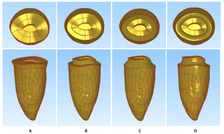
1.2.2 不同边缘设计形式的下颌第1前磨牙髓腔固位冠修复三维数字模型的建立在建立的下颌第一前磨牙三维数字模型基础上,使用PRO/E 2001(Home Page, USA)软件模拟髓腔固位冠修复的牙体预备[3-4, 11](图 1):于釉牙骨质界上1.5 mm截冠,髓腔固位形轴壁外展2~5°,深度5 mm,根管口以下1 mm的牙胶被流动树脂替代。设计一种无冠边缘形式模型:平面对接式边缘,颈缘线位于釉牙骨质界上1.5 mm(图 1A)。设计3种冠边缘形式模型:直角肩台(图 1B)、135°肩台(图 1C)、凹面形边缘(图 1D),颈缘位于釉牙骨质界处,肩台宽1 mm,牙本质肩领高度1.5 mm。

|
图 1 髓腔固位冠修复4种边缘设计模型 Figure 1 Models for endocrown restoration and the 4 marginal designs. The left side was the lingual portion and right was the buccal portion of the teeth. A: Flat margin; B: 90° shoulder; C: 135° shoulder; D: Chamfer shoulder. |
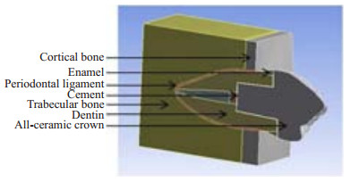
将模型导入ANSYS R 15.0(Swanson Analysis, Houston, PA, USA)软件中建立有限元模型(含牙槽骨)(图 2)。将模型划分为四面体网格。

|
图 2 髓腔固位冠修复含边界条件图 Figure 2 Model for endocrown restoration with boundary conditions. |
假设所有材料均为连续、均质、各向同性的线性弹性体(牙骨质和牙本质弹性模量极为接近,并且牙骨质较薄,故简化为同一物质)[12]。材料受力后的变形为小变形,受力时模型各界面均不产生相对滑动。牙槽骨的颊舌面及底部完全固定,6个方向位移均为0[4]。各部分的弹性模量和泊松比见表 1[4, 13-15]。
| 表 1 牙体组织和修复材料的力学参数 Table 1 Mechanical parameters of tooth tissue and restoration materials |
加载位置为牙齿长轴与牙合面交点;加载力值为100N [4, 9, 16-17];加载方向为与牙齿长轴平行、与牙齿长轴成45° [16-17]。
1.5 分析指标计算不同边缘设计形式的牙齿、修复体在不同加载条件下颈部的最大Von Mises应力值和应力分布情况。
2 结果模型的单元数和节点数如表 2所示。
| 表 2 4种边缘形式模型的节点数与单元数 Table 2 Number of nodes and elements for the 4 marginal designs |
采用有限元软件中几何模型的可视化彩色图像功能显示颈部的Von Mises应力分布情况,如图 3~6所示。

|
图 3 平行牙齿长轴加载4种边缘形式修复体的Von Mises应力分布云图 Figure 3 Von Mises stress distribution nephogram for restoration under the load parallel to the long axis of tooth. The left side was the lingual portion and right was the buccal portion of the teeth. A: Flat margin; B: 90° shoulder; C: 135° shoulder; D: Chamfer shoulder. |

|
图 4 平行牙齿长轴加载4种边缘形式牙体组织的Von Mises应力分布云图 Figure 4 Von Mises stress distribution nephogram for tooth tissue under the load parallel to the long axis of tooth. The left side was the lingual portion and right was the buccal portion of the teeth. A: Flat margin; B: 90° shoulder; C: 135° shoulder; D: Chamfer shoulder. |

|
图 5 与牙齿长轴成45°加载4种边缘形式修复体的Von Mises应力分布云图 Figure 5 Von Mises stress distribution nephogram for restoration under the load at 45° to the long axis of the tooth. The left side was the lingual portion and right was the buccal portion of the teeth. A: Flat margin; B: 90° shoulder; C: 135° shoulder; D: Chamfer shoulder. |

|
图 6 与牙齿长轴成45°加载4种边缘形式牙体组织的Von Mises应力分布云图 Figure 6 Von Mises stress distribution nephogram for tooth tissue under the load at 45° to the long axis of the tooth. The left side was the lingual portion and right was the buccal portion of the teeth. A: Flat margin; B: 90° shoulder; C: 135° shoulder; D: Chamfer shoulder. |
施加与牙齿长轴平行的载荷,4种边缘形式在牙体组织和修复体颈部的舌侧边缘出现应力集中,平面对接式边缘和直角肩台的颈部应力分布要比135°肩台和凹面形边缘的应力分布均匀(图 3,4)。
2.2 与牙齿长轴成45°加载时的应力分布情况施加与牙齿长轴成45°的载荷,牙齿和修复体的最大Von Mises应力值均明显高于与牙齿长轴平行加载情况下的应力值。4种边缘形式的牙体组织和修复体的颈部应力主要集中在颊侧边缘,平面对接式边缘的颈部应力分布较其他3种边缘形式要均匀连续(图 5,6)。
2.3 加载后的应力值无论施加与牙齿长轴平行还是与牙齿长轴成45°的载荷,平面对接式边缘的牙齿和修复体颈部的应力值均最小,直角肩台的颈部应力水平要低于135°肩台和凹面形边缘。修复体颈部的应力值要大于牙齿颈部的应力值(图 7)。

|
图 7 不同边缘形式的牙齿与修复体颈部的Von Mises应力峰值比较 Figure 7 Comparison of the maximum Von Mises values on the cervical region of tooth and the restoration in different marginal designs. |
平面对接式边缘牙釉质的应力水平高于牙本质(图 4A、图 6A)。
3 讨论修复体的边缘设计会影响基牙和修复体的边缘强度与咬合力作用下边缘部位的应力分布。本研究的结果显示在平行牙齿长轴加载的情况下,4种边缘形式在牙体组织和修复体颈部的舌侧边缘出现应力集中;施加与牙齿长轴成45°的载荷,4种边缘形式的应力主要集中在牙体组织和修复体颈部的颊侧边缘。牙颈部的应力集中是牙齿承受咬合力量时的正常反应[6]。平行牙齿长轴加载时,舌侧颈部应力集中可能与下颌前磨牙牙冠舌倾有关,模拟施加的斜向颊侧的载荷可能是导致斜向加载情况下颊侧颈部应力集中的原因。Hassan等[18]通过研究不同边缘形式的高嵌体修复后颈部的应力分布情况发现直角肩台颈部的应力值低于凹面形边缘设计,本研究的结果与其一致,这可能与直角肩台修复体颈部的陶瓷厚度高于凹面形边缘有关。135°肩台设计由于采用斜面设计形式使得应力的分布趋于缓和,但是在牙齿颈部应力集中区域的应力值最高,这可能会导致该部位的牙体组织发生应力疲劳,长期作用可能会导致折裂的发生。凹面形边缘修复体颈部的应力值最高可能与此处陶瓷厚度较薄有关,咬合力作用下亦有可能导致修复体颈部瓷裂以及崩瓷的发生。
目前有关髓腔固位冠的研究文献中,修复体多设计为平面对接式边缘[2-5]。本研究发现采用平面对接式边缘设计,牙齿和修复体颈部的最大Von Mises应力值要低于3种肩台设计形式,且应力分布要更为均匀连续。肩台的设计要磨除更多的健康牙体组织,而平面对接式边缘最大限度地保存了余留牙体组织,单一平面的预备避免了多重平面交界部位对应力传递分散的不良影响使得应力分布情况更为有利。髓腔固位冠的平面对接式边缘设计保留了釉牙骨质界以上1.5 mm的牙釉质结构,由于牙釉质与牙本质弹性模量的不同使得这两者的应力水平存在差异。从图 4A和图 6A也可以看出牙釉质的应力水平明显高于牙本质,这是否会导致牙釉质在咬合力量的长期作用下发生折裂剥脱,继而引起边缘微渗漏、继发龋而最终导致修复的失败,有待进一步的研究。
无论是施加与牙齿长轴平行还是与牙齿长轴成45°的载荷,修复体颈部的应力值都要高于牙齿颈部的应力值。这可能与修复体与牙体组织之间的粘接剂吸收分散了部分应力有关。
当牙齿-修复体复合体承受斜向负载的作用时,颈部的应力水平要高于承受与牙齿长轴平行的负载时的情况,咀嚼运动产生的斜向作用力是威胁修复体使用寿命的重要因素,因此建议临床上进行髓腔固位冠修复时降低牙尖斜度以减少侧向力对基牙牙体组织和修复体的不良影响。
平面对接式边缘的颈部应力峰值要低于有肩台设计形式者,而肩台设计形式中直角肩台的应力峰值要小于135°肩台和凹面形边缘形式。本研究结果从应力分析角度说明髓腔固位冠修复平面对接式边缘要优于3种肩台设计形式,但总体来说各最大Von Mises应力值之间的差距不大。而具体选择何种边缘设计形式还应再综合边缘适合性、微渗漏、抗折强度研究结果以及长期临床随访观察而得出结论。
| [1] |
Lander E, Dietschi D. Endocrown: a clinical report[J].
Quintessence Int,2008, 39 (2) : 99-106.
( 0) 0)
|
| [2] |
Biacchi GR, Basting RT. Comparison of fracture strength of endocrowns and glass fiber post-retained conventional crowns[J].
Oper Dent,2012, 37 (2) : 130-6.
DOI: 10.2341/11-105-L. ( 0) 0)
|
| [3] |
Chang CY, Kuo JS. Fracture resistance and failure modes of CEREC endo-crowns and conventional post and core-supported CEREC crowns[J].
J Dent Sci,2009, 4 (3) : 110-7.
DOI: 10.1016/S1991-7902(09)60016-7. ( 0) 0)
|
| [4] |
Lin CL, Chang YH, Chang CY, et al. Finite element and Weibull analyses to estimate failure risks in the ceramic endocrown and classical crown for endodontically treated maxillary premolar[J].
Eur J Oral Sci,2010, 118 : 87-93.
DOI: 10.1111/eos.2010.118.issue-1. ( 0) 0)
|
| [5] |
HM ED, RN HA, Platt JA. Fracture Resistance and Microleakage of Endocrowns Utilizing Three CAD-CAM Blocks[J].
Oper Dent,2015, 40 (2) : 201-10.
DOI: 10.2341/13-143-L. ( 0) 0)
|
| [6] |
Kierklo A, Tribillo R, Walendziuk A, et al. The influence of cervical lesion on the stress state and strength of tooth with occlusal restoration: a numerical model study[J].
Rocz A kad Med Bialymst,2002, 47 : 95-104.
( 0) 0)
|
| [7] |
白保晶, 曹文, 张振庭. 存留壁和髓室底厚度对磨牙桩冠修复的应力分析[J].
口腔颌面修复学杂志,2010, 11 (2) : 81-3.
( 0) 0)
|
| [8] |
Jelena Juloski, Ivana Radovic, Cecilia Goracci, et al. Vulicevic, Marco Ferrari[J].
Ferrule Effect: A Literature Review,2012, 38 (1) : 11-9.
( 0) 0)
|
| [9] |
Pierrisnard L, Bohin F, Renault P, et al. Coronoradicular reconstruction of pulpless teeth: a mechanical study using finite element analysis[J].
J Prosthet Dent,2002, 88 (4) : 442-8.
DOI: 10.1067/mpr.2002.128376. ( 0) 0)
|
| [10] |
王惠芸. 我国人牙的测量和统计[J].
中华口腔科杂志,1959, 7 (3) : 149-55.
( 0) 0)
|
| [11] |
李鸿波, 刘洪臣. 老年低牙合龈距磨牙牙体缺损的髓腔固位冠修复[J].
中华老年口腔医学杂志,2012, 10 (4) : 251-3.
( 0) 0)
|
| [12] |
缪羽, 吴凯敏, 于蕴之, 等. 不同边缘设计的聚合瓷高嵌体修复后牙体组织的有限元应力分析[J].
现代口腔医学杂志,2011, 25 (6) : 440-5.
( 0) 0)
|
| [13] |
Lin CL, Chang YH, Pa CA. Estimation of the risk of failure for an endodontically treated maxillary premolar with MODP preparation and CAD/CAM ceramic restorations[J].
J Endod,2009, 35 (10) : 1391-5.
DOI: 10.1016/j.joen.2009.06.020. ( 0) 0)
|
| [14] |
Lin CL, Chang YH, Liu PR. Multi-factorial analysis of a cusp-replacing adhesive premolar restoration: a finite element study[J].
J Dent,2008, 36 : 194-203.
DOI: 10.1016/j.jdent.2007.11.016. ( 0) 0)
|
| [15] |
张丹, 白保晶, 张振庭. 不同垫底材料对全瓷嵌体修复应力分布影响的三维有限元研究[J].
北京口腔医学,2011, 19 (2) : 82-4.
( 0) 0)
|
| [16] |
Soares PV, Souza LV, Veríssimo C, et al. Effect of root morphology on biomechanical behaviour of premolars associated with abfraction lesions and different loading types[J].
J Oral Rehabil,2014, 41 (2) : 108-14.
DOI: 10.1111/joor.12113. ( 0) 0)
|
| [17] |
Palamara JE, Palamara D, Messer HH, et al. Tooth morphology and characteristics of non-carious cervical lesions[J].
J Dent,2006, 34 (3) : 185-94.
DOI: 10.1016/j.jdent.2005.05.005. ( 0) 0)
|
| [18] |
Abu Hassan, Abu Hammad, Harrison. Stress distribution associated with loaded ceramic onlay restorations with different designs of marginal preparation.An FEA study[J].
J Oral Rehabil,2000, 27 (4) : 294-8.
DOI: 10.1046/j.1365-2842.2000.00517.x. ( 0) 0)
|
 2016, Vol. 36
