2. 甘肃省骨关节疾病研究重点实验室 ;
3. 兰州大学第二医院血液科,甘肃 兰州730030
2. Orthopedics Key Laboratory of Gansu Province ;
3. Department of Hematology, the Second Hospital of Lanzhou University, Lanzhou 730030, China
膝关节骨性关节炎(osteoarthritis,OA)是由于关节软骨破坏,骨赘大量形成,最终导致关节间隙狭窄的一种疾病,好发于老年人,女性多于男性,会导致严重的关节疼痛甚至丧失运动能力。OA没有较好的治疗方法,轻度病人常保守治疗,严重病人常采用膝关节置换。鉴于OA严重的后果及昂贵的治疗费用,应加强对高危人群的预防治疗。
Wada等[1]和León等[2]研究发现OA患者由于髁间窝内骨赘增生从而会导致髁间窝狭窄。为了准确评价髁间窝狭窄,人们提出了髁间窝宽度指数(notch widthindex,NWI)的概念,NWI=髁间窝宽度/股骨髁宽度,为相对指标,能降低测量误差,平衡个体差异,较准确的反应髁间窝大小[3-4]。Souryal[4]、Keays[5]等在X线,van Eck等[6]在CT 图像上分别测量了冠状位序列的NWI,Domzalski[7]、Sonnery-Cottet[8]、Park[9]等在MRI图像上测量了冠状位NWI,Hoteya等[10]为了更好的说明前交叉韧带(ACL)止点处髁间窝宽度,测量了冠状位ACL股骨止点处图像的NWI-A及其后序列的NWI-P。但以上学者所得数据均有较大差异,而且到目前为止还没有明确的针对OA病人的冠状位NWI界值来确定髁间窝狭窄的标准。因此本研究中我们选取诊断明确的OA病人为研究对象,采用髁间窝软组织成像较好的MRI图像进行研究,统一了测量标准,并增加了样本量。
1 资料与方法 1.1 研究对象我们回顾性分析了2011年1月~2014年12月在兰州大学第二医院拍摄膝关节核磁的1107例门诊和住院病人的病例资料。纳入标准:(1)中老年人(≥45 岁);(2)依据中华医学会风湿病学分会OA诊断标准[11]确诊为膝OA,且无明确外伤或手术史,并选取膝关节完全正常的健康人作为对照;(3)MRI图像清晰,序列完整。排除标准:(1)<45岁;(2)有膝关节外伤或手术史造成髁间窝破坏;(3)诊断不明确;(4)MRI图像不清晰或序列不完整。本研究在伦理委员会的监督下进行,并取得病人的知情同意。依据X线平片和MRI图像,采用K-L评分标准[12-13]分组,0级:正常;Ⅰ级:关节间隙可疑变窄,可能有骨赘;Ⅱ级:有明显骨赘,关节间隙可疑变窄;Ⅲ级:中等量骨赘,关节间隙变窄较明显,有硬化性改变;Ⅳ级:大量骨赘,关节间隙明显变窄,严重硬化性病变及明显畸形。Ⅰ、Ⅱ级为轻度OA,Ⅲ、Ⅳ级为中重度OA。并与Lequesne制定的膝OA严重指数(ISOA)评分[14]相对照,1~4分为轻度OA,大于4分为中重度OA。
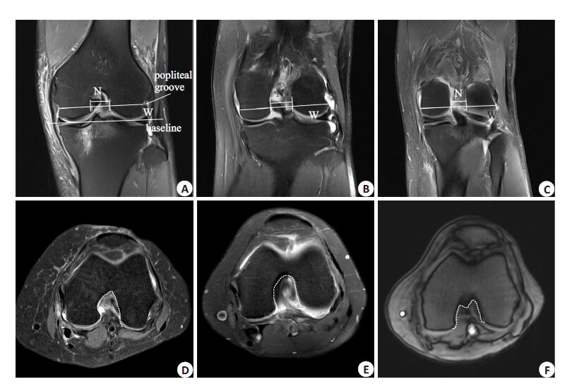
1.2 测量方法采用phillips 3.0-T MRI扫描仪,患者仰卧位,膝关节屈曲10°~15°,线圈中心位于髌骨下极水平。扫面序列:冠状位和轴位,层厚3 mm。选取冠状位序列中显示髁间窝最清楚的图层,用RadiAnt DICOM Viewer 1.9软件进行测量,髁间窝宽度指数(NWI)=髁间窝宽度(N)/股骨髁宽度(W),NWI测量参照Domzalski等[7]的测量标准,NWI-A、NWI-P的测量参照Hoteya等[10]的测量标准(图 1)。选取轴位序列中显示髁间窝最清楚的图层,参照Al-Saeed等[15]的标准进行分型,分为A、U、W三型(图 1)。由3人独立完成,数据取平均值,分型不一致时协商解决。

|
图 1 冠状位不同层面NWI的测量及轴位髁间窝分型 Figure 1 Measurement of NWI at different levels in coronal MR images and intercondylar notch shape typing on axial |
数据用SPSS 22.0 软件进行统计分析,结果以均数±标准差的形式表示。不同层面不同分组间NWI的比较采用单因素方差分析,率的比较采用χ2检验,相关性检验采用Spearman非参数检验。P<0.05为差异有统计学意义。
2 结果 2.1 一般资料如表 1,收集到符合条件的病历有健康组70例,轻度OA组42例,中重度OA组37例。
| 表 1 不同分组的性别、年龄 Table 1 Gender and age (year) in different groups |
如表 2,在冠状位上,轻度OA 组NWI、NWI-A、NWI-P 与健康组相近,无统计学差异(F=0.221/0.064/0.033,P=0.639/0.801/0.856)。中重度OA 组NWI、NWI-A、NWI-P均明显小于健康组,差异有统计学意义(F=9.876/10.010/11.783,P=0.002/0.002/0.001)。中重度OA组各层面NWI均明显小于轻度OA组差异有统计学意义(F=6.657/7.361/9.873,P=0.012/0.008/0.002)。
| 表 2 不同层面不同分组间NWI的比较 Table 2 Comparison of NWI in different sequences between different groups |
由于中重度OA组与健康组各层面NWI均有显著差异,因此做这两组之间的Spearman 相关性检验,NWI、NWI-A、NWI-P 与中重度OA 的相关系数为r=-0.251、-0.276、-0.302,说明NWI、NWI-A、NWI-P 均与是否患中重度OA有显著相关性(P<0.01)。
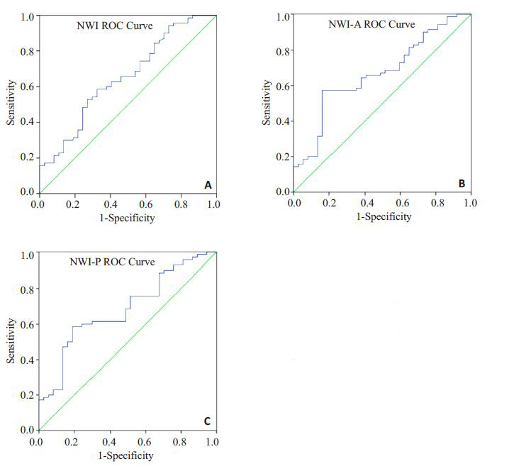
2.4 ROC曲线中重度OA组与健康组得到NWI、NWI-A、NWI-P的ROC 曲线,NWI 的曲线下面积为分别为0.652、0.668、0.683,最佳界值为约登指数(敏感性+特异性-1)最大点对应的值,得出最佳界值为分别为0.248、0.255、0.266(图 2)。

|
图 2 不同层面NWI的ROC曲线 Figure 2 ROC curves of different sequences in moderate to severe OA and healthy groups |
在健康和中重度OA 组,分别以NWI<0.248、NWI-A<0.256、NWI-P<0.266 作为髁间窝狭窄的依据。在健康组中有W型2 人,其NWI、NWI-A、NWI-P均比界值大,被分到髁间窝非狭窄组。因W型较少且髁间窝宽度较宽,NWI较大,与U型接近,所以把W型合并为U型。在冠状位NWI层面,A型在髁间窝狭窄组中的比例77.4%,大于非狭窄组55.6%,U型在髁间窝狭窄组中的比例22.6%,小于非狭窄组44.4%,有统计学差异(P=0.017)。在冠状位NWI-A层面,A型在髁间窝狭窄组中的比例81.7%,远大于非狭窄组46.8%,U型在髁间窝狭窄组中的比例18.3%,远小于非狭窄组53.2%,有显著统计学差异(P<0.05)。在冠状位NWI-P层面,A型在髁间窝狭窄组中的比例83.3%,远大于非狭窄组44.7%,U型在髁间窝狭窄组中的比例16.7%,远小于非狭窄组55.3%,有显著统计学差异(P<0.01,表 3)。
| 表 3 不同层面髁间窝狭窄与非狭窄组髁间窝分型的比较 Table 3 Comparison of intercondylar notch type between intercondylar notch narrowing and non-narrowing groups in different sequences |
众多学者对OA 病人的髁间窝进行了研究,Shepstone等[16]发现,健康人髁间窝内侧壁比较凹而OA患者内侧壁比较直甚至凸出,这种形态差异可能会减小髁间窝宽度。Wada等[1]和León等[2]发现OA患者髁间窝内可出现各种类型的骨赘增生,其中以顶端和开口处增生最常见,这种增生必然会减小髁间窝宽度,导致NWI减小,形成髁间窝狭窄。我们研究发现中重度OA患者NWI、NWI-A、NWI-P 均明显小于健康人和轻度OA患者(P<0.05),说明中重度OA患者有明显的髁间窝狭窄,与以上学者结论一致。而轻度OA患者以上指标均与健康人接近,说明轻度OA患者髁间窝狭窄不明显。
有关冠状位不同层面髁间窝狭窄NWI界值的确定尚有很大争议。Tillman等[17]对200例骨骼标本进行测量发现NWI与性别和人种有关。Souryal等[4]在X线上测量NWI,得出健康人冠状位NWI=0.2338,NWI<0.2为髁间窝狭窄。Keays等[5]发现X线图像会出现重影,正常人的NWI在前面为0.24,后面为0.30,前面NWI<0.2,后面NWI<0.24可认为髁间窝狭窄。van Eck等[6]发现在CT 上,不同拍摄方法所测得的NWI各不相同。Sonnery-Cottet等[8]对髁间窝冠状位MRI图像进行测量得出健康人NWI=0.27±0.02,ACL损伤患者的NWI=0.22±0.02,并认为NWI<0.21 是ACL 损伤的危险因素。Park 等[9]测量健康女性的冠状位MRI 图像得出NWI=0.25±0.02。Domzalski 等[18]测量了76 例未成年人髁间窝冠状位MRI图像,发现15-17 岁未成年人的NWI=0.254±0.032,且随着年龄的增加而减小。Hoteya等[10]为了更准确的测量ACL股骨止点处的NWI,选取ACL股骨止点层面(A)和之后的一个层面(P),得出NWI-A、NWI-P,健康人的NWI-A=0.266 ± 0.030,NWI-P=0.273±0.033,并认为小于0.25为髁间窝狭窄。我们得出健康人NWI=0.252±0.019,NWI-A=0.261±0.024,NWI-P=0.271±0.026,与上述研究结果均存在一定差异,可能与人种、拍摄体位、摄像工具、测量方法、测量平面等众多因素有关。我们以ROC曲线约登指数最大点处对应的值为最佳界值,得出冠状位NWI<0.248、NWI-A<0.256、NWI-P<0.266时为髁间窝狭窄的结论,同时发现以上界值均与健康组95%可信区间下限接近,说明ROC曲线所得界值合理。
对于髁间窝分型,Anderson等[3]对CT图像研究发现U型不宜发生狭窄,而侧壁出现波形的髁间窝容易狭窄,van Eck等[19]在关节镜下发现,A型的髁间窝宽度在全段都比U型小。Sutton等[20]发现女性的髁间窝宽度比男性小,且A型较多,说明A型髁间窝容易狭窄。Al-Saeed 等[15]发现A型髁间窝较窄,且髁间窝形状是ACL损伤的一个危险因素,A型易发生ACL损伤。我们发现在NWI小于界值的髁间窝狭窄组中A型所占的比例均明显高于非狭窄组,说明A型髁间窝容易发生狭窄,与上述结论一致。
大量研究证实髁间窝狭窄是ACL损伤的独立危险因素,ACL损伤患者的NWI比健康人小[4-5, 7-10, 21]。Stijak[22]、Dienst等[23]认为髁间窝狭窄患者的ACL横截面积较小、强度较弱,生物学性能较差,易发生损伤。Everhart等[24]发现ACL在膝关节运动时可与狭窄的髁间窝发生碰撞,产生磨损,导致ACL损伤。ACL损伤会改变下肢力线,破坏关节软骨,产生关节炎症,导致骨赘增生,骨赘增生后会进一步加重关节磨损,最终导致OA[25]。即使在ACL损伤后进行重建仍不能有效阻止OA的发生[26]。Hernigou[27]和Stein等[28]发现在OA患者中髁间窝宽度或NWI越小,越容易发生ACL损伤。鉴于ACL损伤与OA的这种关系,对髁间窝狭窄的健康人群提前进行预防治疗,不仅能预防ACL损伤,还能预防OA。
综上所述,中重度OA患者存在明显的髁间窝狭窄,冠状位NWI、NWI-A、NWI-P均明显比健康人小,在MRI图像上NWI<0.248、NWI-A<0.256、NWI-P<0.266可以作为髁间窝狭窄的依据,A型的髁间窝比U型更容易发生狭窄,对髁间窝狭窄者采取措施提前预防对OA的发生可起到一定的防治作用。可采取的预防措施主要有定时体检、及时提供防治建议、减轻体质量、限制剧烈体育运动等。但由于本研究仅选取了一家医院的病例进行研究,病例取材范围较窄,样本量不够大,且未考虑是否并发膝关节韧带损伤、半月板损伤等疾病及既往是否关节腔内注射玻璃酸钠等用药情况,因此相关结论需要用多中心大样本研究,全面考虑可能的干扰因素,进一步深入探讨。
| [1] | Wada M, Tatsuo H, Baba H, et al. Femoral intercondylar notchmeasurements in osteoarthritic knees[J]. Rheumatology (Oxford),1999, 38 (6) : 554-8. DOI: 10.1093/rheumatology/38.6.554. |
| [2] | León HO, Blanco CE, Guthrie TB, et al. Intercondylar notch stenosis in degenerative arthritis of the knee[J]. Arthroscopy,2005, 21 (3) : 294-302. DOI: 10.1016/j.arthro.2004.11.019. |
| [3] | Anderson AF, Lipscomb AB, Liudahl KJ, et al. Analysis of the intercondylar notch by computed tomography[J]. Am J Sports Med,1988, 15 (6) : 547-52. |
| [4] | Souryal TO, Moore HA, Evans JP. Bilaterality in anterior cruciate ligament injuries: associated intercondylar notch stenosis[J]. Am J Sports Med,1988, 16 (5) : 449-54. DOI: 10.1177/036354658801600504. |
| [5] | Keays SL, Keays R, Newcombe PA. Femoral intercondylar notch width size: a comparison between siblings with and without anterior cruciate ligament injuries[J]. Knee Surg Sports Traumatol Arthrosc,2014 . |
| [6] | Van Eck CF, Martins CA, Lorenz SG, et al. Assessment of correlation between knee notch width index and the three-dimensional notch volume[J]. Knee Surg Sports Traumatol Arthrosc,2010, 18 (9) : 1239-44. DOI: 10.1007/s00167-010-1131-3. |
| [7] | Domzalski M, Grzelak P, Gabos P. Risk factors for Anterior Cruciate Ligament injury in skeletally immature patients: analysis of intercondylar notch width using Magnetic Resonance Imaging[J]. Int Orthop,2010, 34 (5) : 703-7. DOI: 10.1007/s00264-010-0987-7. |
| [8] | Sonnery-Cottet B, Archbold P, Cucurulo T, et al. The influence of the tibial slope and the size of the intercondylar notch on rupture of the anterior cruciate ligament[J]. J Bone Joint Surg Br,2011, 93 (11) : 1475-8. |
| [9] | Park JS, Nam DC, Kim DH, et al. Measurement of knee morphometrics using MRI: A comparative study between ACLInjured and Non-Injured knees[J]. Knee Surg Relat Res,2012, 24 (3) : 180-5. DOI: 10.5792/ksrr.2012.24.3.180. |
| [10] | Hoteya K, Kato Y, Motojima S, et al. Association between intercondylar notch narrowing and bilateral anterior cruciate ligament injuries in athletes[J]. Arch Orthop Trauma Surg,2011, 131 (3) : 371-6. DOI: 10.1007/s00402-010-1254-5. |
| [11] | 中华医学会风湿病学分会. 骨关节炎诊断及治疗指南[J]. 中华风湿病 学杂志,2010, 14 (6) : 416-9. |
| [12] | Kellgren JH, Lawrence JS. Radiological assessment of osteoarthrosis[J]. Ann Rheum Dis,1957, 16 (4) : 494-502. DOI: 10.1136/ard.16.4.494. |
| [13] | Kim HJ, Lee JY, Kim TJ, et al. Association between serum vitamin D status and health-related quality of Life (HRQOL) in an older Korean population with radiographic knee osteoarthritis: data from the Korean National health and nutrition examination survey (2010-2011)[J]. Health Qual Life Outcomes,2015, 13 (1) : 48. DOI: 10.1186/s12955-015-0245-1. |
| [14] | Lequesne M. Indices of severity and disease activity for osteoarthritis[J]. Semin Arthritis Rheum,1991, 20 (Suppl 2) : 48-54. |
| [15] | Al-Saeed O, Brown M, Athyal R, et al. Association of femoral intercondylar notch morphology, width index and the risk of anterior cruciate ligament injury[J]. Knee Surg Sports Traumatol Arthrosc,2013, 21 (3) : 678-82. DOI: 10.1007/s00167-012-2038-y. |
| [16] | Shepstone L, Rogers J, Kirwan JR, et al. Shape of the intercondylar notch of the human femur: a comparison of osteoarthritic and non-osteoarthritic bones from a skeletal sample[J]. Ann Rheum Dis,2001, 60 (10) : 968-73. DOI: 10.1136/ard.60.10.968. |
| [17] | Tillman MD, Smith KR, Bauer JA, et al. Differences in three intercondylar notch geometry indices between males and females: a cadaver study[J]. Knee,2002, 9 (1) : 41-6. DOI: 10.1016/S0968-0160(01)00135-1. |
| [18] | Domzalski ME, Keller MS, Grzelak P, et al. MRI evaluation of the development of intercondylar notch width in children[J]. Surg Radiol Anat,2015, 37 (6) : 609-15. DOI: 10.1007/s00276-015-1433-8. |
| [19] | Van Eck CF, Martins CA, Vyas SM, et al. Femoral intercondylar notch shape and dimensions in ACL-injured patients[J]. Knee Surg Sports Traumatol Arthrosc,2010, 18 (9) : 1257-62. DOI: 10.1007/s00167-010-1135-z. |
| [20] | Sutton KM, Bullock JM. Anterior cruciate ligament rupture: differences between males and females[J]. J Am Acad Orthop Surg,2013, 21 (1) : 41-50. DOI: 10.5435/JAAOS-21-01-41. |
| [21] | Sw?rd P, Kostogiannis I, Roos H. Risk factors for a contralateral anterior cruciate ligament injury[J]. Knee Surg Sports Traumatol Arthrosc,2010, 18 (3) : 277-91. DOI: 10.1007/s00167-009-1026-3. |
| [22] | Stijak L, Bumbasirević M, Kadija M, et al. Morphometric parameters as risk factors for anterior cruciate ligament injuries - a MRI case-control study[J]. Vojnosanitetski Pregled,2014, 71 (3) : 271-6. DOI: 10.2298/VSP1403271S. |
| [23] | Dienst M, Schneider G, Altmeyer K, et al. Correlation of intercondylar notch cross sections to the ACL size: a high resolution Mr tomographic in vivo analysis[J]. Arch Orthop TraumaSurg,2007, 127 (4) : 253-60. DOI: 10.1007/s00402-006-0177-7. |
| [24] | Everhart JS, Flanigan DC, Simon RA. Association of noncontact anterior cruciate ligament injury with presence and thickness of a bony ridge on the anteromedial aspect of the femoral intercondylar notch[J]. Am J Sports Med,2010, 38 (8) : 1667-73. DOI: 10.1177/0363546510367424. |
| [25] | Simon D, Mascarenhas R, Saltzman BM, et al. The relationship between anterior cruciate ligament injury and osteoarthritis of the knee[J]. Adv Orthop,2015 : 928301. |
| [26] | Barenius B, Ponzer S, Shalabi A, et al. Increased risk of osteoarthritis after anterior cruciate ligament Reconstruction: a 14-year follow-up study of a randomized controlled trial[J]. Am J Sports Med,2014, 42 (5) : 1049-57. DOI: 10.1177/0363546514526139. |
| [27] | Hernigou P, Garabedian JM. Intercondylar notch width and the risk for anterior cruciate ligament rupture in the osteoarthritic knee: evaluation by plain radiography and CT scan[J]. Knee,2002, 9 (4) : 313-6. DOI: 10.1016/S0968-0160(02)00053-4. |
| [28] | Stein V, Li L, Guermazi A, et al. The relation of femoral notch stenosis to ACL tears in persons with knee osteoarthritis[J]. Osteoarthritis Cartilage,2010, 18 (2) : 192-9. DOI: 10.1016/j.joca.2009.09.006. |
 2015, Vol. 35

